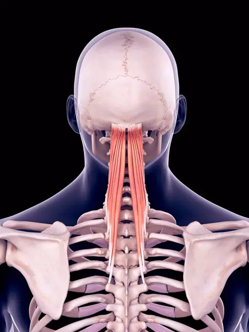
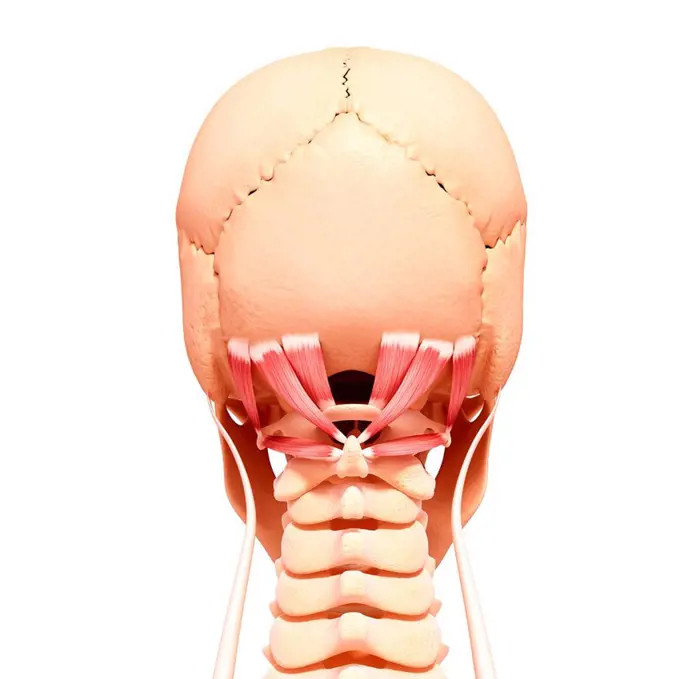
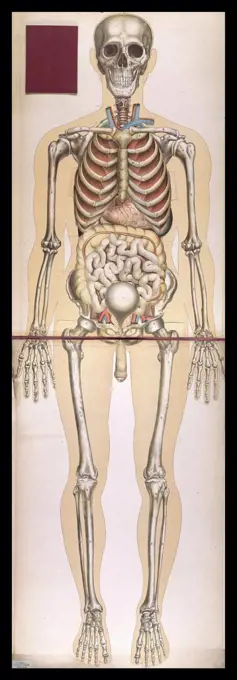
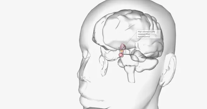
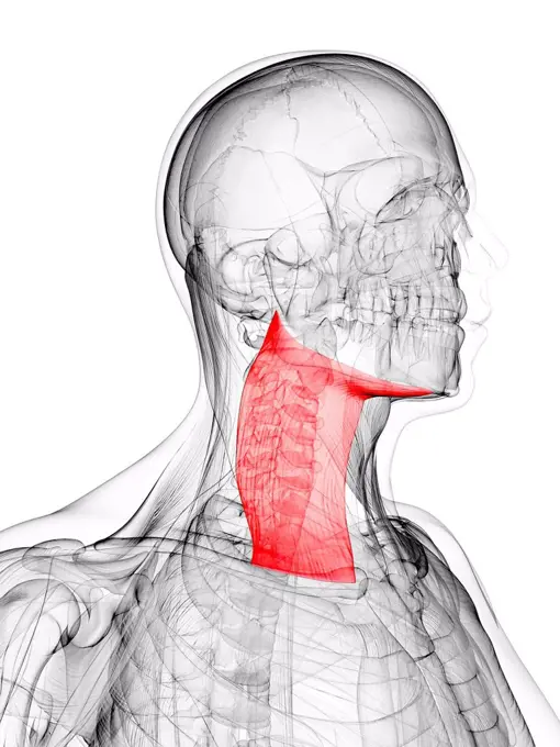
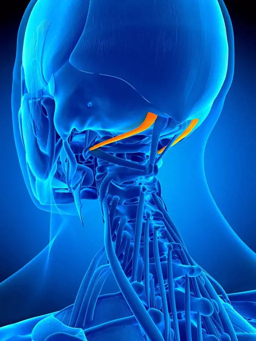
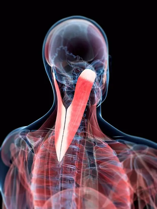
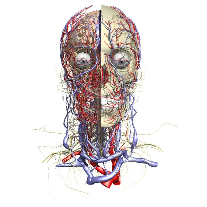
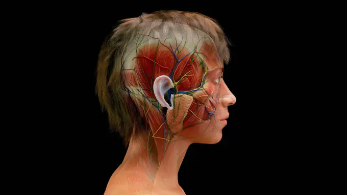
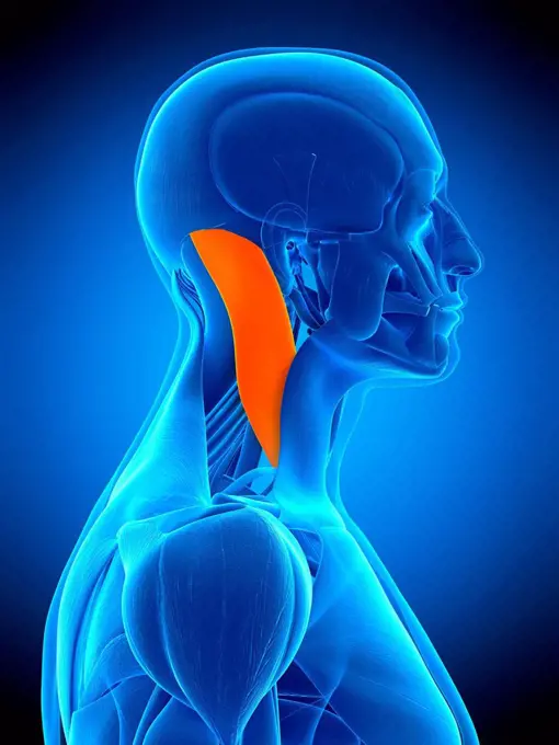
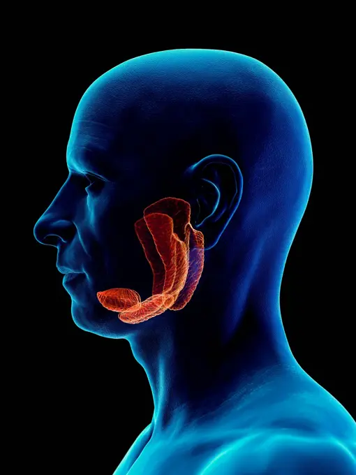
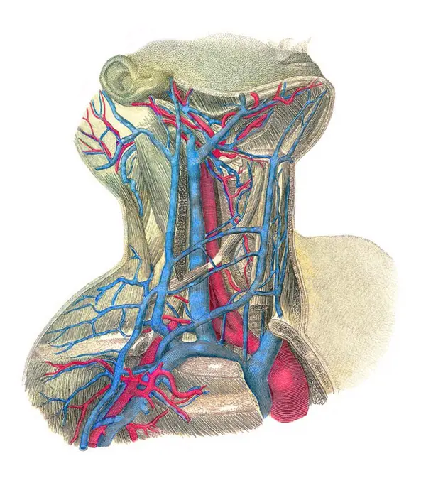
My Boards
View Pictures
  • My Boards

Human Vascular Anatomy Illustrations

Detailed computer artwork and illustrations showcasing the human vascular system, highlighting muscles and arteries in a semi-transparent style.

Illustration of the semispinalis capitis muscles.
275 assets in this story
4128R-26224
Teeth, computer artwork.
4239-20481101
Endorphins released in the hypothalamus travel down the spinal cord to block pain messages.
4128-28682490
Spinal cord anatomy, illustration
4128-15665533
Jaw bone, illustration
4128R-11472960
Human vascular system, computer artwork.
4378-348
Front view of the renal system and it's blood supply. The vertebral column and pelvis is also included.
4128R-19631
Zygomatic bone, computer artwork.
1525-56198341
Human anatomy of a male face, neck and shoulder muscle anatomy, medical image of human anatomy 3D illustration. Anatomy of male head muscular system
4128-30417076
Male lymphatic system, illustration
4128R-11322358
Skull. Coloured X-ray of the skull of a 30 year old male.
4128R-12580727
Human artery, computer illustration.
4269-39015
3D computed tomographic (CT) scan reconstruction of a skull showing an intracranial aneurysm (shaped swelling) located in the arterial circle of Willis (where the basilar artery and internal carotid arteries meet).
4128R-13572823
Illustration of the levator anguli oris muscle.
4128-30420018
Head and neck bones, X-ray
4128R-12578455
Human neck pain, computer illustration.
4128R-34424
Human neck musculature, computer artwork.
1788-39795
Medicine: Human anatomy, muscular system, head. Muscles, nerves, glands. Drawing
1746-21105553
Anatomical illustrationfrom 'Le corps humain et grandeur naturelle', published by Bougle (Paris 1899)
4128-30417068
Male neck organs, illustration
824-63163827
Bone system: human skull with jawbone, eye sockets.
1525-56234228
High volume of CRH released from hypothalamus 3D rendering. High volume of CRH released from hypothalamus
4128R-11544436
Person using a mobile phone, computer illustration.
4128R-26598
Neck muscle. Computer artwork showing the platysma muscle.
4378-1576
A human model showing the deltoid and pectoralis major as well as muscles in the neck and face.
1899-54028620
Illustration of the areas in the brain involved in the process of depression. Lessening in volume and activity of the prefrontal cortex (emotions, planning and social behavior) and the hippocampus (memorization and mood control), hyperactivity of the amygdala (controlling anxiety and hypersensitivity). (Photo by: BSIP/UIG via Getty Images)
4128R-11544126
Human brain, computer illustration.
4128R-13572959
Illustration of the obliquus superior capitis muscle.
4128R-12580761
Human nerves, computer illustration.
4128-30417573
Neck anatomy, illustration
4128-15665394
Thyroid gland, illustration
4378-3504
A sectional view of the human head revealing the anatomy of the ear canal and inner ear anatomy.
4128-30416644
Muscles of the head and torso, illustration
4128R-21970
Neck muscles. Computer artwork of the spelnius capitis muscles.
4128R-33444
Male brain, computer artwork.
4128R-13572968
Illustration of the omohyoid muscle.
4128R-15515818
Illustration of an obese woman's thyroid gland
1525-56183861
The auditory cortex is the sensory system for the sense of hearing. It includes both the sensory organs (the ears) and the auditory parts of the senso 3D illustration. Sound passes through the ear and carries signals to the brain stem through the auditory nerve.
1525-27039752
Realistic head muscles of man isolated in white background. Head muscles of man - 3D render
4128R-33108
Human skeleton, computer artwork.
6188-67688161
Menschliche Kopfmuskeln, Augen und Nasenknorpel. Human anatomy 3d illustration of head muscular system, front view, on white background. Copyright: xZoonar.com/Pixelchaosx 23157709
824-63147253
Depiction of an ischemic stroke, or cerebrovascular accident (CVA).
4128R-12576985
Human face anatomy, computer illustration.
4378-580
Rear view of the larynx showing it's structure in relation to the trachea.
4239-69152021
Medical concept shows swelling of the optic nerve caused by increased intracranial pressure.
4239R-8270
Medical illustration of male facial muscles, front view.
4409-66101
Lymph nodes and vessels in the head, neck and chest. Handcolored steel engraving by Corbie of a drawing by Leveille from Dr. Joseph Nicolas Masse's "Petit Atlas complet d'Anatomie descriptive du Corps Humain," Paris, 1864, published by Mequignon-Marvis. Masse's "Pocket Anatomy of the Human Body" was first published in 1848 and went through many editions.
1899-12802
Neurovascular structures of the head and neck Anterior view
4128-30416546
Female facial muscles, illustration
1788-39553
Medicine: lumbago, lumbar and pelvic regions. Drawing
1848-49200656
Medicine, human anatomy, the circulatory system, blood circulation: partial dissection of the neck, jaw and face of a man, with arteries, blood vessels and veins shown in red and blue. Coloured lithograph by J. Maclise, 1841, 1844, Historic, digitally restored reproduction from a 19th century original
4128R-13572990
Illustration of the sternocleidomastoid muscle.
4269-39016
3D computed tomographic (CT) scan reconstruction of a skull showing an intracranial aneurysm (shaped swelling) located in the arterial circle of Willis (where the basilar artery and internal carotid arteries meet).
4128-30416738
Male salivary gland, illustration
4128R-12577434
Human artery, computer illustration.
4128R-11322976
Human facial muscles and nerves, computer artwork.
4128R-13586527
Illustration of the caudate nucleus of the human brain.
4128R-8454
Blood vessels of the neck, artwork.
1525-56190058
Full anatomy upper and lower teeth 3D illustration. Full anatomy upper and lower teeth
6188-60962754
Human Body Muscular System Head Muscles Orbicularis Oculi Muscle Anatomy 3D Illustration Concept of Human Body Muscular System Head Muscles Orbicularis Oculi Muscle Anatomy Copyright: xZoonar.com/magicminex 21748200
4128-30417072
Male thyroid gland, illustration
4239-69152033
Medical illustration depicting the anatomy of a sagittal section of the human cranium.
4128-19490802
Olfactory system, illustration
4128R-15516683
Illustration of an old woman's larynx
4128R-21856
Medial globus pallidus. Computer artwork of the brain showing the medial or internal globus pallidus red, which is helps to control movement.
1525-56211421
Full anatomy upper and lower teeth 3D illustration. Full anatomy upper and lower teeth
4128R-21868
Third ventricle. Computer artwork of the brain showing the third ventricle red, which is filled with cerebrospinal fluid and cushions the brain.
1746-30002639
Muscles of the head. Pterygoid region.
4128R-13723595
Illustration of the anatomy of the human brain and its veins.
824-63151086
Anatomy, eye
4128R-11297167
Molar tooth, computer artwork.
4128R-13412553
Human sinuses
824-63183445
A cross-section illustration of the left maxillary sinus, showing the mucous membrane and cilia.
4128-30419478
Female ear, illustration
4239-18641785
Medical illustration showing the structure of a motor neuron and sensory neuron.
1848-77189242
3D Illustration Concept of Human Skeleton System Rib Cage Described with Labels Anatomy
PREVIOUS
of 3
NEXT
2401 S. Ervay, Suite 206
Dallas, Texas 75215
United States
Get Started
Free ResearchMy BoardsMy Cart
For Creators
How To License Your ContentContributor PortalFrame of Mind
Resources
API accessPricing
Contact
+1 866 236 0087help@viewpictures.co.uk Contact form
©2026 View Pictures. All Rights Reserved. -A
This site is protected by reCAPTCHA and Google Privacy Policy and Terms of Service apply.