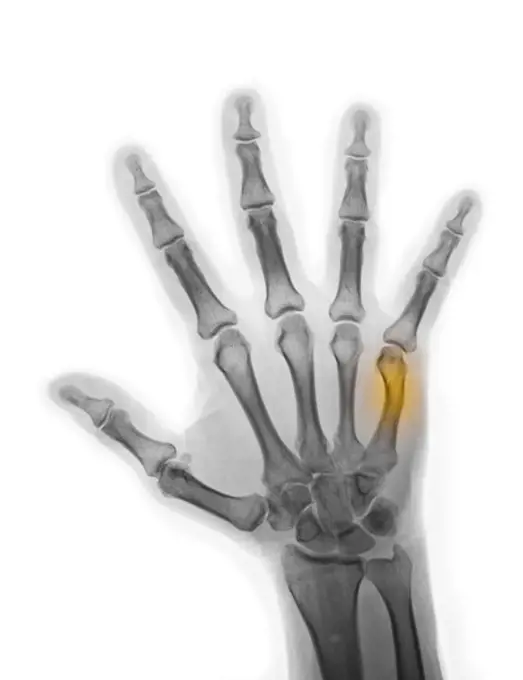
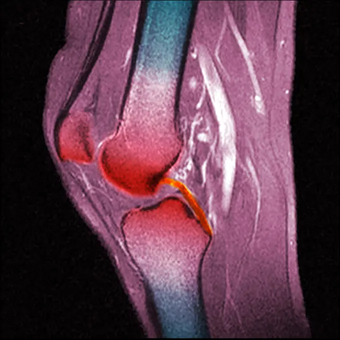
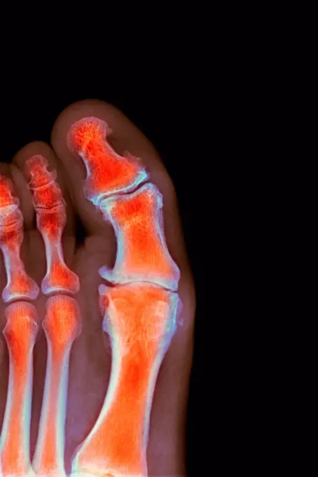
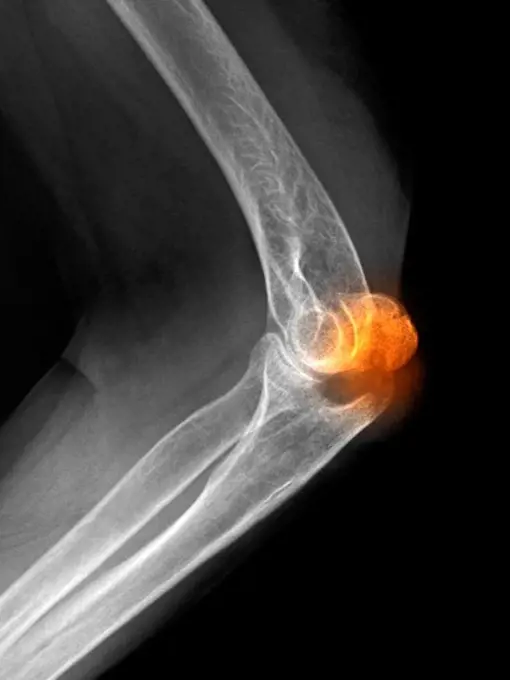
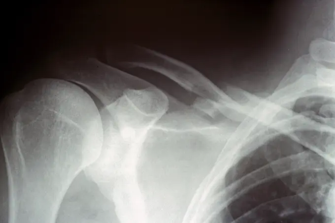
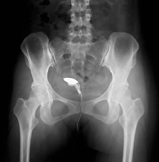
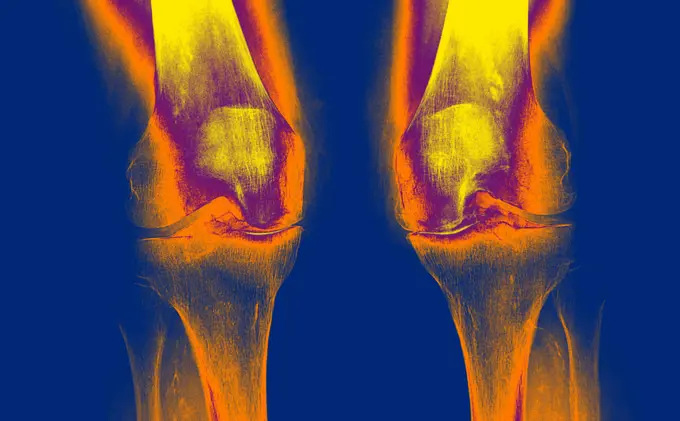
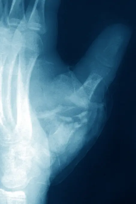
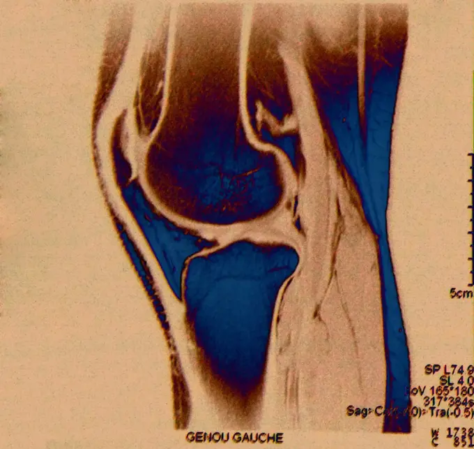
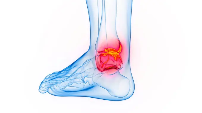
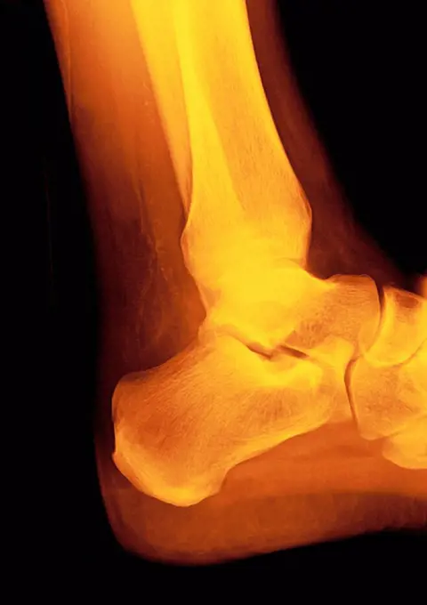
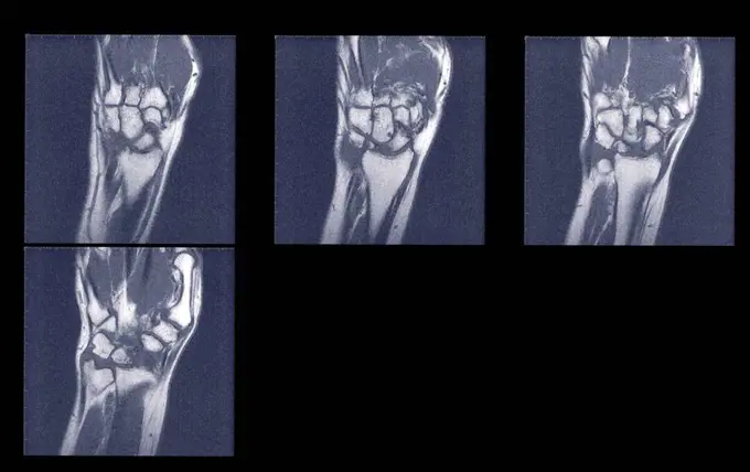
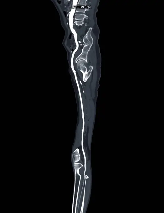
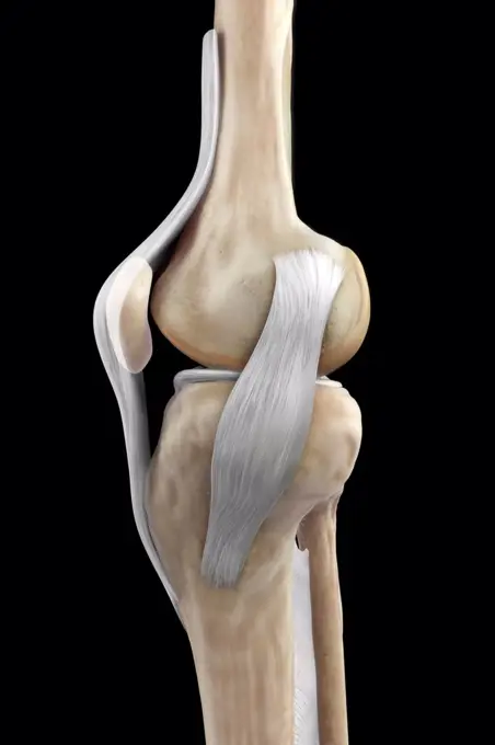
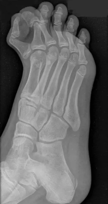
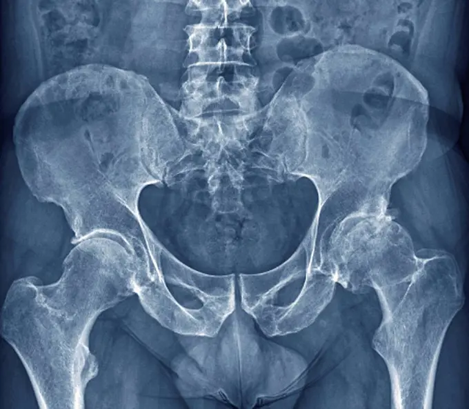
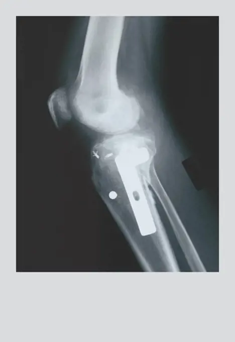
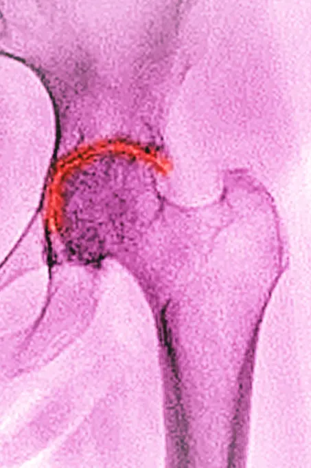
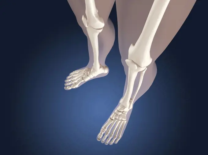
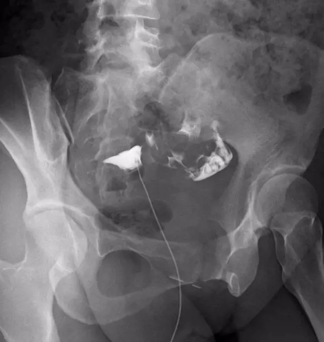
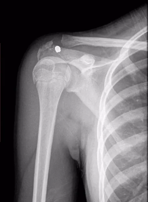
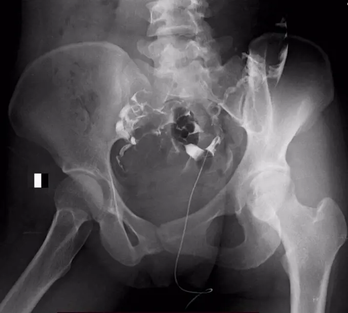
My Boards
View Pictures
  • My Boards

Knee X-Rays & Conditions

Various X-ray images displaying knee health, including normal conditions and degenerative arthritis, with colored effects.

Normal knee. Coloured X_ray of the knee of a 44 year old woman.
218 assets in this story
1841R-111792
4297-1516
Colorized x-ray of the hand of a 57 year old man showing a healing boxer's fracture. This is a break in the fifth metacarpal caused when the hand punches a hard object, such as a wall
6188-56017710
X-ray of a broken leg X-ray of a broken leg Copyright: xZoonar.com/OlgaxPopovax 5153593
4128-19489692
Humerus fracture, X-ray
824-63178651
Trauma-contusion of the right thumb after a shock. No fracture. Bone tearing
1899-86323
Knee MRI
4128-30420001
Fractured pelvis, CT scan
4128-18929599
Smith fracture, X-ray
1841R-111810
4128-30419999
Fractured pelvis, CT scan
1525-56722427
Rheumatoid arthritis (RA) is a chronic, autoimmune, inflammatory condition. 3D rendering. Rheumatoid arthritis (RA) is a chronic, autoimmune, inflammatory condition.
4297-1372
X-ray of a hand holding an iPod
1525-19878752
X-ray picture of human knee joint isolated on white background
4128-19246788
Inflamed ankle, illustration.
824-63198627
Representation of a hallux valgus. Also called bunion, hallux valgus corresponds to an abnormal deviation of the big toe, which causes a deformation of the forefoot at the level of the first metatarsal and the big toe.
4128-28514548
Foot, 3D CT scan
824-63215602
Ankle x-ray.
4128-30420329
Arthritis of the foot, conceptual illustration
4128-30422282
Inflamed ankle, illustration
4128-30420322
Arthritis of the foot, conceptual illustration
4128R-12922503
Hip before hip replacement surgery, X-ray
4128-19357590
X-ray of a patient with osteosarcoma of the femur bone, showing an ill-defined lytic lesion with a wide zone transition and irregular periosteal react...
824-63217798
Normal knee seen on a sagittal section MRI scan.
4128R-15466
Normal shoulder. Coloured X_ray of the healthy left shoulder of a 28 year old patient.
4378-20013910
Right Knee Ligaments
4269-27207
Knee. Sagittal (side view) Magnetic Resonance Image (MRI) of the knee showing the posterior cruciate ligament (in orange). The distal femur (above), the proximal tibia (below), and the patella or knee cap (left) are in red.
4128-19248861
Painful ankle joint, illustration.
1899-54027130
Arteritis seen in an angiogram of the left hand.
824-63188649
Osteosarcoma in the tibia, seen in a frontal MRI scan.
4128-20043136
Hand polydactyly, X-ray
4128R-29805
Hip pain, conceptual computer artwork.
4128R-12922483
Degenerative foot deformation, X-ray
4297-1084
X-ray of an olecranon fracture in an 82 year old woman
4378-20013751
Navicular Bone
4128-V58562403
4128-30420333
Arthritis of the foot, conceptual illustration
6114-50976697
An x-ray of a fracture of the clavicle collar bone.
4128R-13374640
Human knee joint
4128-19489651
Obstructed fallopian tubes, X-ray
4269-25686
Knee arthrosis. X-ray of knees affected by arthosis (green), also called osteoarthritis or gonarthrosis. It is a joint disease that can cause cartilage destruction (seen as a reduction in the joint space), bone erosions, and tendon inflammation.
1899-53508074
In a radiology center, Telos dynamic radiography of a knee, following a ruptured ligament.
1525-26344984
Foot and hand icon of radiography and x-ray concept vector on white background. Phalanges of hand and foot. Leg trauma, pain, osteoporosis illustration.. Foot and hand icon of radiography and x-ray concept vector on white background. Phalanges of hand and foot.
824-63206001
Thumb fracture seen on an x-ray of the right hand.
1899-86317
Knee MRI
1795R-53402
Studio shot of digital tablet with x-ray of human foot
1899-65662233
Rheumatoid arthritis Chronic rheumatic arthritis - Chronic progressive polyarthritis This disease is the cause of ankylosis and bone deformities. This condition preferably affects women, it is responsible for cartilage, bone and ligament destruction, its origin is unknown. Hand x-ray.
4297-1200
Colorized x-ray of the lower leg of an 8 year old girl showing fractures of the tibia and fibula
4129-122
forearm x-ray
6114-50976708
An x-ray of a lower leg fracture tibia and fibula the classic boot top fracture.
4128-V58576524
1525-22162389
Medical Article
4128-30422280
Inflamed ankle, illustration
4128R-3348
Normal ankle joint, X_ray
4269-25503
Rheumatoid polyarthritis. X-ray of hand with Rheumatoid polyarthritis. Rheumatoid arthritis is a chronic joint disease that can cause cartilage destruction, bone erosions, and tendon inflammation and rupture. It can lead to polyarthritis which happens when inflammation spread to multiple joints by.
4128-16073302
Scans of wrists, MRI
4128-28766992
Femoral artery, CT scan
824-63126785
Calcaneum fracture.
4378-5573
Right Knee Ligaments
1525-26870452
Foot Bone Anatomy Vector Illustration
824-63123125
Hip prosthesis, x-ray
1795R-53406
Studio shot of digital tablet with x-ray of human knee
4128-15980491
Hip replacement, computer illustration.
824-63178672
X-ray of the fourth finger of the left hand of a 12-year-old boy. Contusion with no visible bone lesion.
1525R-14840702
Spine diagnostics symbol design
1848-49516151
Mid 20th century X-ray photograph, radiograph showing bursitis in the knee joint, inflammation of one or more bursae of synovial fluid in the body
4128-20239778
Extra toe, X-ray
4128-30420318
Arthritis of the foot, conceptual illustration
4128-18929555
Fibrous dysplasia, X-ray
4128R-26911
Osteoarthritis of the hip. Coloured frontal X-ray (front view) of the pelvis in a 59 year old patient showing osteoarthritis of both hip joints. The b...
1525-24976946
Close-up of xray of hand.
4408-1353
Knee Replacement, Medical - Xray / Radiology
5507-39365198
Gypsum on leg and crutches
1899-53511738
Osteoarthritis of the left hip (coxarthrosis) visualized by a pelvic x-ray of the front. (Photo by: CAVALLINI JAMES/BSIP/UIG)
4128-V58575349
1525-22236364
Medical knee model.
1428R-462
X-ray view of knees
824-63163847
Bone joint of the hip without and with the coxofemoral joint capsule.
1525-22197439
human knee
4128R-12576677
X-rays of hands.
4128-18929611
Blocked fallopian tube, X-ray
4252-4264
Knee articulation
1838-48084394
Before and after medical x-rays of mid-adult woman's surgically-repaired knee
4128-18929595
Gun shot in shoulder, X-ray
4239R-7992
X-ray view of human foot.
1525-26442571
Foot Bone Anatomy Vector Illustration
4128-18929623
Healthy female reproductive system, X-ray
6188-56013067
Watercolor Illustration of firs aid person with injured hand, bandaged arm. Physiotherapy clipart. Watercolor Illustration of firs aid person with injured hand with wrist, bandaged arm. Physiotherapy clipart Copyright: xZoonar.com/LiubovxPonomarchukx 20352773
6188-66528735
Finger osteoarthritis. Left: healthy joint. Middle: moderate osteoarthritis. The cartilage is worn, the cartilaginous debris causes inflammation of the joint. Right: erosive osteoarthritis. The cartilage has completely disappeared, there is disc pinching with bone erosion. The joint is very inflamed and swollen. Bone remodeling occurs: condensation of the bone osteocondensation in places and geodes cavities in the contiguous regions. Osteophytes bony growths appear causing a Heberden s node cyst. Arthrosis hand drawing 016238_057
4128-V58567346
4269-36641
X-ray of a foot affected by bunion (hallux valgus) after surgical treatment.
824-63178656
Trauma-contusion of the right thumb after a shock. No fracture. Bone avulsion from the base P1 of the thumb
1525-56234400
A tear in the anterior cruciate ligament is one of the most common knee injuries. 3D rendering. A tear in the anterior cruciate ligament is one of the most common knee injuries.
4378-5655
Right Knee Ligaments
4128R-13411865
Foot bones
824-63178581
Nose-forehead x-ray of the sinuses of a 42-year-old woman. Normal transparency of all the sinuses of the face. Bilateral hypertrophy of the inferior turbinates.
1525-75819425
Pelvis x ray, healthcare in clinics or hospital, diagnostics and analysis of patient health state. Roentgen scan of human body with bones and joints, examination and care. Vector in flat style. X ray of human pelvis roentgen scan of body vector
824-63178605
Sinus X-ray of a 35-year-old woman. No anomaly noted
4269-24794
Rheumatoid polyarthritis. Scintigram of the arms showing rheumatoid polyarthritis in the elbows and wrists.
4378-5626
Right Knee Ligaments
4128-18929460
Fractured leg in cast, X-ray
PREVIOUS
of 3
NEXT
2401 S. Ervay, Suite 206
Dallas, Texas 75215
United States
Get Started
Free ResearchMy BoardsMy Cart
For Creators
How To License Your ContentContributor PortalFrame of Mind
Resources
API accessPricing
Contact
+1 866 236 0087help@viewpictures.co.uk Contact form
©2026 View Pictures. All Rights Reserved. -B
This site is protected by reCAPTCHA and Google Privacy Policy and Terms of Service apply.