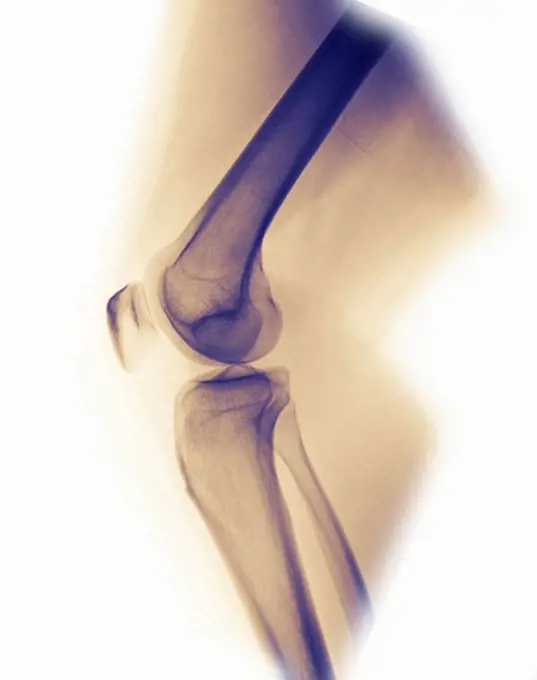
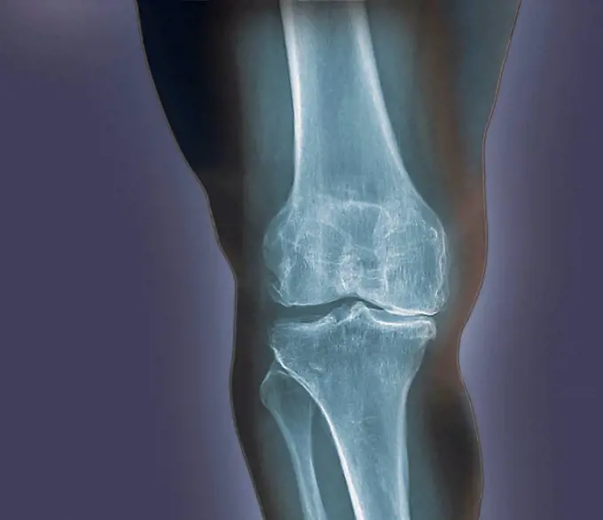
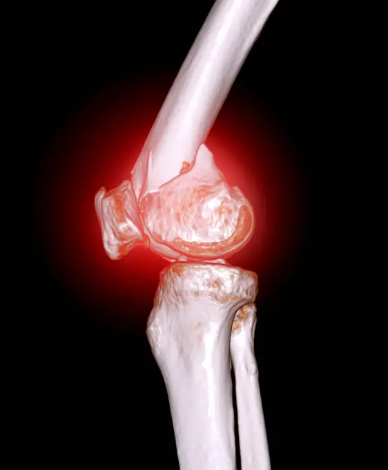
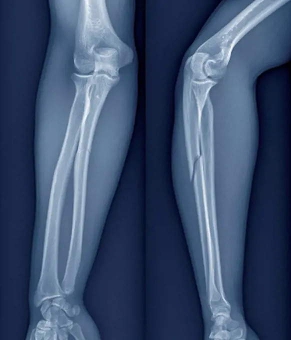
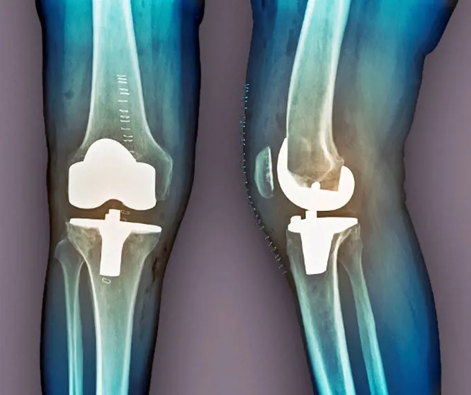
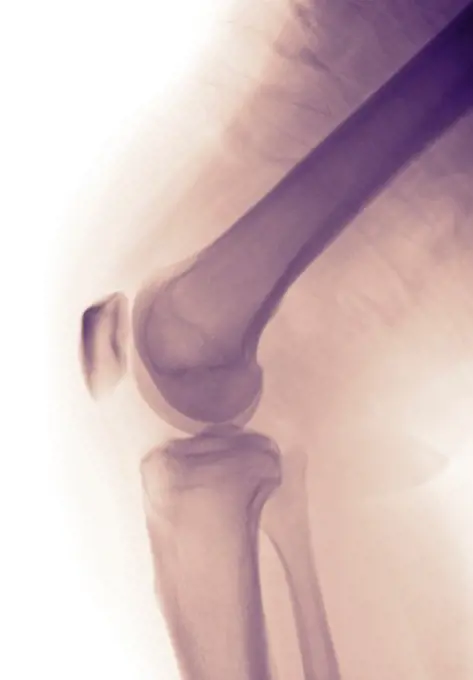
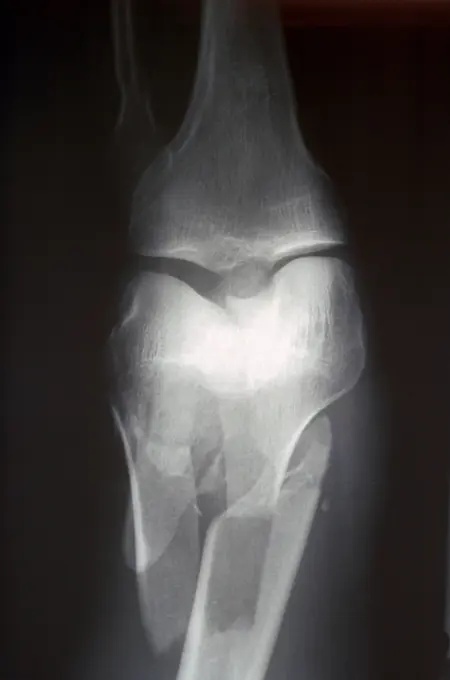
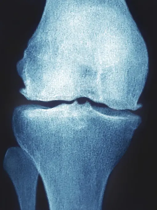
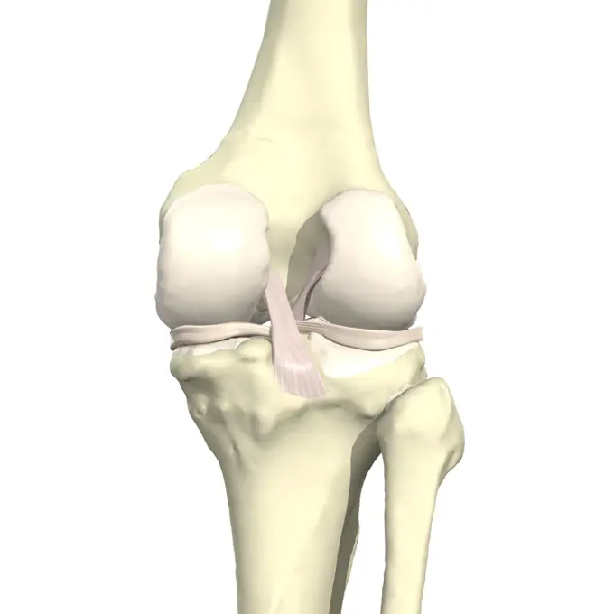
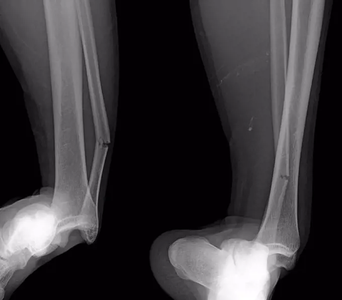
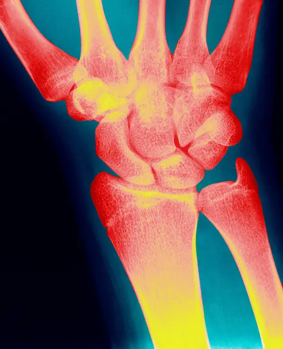
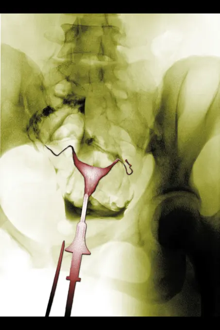
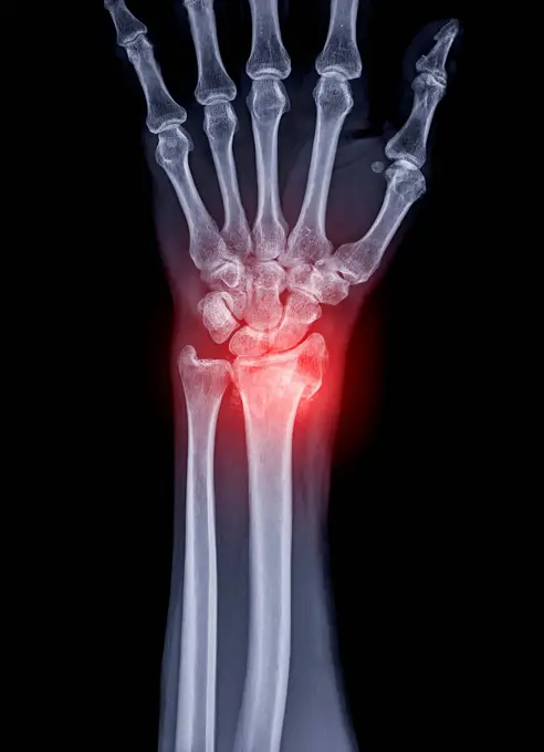
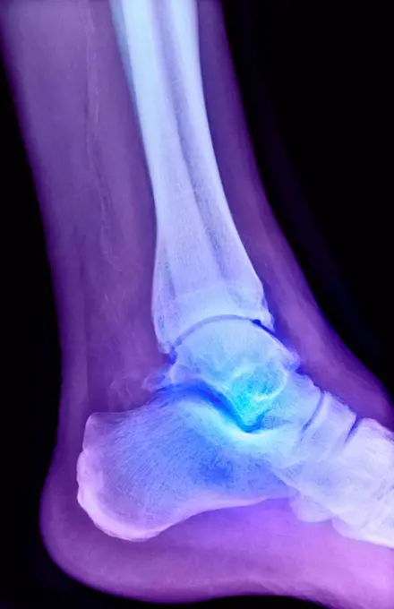
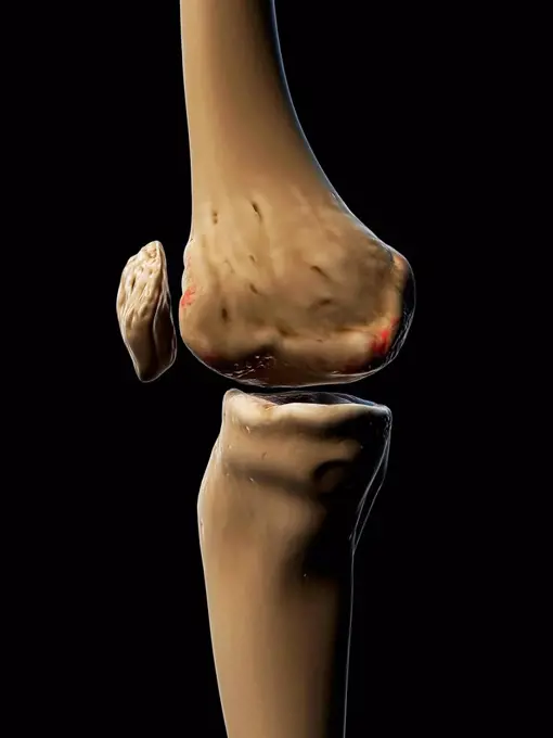
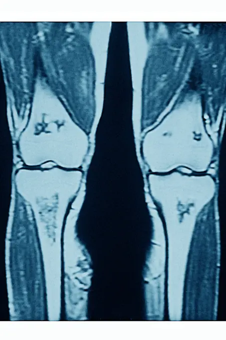
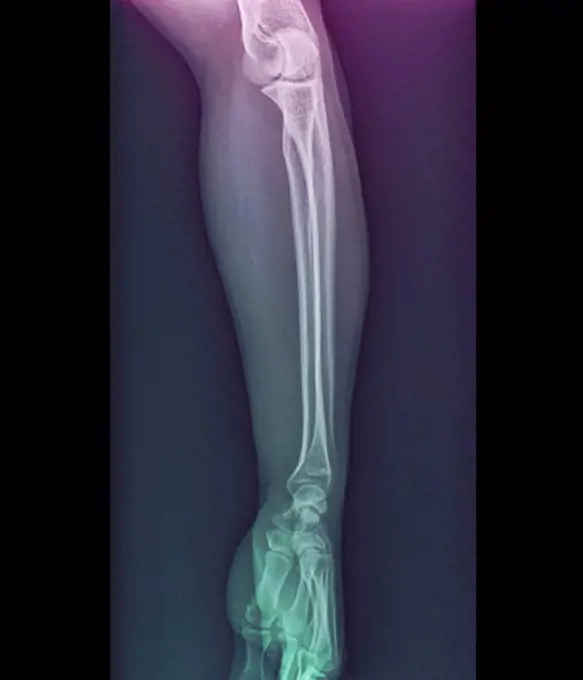
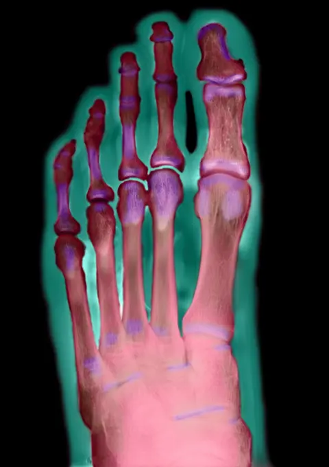
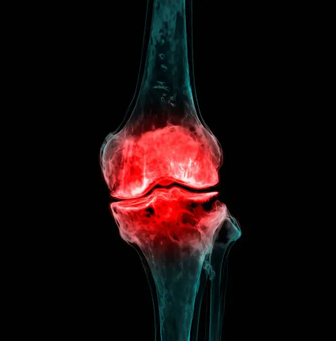
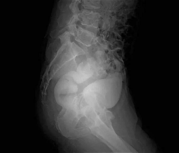
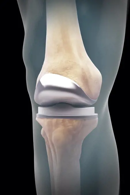
My Boards
View Pictures
  • My Boards

Knee X-Rays & Conditions

Various X-ray images displaying knee health, including normal conditions and degenerative arthritis, with colored effects.

Normal knee. Coloured X_ray of the knee of a 44 year old woman.
218 assets in this story
4297-1065
Normal knee x-ray of a 33 year old woman
4128R-30929
Arthritis of the knee. Coloured X-ray of the knee of an obese 48 year old patient showing degenerative arthritis caused by the extreme stress on the k...
1899-85937
Knee X Ray
1899-85883
Foot X Ray
4128-V58575852
4128R-12577987
Normal knee. Frontal X-ray of the healthy left knee of a 28 year old.
824-63194041
Knee x-ray on a 73-year old woman. The exam shows widespread bone demineralisation, a discrete pinching in the internal femorotibial joint space and an osteosynthesis of the left tibial plateau (screw).
4128R-12797
Normal knee. Coloured X_ray of the knee of a 44 year old woman.
4128-30419975
Fractured knee, CT scan
824-63194036
Knee x-ray on a 73-year old woman. The exam shows widespread bone demineralisation, a discrete pinching in the internal femorotibial joint space and an osteosynthesis of the left tibial plateau (screw).
4128-28766900
Fractured ulna, X-ray
4128R-15737
Broken arm. X_ray of the arm of a 20 year old patient with a fractured radius lower arm bone.
4128-19489593
Broken knee cap, X-ray
4128R-30947
Total knee replacement. Coloured frontal (left) and profile (right) X-rays of the right knee of a 69 year old patient after total knee replacement sur...
4128R-12656
Normal child´s knee. Frontal coloured X_ray of the knee of a 10 year old child.
1899-85881
Foot X Ray
4297-1070
Normal knee x-ray of a 58 year old man
1525-28448576
X-ray film with doctor’s hand
824-63128351
Ankle, x-ray
4297-1186
Colorized x-ray of the foot of a 37 year old man showing fractured metatarsals. This person had his foot run over by a vehicle
4128R-12671
Normal knees. Frontal X_ray of the flexed knees of a 24 year old patient.
4128R-11544369
Broken thigh bone, computer illustration.
1899-85846
Knee X Ray
1899-85829
Flat Foot X Ray
4269-27213
Osteoarthritis of the knee. Colored X-ray of the knee showing osteoarthritis or gonarthosis. It has caused a lack of space between the femur (thigh bone, above) and the tibia (shin bone, below) in blue due to erosion of the cartilage lining the joint.
4128-15665832
Healthy knee joint, illustration
4297-1524
Colorized x-ray showing a severely angulated fracture of the distal radius and ulna in a 12 year old girl
6114-50976766
An x-ray  of a fracture lower third femur.
4128R-6822
Normal elbow joint, X_ray
824-63188617
Osteoarthritis of the knee seen on a frontal x-ray.
824-63178681
X ray of the left foot of a 29 years old male patient. Fracture of the big toe
824-63178626
Child of 15 years measuring 1.48 m which corresponds to 13 years and 4 months. Bone age is calculated on both the hand and the elbow.
4128-111581754
Hip replacement, computer illustration.
1525-28452936
X ray film with doctor’s hand to examine
4128-19248932
Painful knee joints, illustration.
4128R-30130
Knee pain, conceptual computer artwork.
4128R-12964776
Broken upper leg bone, Illustration
1899-13094
Ligaments of the knee Posterior view III
4128-28766918
Wrist pain, conceptual X-ray
4239-18641992
Detail of hip joint showing osteoarthritis.
6188-55645103
Hand, Mann Röntgenbild, Mann, Hand Copyright: xZoonar.com/Dr.xNorbertxLangex 13793118
824-63178618
Delay bone age 15 years old child mesearing 1meter 48 high which correspond to the size of a 13 years and 4 months old child. The bone age is calculated on the hand or the elbow radiography
1841R-111805
1899-54027172
Rheumatoid arthritis seen on an x-ray of the hand.
4128-18680900
Dislocated ankle, X-ray
824-63178426
Normal X-ray of the knee of a young child
4128-19247915
Knee joint, illustration.
4128-19489622
Broken ankle, X-ray
4128-19489777
Supracondylar fracture, X-ray
824-63128435
X-ray of a normal right wrist of a 17-year-old young woman. See image n1082105 on the site.
4378-20013501
Right Knee Ligaments
4128R-15516619
Illustration of an elbow replacement
4128R-29995
Joint pain, conceptual computer artwork.
824-63123167
Hysterography.
4128-V58575839
4128-28769205
Fractured ulna, X-ray
824-63170784
Post-traumatic wrist arthritis. X-ray of left hand.
4128-30420331
Arthritis of the foot, conceptual illustration
4297-1039
Foot x-ray of a 52 year old man with a fracture of his great toe, highlighted in color
4128R-11287824
Osteoarthritis of the ankle. Coloured X-ray of the ankle of an 80 year old female patient with severe osteoarthritis.
824-57655649
X-ray of rheumatism in the hands.
4128-V58576818
4297-1348
Normal x-ray of the ankle of a 12 year old girl
255-28641408
Vintage Photograph. X Ray images of Elbow, Anterior Posterior view and Lateral View
4128R-10598
Arthritic ankle, X_ray
4378-20013929
Right Knee Ligaments
4128-19357510
X-ray showing the dislocated ankle of a patient in a traffic accident.
4128-19489613
Broken knee cap, X-ray
4239-18641382
Conceptual image of rheumatoid arthritis (RA) in the human hand. RA is a chronic inflammatory disorder that typically affects the small joints in your hands and feet.
4128R-11298412
Arthritic knee, computer artwork.
4128-20041813
Fractured femur, X-ray
4128-19052615
Knee joints, illustration
824-63218197
Osteoarthritis of the knee. Wear then destruction of cartilage in various areas of the knee joint. Frontal MRI scan of the knees.
4128R-13410640
Foot nerves
4128R-11472879
Human hip replacement, computer artwork.
1899-54028016
Screw and plate osteosynthesis on a fracture of the fibula. Frontal x-ray: tibia, fibula, malleolus and astragalus.
4297-1197
Colorized x-ray of the hand of a 16 year old boy who dislocated his thumb at the
4128R-13572577
Illustration of the superior peroneal retinaculum.
4128R-12964770
Broken lower leg bones, Illustration
4378-5328
Medial Tibial Stress Syndrome
4128R-3915
Normal elbow and wrist joints, X_ray
4128-20045707
Fractured femur, illustration
1848-61113907
Shoulder joint, X-ray
4378-20014021
Right Knee Ligaments
824-63178683
X ray of the left foot of a 29 years old male patient. Normal
1899-85943
Knee X Ray
4128-28514562
Knee pain, conceptual image
4128-20043269
Rectovesical fistula, X-ray
4128-19357525
Fractured heel X-ray of a patient's fall from height showing a comminutive fracture of the heel, the calcaneus.
824-63190436
Illustration of areas affected by gout. It most commonly affects the joint between the metatarsal and the first phalanx in the big toe, but can also affect the ankle, heel or knee joints.
6188-58132698
human radiography scan human radiography scan on blue background Copyright: xZoonar.com/OxanaxRishnyakx 5667514
4378-5125
Right Knee Ligaments
4128R-11472892
Human hip replacement, computer artwork.
4128-30420306
Arthritis of the foot, conceptual illustration
4297-1202
Wrist and Forearm X-ray of a buckle fracture of a 9 year old boy
4128-18929570
Monteggia fracture, X-ray
6188-54821818
steel plate holding together the tibia of the leg. x-ray of the leg. A steel plate holding together the tibia of the leg. Copyright: xZoonar.com/LeonidxEremeychukx 14369274
4378-20393408
Knee Replacement
824-57655652
Normal left hand radiograph.
4297-1365
Normal hand x-ray of a 68 year old man who is wearing a wedding ring
PREVIOUS
of 3
NEXT
2401 S. Ervay, Suite 206
Dallas, Texas 75215
United States
Get Started
Free ResearchMy BoardsMy Cart
For Creators
How To License Your ContentContributor PortalFrame of Mind
Resources
API accessPricing
Contact
+1 866 236 0087help@viewpictures.co.uk Contact form
©2026 View Pictures. All Rights Reserved. -A
This site is protected by reCAPTCHA and Google Privacy Policy and Terms of Service apply.