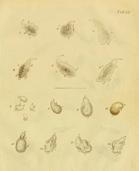
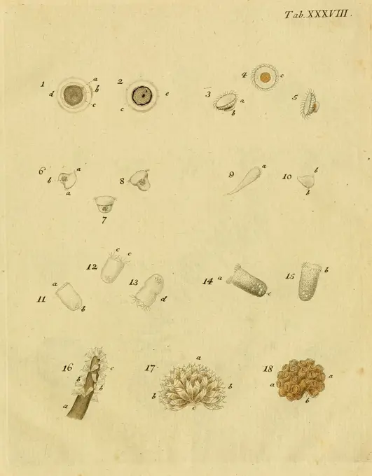
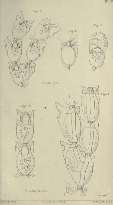
Marine and Fungal Anatomy Illustrations

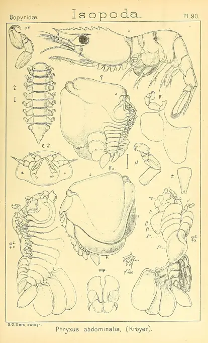
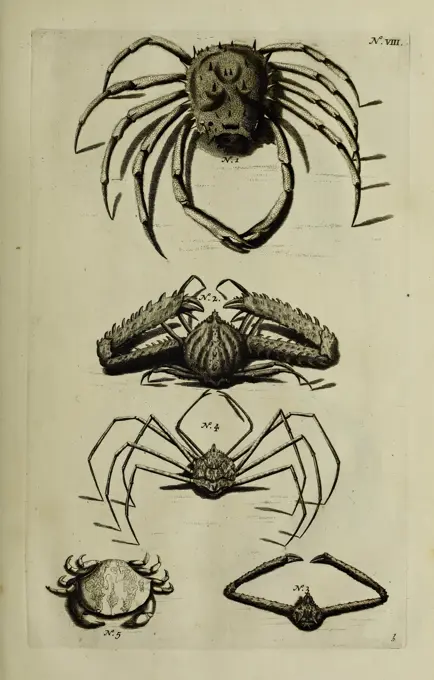
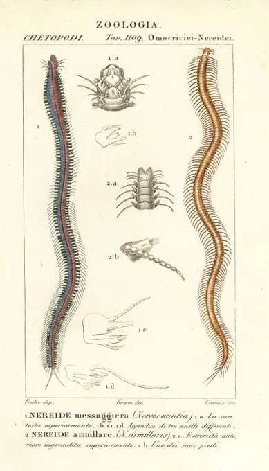
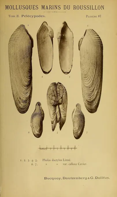
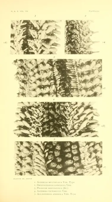
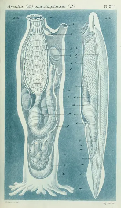
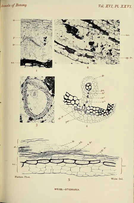
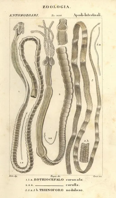
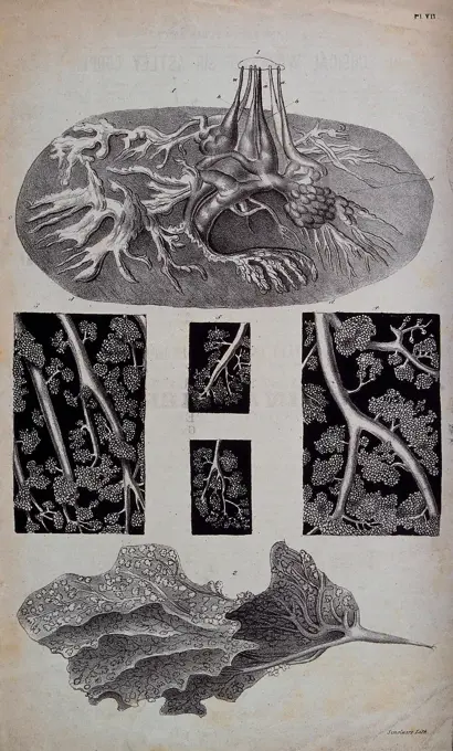
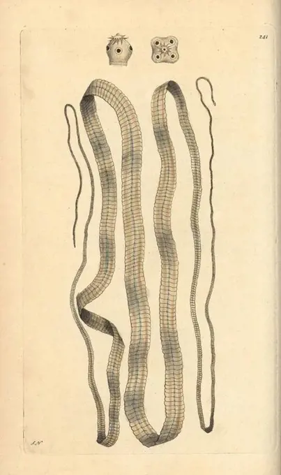
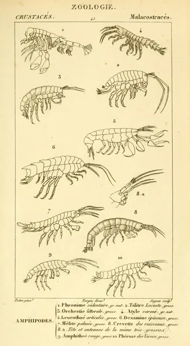
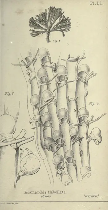
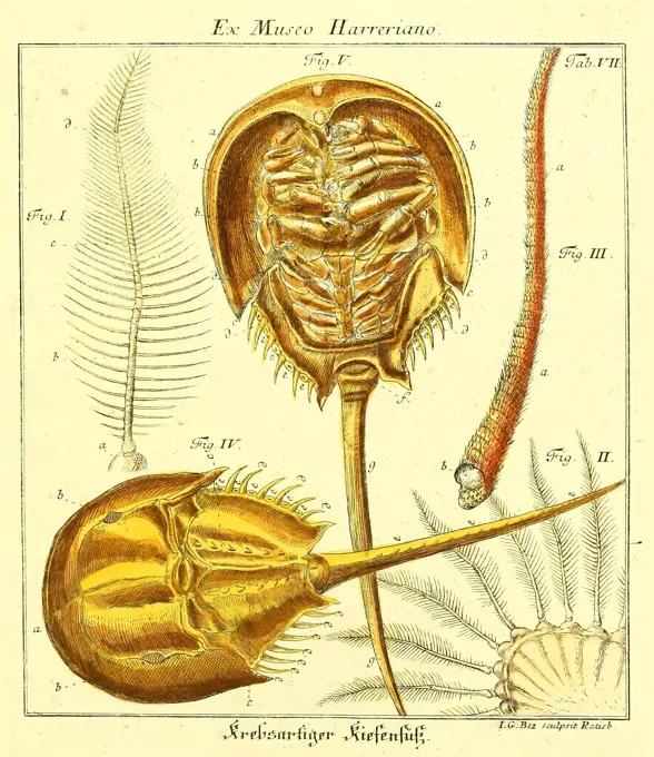
Scientific illustrations of marine invertebrates and fungi, displaying detailed anatomical features and classifications for educational purposes.

An account of the Crustacea of Norway Christiania, A. Cammermeyer, 1895-1928, Crustacea, Norway, Crustaceans, This illustration features a detailed scientific study of various isopod species, specifically focusing on *Phryxus abdominals*. It includes labeled diagrams showcasing different anatomical features such as the head, thorax, and abdomen, displaying their segmented bodies and appendages. Various views highlight the morphology of these crustaceans, with specifics on limb structures and carapace details. The background appears simplistic, emphasizing the intricacy and precision of the depicted specimens intended for educational purposes in zoology.
Thesaurus imaginum piscium testaceorum Lugduni Batavorum apud P. van der Aa 1711 Indonesia Maluku Crustacea Mollusks Mineralogy Museum of Comparative Zoology, A detailed illustration showcasing various species of crabs and spiders, arranged methodically for comparative study. At the top, a large spider is depicted with prominent body and leg features, followed by a representation of a more robust crab below it. The image continues with two additional crabs and a spider, displaying their unique physical attributes. Each creature is labeled with identifiers, emphasizing the distinct morphology and anatomical characteristics of these arachnids and crustaceans, providing valuable insight for entomological and marine biology studies.Abdrucke und Versteinerungen des Zwickauer Schwarzkohlengebirges und seiner Umgebungen, Zwickau, G. Richter'schen Buchhandlung, 1835, carboniferous, Germany, paleontology, Zwickau, This illustration showcases a collection of geological specimens, specifically fossilized corals. Each labeled section highlights distinct features, such as the ribbed textures and unique shapes of the corals. The arrangement includes elongated cylindrical forms, flattened modules, and various cross-sections that demonstrate the intricate patterns and organic structures inherent in these ancient marine organisms. The labeled identification numbers guide the viewer through the different specimens, emphasizing the diversity and detail found in paleontological studies.Magazine, of, zoology, and, botany, Edinburgh, Dublin, botany, natural, history, periodicals, zoology., This illustration showcases a variety of botanical specimens, focusing on different forms of liverworts and mosses. Each labeled figure demonstrates distinct morphological features, highlighting diverse leaf shapes, structures, and growth patterns. The collection includes various species, emphasizing their unique characteristics such as lobed leaves, branching patterns, and reproductive structures. The arrangement allows for a comparative study of these plants, providing insight into their classification and morphology. The detailed representation aids in understanding the complexity and variability of these lesser-known flora.Die Versteinerungen des rheinischen Schichtensystems in Nassau, Wiesbaden, Kreidel & Niedner, 1850-1856. geology Germany Hesse Nassau Province paleontology Museum of Comparative Zoology, A collection of various ammonite fossils and their detailed illustrations is showcased. The arrangement features several different species, each exhibiting unique shell structures and patterns. Among the fossils, one prominent specimen displays a detailed spiral shape, indicating its age and the intricate design of its shell. Surrounding illustrations provide a scientific representation of the fossils, highlighting the diverse forms and textures. Additionally, waveforms at the bottom suggest a correlation to the geological or evolutionary study of these creatures, emphasizing their historical significance in paleontology. This visual compilation offers insight into the diverse morphology of ammonites and their importance as indicators of ancient marine environments.Bar-toothed nereid, Perinereis nuntia 1 and nereid, Syllis armillaris 2. Handcoloured copperplate stipple engraving from Antoine Laurent de Jussieu's Dizionario delle Scienze Naturali, Dictionary of Natural Science, Florence, Italy, 1837. Illustration engraved by Carini, drawn by Jean Gabriel Pretre and directed by Pierre Jean-Francois Turpin, and published by Batelli e Figli. Turpin (1775-1840) is considered one of the greatest French botanical illustrators of the 19th century.Les mollusques marins du Roussillon Paris, J.-B. Baillière, 1882-1898 France Mollusks Roussillon province, This illustration showcases various species of marine mollusks from the Roussillon region, specifically focusing on bivalve shells. The composition features multiple views of these shells, highlighting their distinctive shapes and characteristics. Each shell is meticulously numbered for identification, with species names listed beneath. The work is attributed to researchers Bucquoy, Dautzenberg, and Dollfus, contributing to the scientific study of marine life. Such visual documentation provides valuable insight into the diversity of mollusks and their anatomical features.Symbolæ ad monographiam Cymothoarum Crustaceorum Isopodum familiae, Hauniæ, Typis H. H. Thiele, 1879-84, Isopoda, A detailed scientific illustration showcasing various species of marine polychaete worms. Each specimen is meticulously labeled with numbers, indicating different types and anatomical features. The image displays the intricate body structures and segmentations characteristic of these organisms, highlighting their diverse forms. The arrangement includes multiple angles and cross-sections to provide a comprehensive view of their morphology, emphasizing the complexity and beauty of marine life.Indian, Tertiary, and, Tertiary, Vertebrata, Calcutta, Geological, Survey, Office, 1874-1902, India, paleontology, vertebrates, fossil, A collection of fossilized vertebrate bones is displayed, showcasing various jaw bones and other skeletal elements. Each specimen is meticulously labeled with numbers indicating their specific identification. The arrangement highlights the complexity of anatomical structures, illustrating features like dental patterns and morphology. These fossils are likely from a historical geological period, offering insights into the evolutionary history of vertebrates.Dreer's garden calendar, Philadelphia, Pa, Henry A. Dreer, 1884, flowers, gardening, seeds, catalogs, nursery stock, Henry A. Dreer firm, garden stories, internet resource, This illustration presents a botanical guide from Dreer's Garden Calendar, showcasing various types of vegetables. The first section features two distinct early cabbage varieties: 'Early Oxheart Cabbage' and 'Large Late Dutchhead Cabbage.' Both are depicted with detailed shading and labels to differentiate them. The central part highlights 'Lindeman's Short-Stemmed Cauliflower,' illustrating its unique shape and texture. Finally, the rightmost section presents a long, slender cucumber, labeled simply as 'English Frame Cucumber.' The layout emphasizes the importance of these vegetables in gardening, with each entry providing clear visual identification for gardeners and enthusiasts alike.Animalcula, infusoria, fluvia tilia et marina, Hauniae, Typis N. Mölleri, 1786, infusoria, animalcules, This illustration features a scientific representation of various single-celled organisms, displaying their diverse shapes and forms. The organisms, categorized in a grid format, showcase differences in size, surface texture, and cellular structures. Each specimen is numbered for reference, indicating a systematic approach to their study. The detailed rendering highlights their biological features, making it an educational reference for understanding protozoan diversity and classification.Animalcula, Infusoria, fluvia, tilia, et, marina, Hauniae, Typis, N. Mölleri, 1786, Infusoria, animalcules, An array of intricate microorganisms and their structures is depicted, showcasing various forms under scientific examination. The composition includes multiple numbered illustrations highlighting different shapes and details of these tiny life forms, ranging from circular and elliptical to more complex configurations. Notable features include various textures, sizes, and arrangements that emphasize the diversity of microscopic life. This collection serves as a visual representation of the biological complexity within a micro-ecosystem, accompanied by a designated table number, indicating its classification within a larger study.Insecta Britanica, Diptera, by Francis Walker. London, Reeve and Benham, 1851-1856, Diptera, Great Britain, insect, The image features a detailed scientific illustration depicting various species of dipterans, commonly known as flies. It includes multiple views of each specimen, showcasing their anatomical features such as wings, legs, and body structure. The illustration is meticulously labeled, indicating specific parts of the insects, with corresponding reference numbers for clarity. This representation highlights the diversity within the dipteran order, emphasizing their unique physical traits and aiding in the study of entomology. The delicate lines and intricate details reflect the precision of scientific illustration in documenting and classifying insect species.Unterhaltungen aus der Naturgeschichte, Augsburg, Schlosser, 1834, mollusks, A collection of intricately detailed botanical illustrations showcasing various plant species. These illustrations feature elongated leaves and unique forms, with some displaying finely detailed veins and textures. There are also a few floral elements depicted, adding an organic variety to the arrangement. Each specimen is numbered, suggesting a systematic classification, likely for scientific study or educational purposes. The overall composition emphasizes the diversity of plant morphology, highlighting the beauty and complexity of nature.ATLAS OF THE PHYSICAL AND POLITICAL HISTORY OF CHILE, 1854. Author: GAY CLAUDIO. Location: BIBLIOTECA NACIONAL-COLECCION. MADRID. SPAIN.An account of the Crustacea of Norway Christiania, A. Cammermeyer, 1895-1928, crustacea, Norway, crustaceans, The illustration showcases various species from the order Isopoda, specifically focusing on the Desmosomidae family. It features detailed anatomical drawings of different isopod forms, highlighting their exoskeleton structure and appendages. Each figure is meticulously labeled with numbers, providing a clear reference to unique characteristics such as limbs and body segments. The artwork emphasizes the diversity and complexity of isopods, serving as an informative guide for biological study.Animalcula, Infusoria, Fluvia, Tilia, Et, Marina, Hauniae, Typis, N. Mölleri, 1786, Infusoria, Animalcules, This illustration features a series of scientifically rendered microorganisms, including various types of single-celled organisms and algae. The composition is divided into numbered sections, each depicting distinct forms and structures. Some exhibit intricate patterns with radiating spines, while others showcase unique shapes, such as spherical or oval bodies. The diversity of sizes and forms highlights the complexity of microscopic life, with textures and details that suggest movement or interaction within their aquatic environments. The plate serves as an educational resource, emphasizing the vast variety and beauty of microscopic organisms.Plate 53. Pathology of the male genitalia, urinary organs.Memoires pour servir a l'histoire des insectes Stockholm, L.L. Grefing, 1752-1778. Arthropoda, Crustacea, Insects, This illustration features a collection of detailed scientific sketches depicting various species of spiders and arachnids. The arrangement includes numbered figures, each labeled with a specific identifier. The drawings showcase different anatomical structures, including body shapes, leg formations, and unique features such as webs and egg sacs. The diversity of forms highlights the intricate details of these creatures, making it a valuable reference for entomological studies.Journal of Botany, British and Foreign. London, Robert Hardwicke, 1863-1942. Botany, Great Britain, Periodicals, Plants, This illustration features various botanical studies focused on the Euphrasia genus, showcasing detailed line drawings of individual leaves and flowers. Each entry is numbered, providing a clear reference for the distinct shapes and structures observed in the specimens. The drawings highlight various characteristics, such as leaf textures and floral arrangements, emphasizing the diversity within the species. The meticulous detail and precision reflect a scientific approach to studying plant morphology and classification.Flore des serres et des jardins de l'Europe, A Gand, chez Louis van Houtte, éditeur, 1845-1880, This illustration features detailed anatomical studies of the Corynanthe Albertinae, showcasing the peculiar morphology of its reproductive structures. The top section displays multiple views of the organism's unique forms, highlighting aspects such as curvature and symmetry. Below, intricate depictions of branching structures resemble a network, labeled with a diagrammatic representation of their arrangement, emphasizing the complexity of their growth patterns. The scientific annotations provide insight into the study of these organisms, reflecting a focus on botanical or biological research.Memoires pour servir a l'histoire des insectes Stockholm, L.L. Grefing, 1752-1778, Arthropoda, Crustacea, Insects, The illustration features a series of labeled diagrams showcasing various arachnids and related organisms. Each figure is meticulously detailed, providing a clear representation of their anatomical features. Figure 1 displays a spider with annotated parts, while Figure 2 illustrates a different arachnid species, labeled for identification. The series continues with various forms, including mites and ticks, highlighting specific details such as legs, body segmentation, and appendages. The illustrations are arranged methodically, allowing for comparative study among the different types represented, each contributing to the understanding of arachnid diversity and morphology.Recherches sur les ossemens fossiles de quadrupèdes Paris Deterville 1812 France Paleontology Geology Paris region Mammal fossils Vertebrate fossils Fossil stories, This illustration features a collection of detailed anatomical drawings showcasing different views and structures of bones or fossils. The first section depicts the intricate contours of a foot, highlighting distinct features such as the arches and toes. Nearby, additional segments illustrate various bone fragments, each labeled with reference numbers for identification. The drawings emphasize the texture and structure of the materials, conveying an understanding of skeletal anatomy and its variations, possibly for educational or research purposes in the study of anatomy or paleontology.Zoological, lectures, delivered, at, the, Royal, Institution, London, Kearsley, 1809, zoology, cuttlefish, An intricate illustration depicting an eight-armed cuttlefish, prominently shown from a frontal perspective. The creature's elongated body and a prominent bulbous mantle support its eight distinct, flexible arms, all adorned with detailed suckers. The arrangement emphasizes the fluidity and grace of this marine animal, while the illustration provides an educational glimpse into its anatomy. The text below identifies the species as 'Eight Armed Cuttle Fish,' indicating its scientific significance. The overall composition highlights both the beauty and complexity of marine life.Lib. VIII, Tabula XVII, Lib. VIII, Adriani Spigelii Bruxellensis equitis D. Marci, olim in Patavino gymnasio anatomiae et chirurgiae professoris primarii Opera quae extant, omnia, Aselli, Gaspare, 1581-1626, Bucretius, Daniel, d. 1631, Fialetti, Odoardo, 1573-1638, Harvey, William, 1578-1657, Spiegel, Adriaan van de, 1578-1625, Valesio, Francesco, b. ca. 1560, Walaeus, Johannes, 1604-1649, van der Linden, Johannes Antonides, 1609-1664, Engraving, 1645, Plate 17, page 165.Prodromus of the Paleontology of Victoria Melbourne G. Skinner Acting Government Printer 1874 Australia Victoria Paleontology, A detailed illustration showcasing several views of marine shells, specifically linked to the paleontology of Victoria. The shells exhibit a variety of shapes and sizes, highlighting their intricate texture and structure. The depiction includes both a large, prominent shell with a spiraled apex and smaller, delicately shaped shells, emphasizing the diversity found within this natural history context. The labels indicate the significance of these specimens within paleontological studies.Die Klassen und Ordnungen des Thier-Reichs, Leipzig, C.F. Winter, 1859-1901, Woodshole, A detailed illustration featuring various specimens of amphibians, showcasing their anatomical structures and characteristics. The numbered figures display different views and components, such as skeletal features, reproductive organs, and unique adaptations of each species. The composition highlights the diversity among amphibians, providing educational insights into their biology and morphology, with labels potentially indicating specific parts or variations. This work serves as an important resource for studying amphibian anatomy and classification.Dictionnaire des sciences naturelles Strasbourg Paris F. G. Levrault Le Normant 1816-1830, The illustration features two detailed depictions of serpentine creatures. The first is labeled '1. ORVET commun,' showcasing a slender, elongated body with a smooth texture and a pointed tail, emphasizing its streamlined form. The second depiction, labeled '2. OPHISAURE ventral,' presents a slightly thicker body with distinct patterns and scales along its length, suggesting a different species. Both specimens are oriented to highlight their lengths and are accompanied by a smaller inset image, possibly providing additional anatomical details or a close-up view of a specific feature. The precision of the lines and the clarity of the labels reflect a focus on biological study, typical of zoological illustrations from earlier eras.Manuel d'actinologie ou de zoophytologie, Paris, F.G. Levrault, 1834-1836, corals, sea anemones, cnidaria, ctenophora, The illustration presents a scientific depiction of marine organisms. At the top, a series of labeled drawings showcases different stages or types of the anatomy of a jellyfish, highlighting its bell shape and tentacles. Below are detailed representations of two distinct forms of bivalve shells, emphasizing their oval and rounded structures. The meticulous lines and soft colors illustrate the intricate patterns and textures typical of these aquatic creatures, demonstrating their biological diversity and elegance in design. Each section is carefully annotated, providing a clear reference for study in marine biology or taxonomy.1. tubes of Hermella alveolata, sandworm, 2. hermella, 3. terebella emmalina, Historic, digitally restored reproduction from a 19th century originalSix figures showing blood vessels and capillaries (). Lithograph.Broad tapeworm, Diphyllobothrium latum. Illustration signed SN (George Shaw and Frederick Nodder).. Handcolored copperplate engraving from George Shaw and Frederick Nodder's "The Naturalist's Miscellany" 1796.. Frederick Polydore Nodder (1751~1801) was a gifted natural history artist and engraver. Nodder honed his draftsmanship working on Captain Cook and Joseph Banks' Florilegium and engraving Sydney Parkinson's sketches of Australian plants. He was made "botanic painter to her majesty" Queen Charlotte in 1785. Nodder also drew the botanical studies in Thomas Martyn's Flora Rustica (1792) and 38 Plates (1799). Most of the 1,064 illustrations of animals, birds, insects, crustaceans, fishes, marine life and microscopic creatures for the Naturalist's Miscellany were drawn, engraved and published by Frederick Nodder's family. Frederick himself drew and engraved many of the copperplates until his death. His wife Elizabeth is credited as publisher on the volumes after 1801. Their son RichaTenants of an old farm, New York, Fords, Howard & Hulbert, 1885 1884, insects, juvenile literature, The illustration depicts a cross-sectional view of a hornet's nest, highlighting its intricate internal structure. The nest is composed of tightly woven hexagonal cells, resembling a honeycomb pattern, which serve as individual chambers for the hornets. These cells are arranged in a layered formation, giving a sense of depth and complexity to the nests architecture. The detailed lines capture the texture and craftsmanship of the hornets' work, emphasizing the natural engineering skills of these insects. The caption references the scale and organization of the nest, illustrating the communal aspect of hornet life within this carefully constructed habitat.American, Marine, Conchology, Philadelphia: Printed for the author 1831, Mollusks, United States, Women in Science, The illustration presents a detailed study of various shell types from the genus Sanguinolima, showcasing three distinct forms. At the top, the first specimen is labeled 'S. florens,' followed by the identification 'A. bicolor' for the middle shell. The third illustration features 'Pethricola,' providing a closer look at its textured surface. Each shell is carefully rendered, highlighting unique shapes and patterns that contribute to their identification in malacology. The scientific taxonomy is indicated alongside each specimen.Considerations generals on the class of crustaceans, Paris, F.G. Levrault, 1825, France, Crustacea, Zoology, A detailed illustration featuring a series of labeled diagrams showcasing various species of crustaceans, specifically within the Malacostraca class. The depiction includes ten distinct figures, each highlighting different anatomical features such as tail structure, limb arrangement, and body segmentation. The figures are labeled numerically from 1 to 10, with annotations identifying specific characteristics and species. The names include terms related to various types of amphipods, emphasizing their diverse morphological traits. The arrangement provides a clear reference for studying the classification and anatomy of these marine organisms.Transactions of the Entomological Society of London, London, The Society, entomology, periodicals, insect, The illustration showcases two detailed views of the larva of Latiroina orbiculus, a type of marine organism. The first panel provides a close-up of the larvas structure, emphasizing its segmented body and distinctive clasping appendages. The second panel offers a broader view, highlighting additional anatomical features and textures that contribute to its functionality and adaptation in the aquatic environment. Both visuals are labeled to identify specific parts, demonstrating the larva's developmental stages and morphological characteristics essential for scientific study.Symbolæ, ad, monographiam, Cymothoarum, Crustaceorum, Isopoda, familia, Hauniæ, Typis, H. H. Thiele, 1879-84, isopoda, The illustration features a series of detailed sketches showcasing various anatomical views of crustaceans from the order Isopoda. Each labeled figure presents a different aspect of the creatures, highlighting their segmented bodies and distinct features such as limbs and appendages. The arrangement is systematic, with side, dorsal, and ventral perspectives represented. The intricate line work emphasizes the biological structure and complexity of these organisms, providing a visual reference for their classification and morphological study. The accompanying labels serve to identify the specific types and variations within this group, offering insight into their diversity.Studies, comparative, on, fossil, fishes, and, living, of, the, genera, Carcharodon, Oxyrhina, and, Galeocerdo, Pisa, T. Nistri, 1881 Shark, Carcharodon, Galeocerdo, fossil, fishes, sharks, fossil, fossil, stories, This illustration showcases several teeth of the prehistoric shark species Carcharocles megalodon, a notable predator that roamed the oceans millions of years ago. The detailed renderings highlight the distinctive triangular shape and serrated edges of the teeth, which are representative of the species' formidable feeding capabilities. Each tooth is labeled with a unique identifier, suggesting different sizes or variations. The scientific precision of the design emphasizes the significance of the megalodon in paleontological studies, reflecting its crucial role in understanding ancient marine ecosystems.The ivory king New York C. Scribner's sons 1886 Elephants Taxonomy: Family= Elephantidae Elephant, This illustration showcases various anatomical features related to elephants. The top section displays a series of molar teeth from different species, including the African elephant, Asiatic elephant, and Mastodon. Each molar is labeled with a corresponding number for identification. Below, notable details include a head illustration of an elephant, highlighting anatomical structures such as the skull, tusks, and jaw. The arrangement emphasizes the unique dental adaptations and skeletal characteristics that distinguish these majestic animals from one another, providing insight into their feeding habits and evolutionary history.Mosquito life New York, G. P. Putnams sons, 1907. Mosquito, Insects, Eggs, Ochlerotatus infirmatus, Ochlerotatus taeniorhynchus, Ochlerotatus pretans, Ochlerotatus sollicitans, Ochlerotatus canadensis, Lepidopla sylvicola, The illustration presents a detailed study of various types of egg specimens from different species. Each specimen is displayed in a systematic arrangement, showcasing distinct shapes and textures. The first specimen, labeled '1,' features a smooth surface with a narrow, elongated form, while '2' displays a more intricately patterned texture. The eggs labeled '3' and '4' exhibit slight variations in size and surface details, reflecting their biological diversity. Specimen '5' presents a wider structure with a unique mesh-like pattern, and '6' features a rounded form, emphasizing the range of morphological characteristics found within this collection. The careful organization and labeling indicate a scientific exploration of these specimens' classifications.Ibis London Published for the British Ornithologists' Union by Academic Press birds ornithology periodicals bones, The illustration showcases a detailed anatomical study of various skeletal structures, featuring both the complete skeleton and isolated bones. Each labeled section represents different aspects of the anatomy, including the skull, vertebrae, limb bones, and other skeletal components, enhancing the understanding of structural relationships within a specific species. The meticulous labeling provides context for each part, making it a valuable resource for students and professionals in the fields of anatomy, biology, and medicine.Abdrucke und Versteinerungen des Zwickauer Schwarzkohlengebirges und seiner Umgebungen, Zwickau, G. Richter'schen Buchhandlung, 1835, carboniferous, Germany, paleontology, Zwickau, An intricate display of various botanical illustrations, showcasing detailed representations of ferns and other plant forms. The layout features numerous leaf patterns and structures, each meticulously labeled with numbers corresponding to their specific characteristics. The design includes a variety of shapes, including fronds, branches, and intricate foliage, providing a rich visual study of plant morphology. The diversity of forms captures the elegance of plant life, presenting a blend of delicate and robust features that highlight the beauty and complexity of the natural world.Les mollusques Paris Fortin Masson et cie 1836-1845 classification mollusks pictorial works shells sea life, This illustration features a detailed study of the anatomy of a marine organism, specifically focusing on various structures and forms. The central portion illustrates a elongated body with circular orange markings, possibly representing reproductive or anatomical features. Surrounding images highlight different morphological aspects, including a cross-sectional view, a close-up of a section of the organism, and an outline of the upper anatomy. The annotations and numbered references indicate specific parts and different stages of the organisms development, showcasing the intricacies of its biological form. The overall composition emphasizes scientific exploration and detailed observation in marine biology.A book of whales, London, J. Murray, 1900, whales, Wood's Hole, porpoise, finless porpoise, Neophocaena, taxonomy: family= Phocoenidae, This detailed scientific illustration features various anatomical studies related to marine life. The top section depicts the embryo of a species related to the genus *Nomeni*, highlighting the structure of dorsal dermal plates. The middle section showcases the dorsal fin of a porpoise, emphasizing dermal ossicles, which are bony structures that contribute to the fin's robustness. The bottom section includes another portion of skin from the same genus, again illustrating dermal ossicles, providing insight into the protective features of the species. Each figure is meticulously labeled, indicating the source as a work by Kükenthal, which signifies a focus on marine biology and the study of aquatic organisms.Zoological, lectures, delivered, at, the, Royal, Institution, London, Kearsley, 1809, zoology, The illustration features several intricate specimens of Holothuria utricularis, a type of sea cucumber known for its distinctive shape and structure. Each specimen showcases elongated bodies resembling shells, adorned with delicate tentacle-like appendages that extend gracefully downward. The detailed line work highlights the unique features, including the intricate textures and patterns on the surface of the organisms. Near the bottom, there is a label identifying the species and providing a brief description. The overall composition emphasizes the beauty and complexity of marine life in a scientific context.Harmonia ruralis, or An Essay Towards A Natural History Of British Song Birds, London, Printed And Sold By The Author 1794-1796, England, Birds, Eggs, Nests, Excellent, Pictorial Works, A intricately woven nest rests amid slender, bending stalks, showcasing delicate craftsmanship in its construction. Within the nest, several speckled eggs are nestled securely, hinting at the promise of new life. The surrounding elements, including the thin, tall grasses and the strategic placement of branches, create a serene and natural habitat, illustrating the harmony of natures design. The overall composition emphasizes both the fragility and beauty of wildlife, capturing a moment of expectation and nurturing in the avian world.Arcana entomologica, or Illustrations of new, rare, and interesting insects, London, William Smith, 1845 i.e. 1841-1845. insects, arthropoda, entomology, classification, pictorial works, arthropod, A detailed illustration showcasing a variety of insects and a flowering plant. The composition includes multiple species of beetles and other insects, each labeled with their respective numbers for identification. The central focus is on an intricately drawn flower with elongated petals, providing a natural context for the insects. Surrounding the flower are several beetles, exhibiting distinct patterns and body shapes, along with a few other insect types depicted in various positions, highlighting the diversity of pollinators and their interaction with the flora. The carefully rendered details capture the unique characteristics and anatomical features of each insect.Tableau, élémentaire, de, l'histoire, naturelle, des, animaux, Paris, Baudouin, imprimeur, an 6 1797 or 1798, zoology, A detailed study of various bird feet, showcasing the distinct anatomical structures and adaptations suited for different species. The illustration is organized into a grid format, with each square displaying a specific type of bird foot, such as the clawed feet of raptors, the webbed toes of aquatic birds, and the perching adaptations of songbirds. Labels accompany each example, indicating the species represented, highlighting the diversity of avian morphology and their specialized functions in their respective environments. The artwork serves as an educational reference for ornithology enthusiasts and practitioners alike.Arcana entomologica, or Illustrations of new, rare, and interesting insects, London, William Smith, 1845 i.e. 1841-1845, insects, arthropoda, entomology, classification, pictorial works, arthropod, The illustration features a detailed study of various insect specimens, primarily focusing on types of wasps. At the center, two larger wasps are depicted with their intricate anatomical features visible, including elongated bodies and distinct wing structures. Surrounding these central figures are smaller, annotated sketches displaying various parts of the insects, such as antennae, legs, and heads, each labeled for identification. Additionally, a plant branch is illustrated, alongside the insects, emphasizing the ecological context. This comprehensive depiction reflects a scientific approach to entomology, showcasing both the insects' morphology and their relationship with plant life.Neck and spinal column. Handcolored steel engraving by Corbie of a drawing by Corbie from Dr. Joseph Nicolas Masse's "Petit Atlas complet d'Anatomie descriptive du Corps Humain," Paris, 1864, published by Mequignon-Marvis. Masse's "Pocket Anatomy of the Human Body" was first published in 1848 and went through many editions.Ragworm, Perinereis cultrifera. The Korean earthworm (Nereis Perinereis aibuhitensis) is a marine polychaete highly appreciated in recreational fishing. It was listed by Grübe in 1878. Old 19th century engraved illustration, El Mundo Ilustrado 1880Tenants of an old farm New York Fords Howard & Hulbert 1885 1884 insects juvenile literature, The illustration depicts a scientific diagram showcasing various structures resembling elongated, cylindrical shapes. At the center, a magnified view highlights a detailed cross-section of one of these forms, indicating a hollow interior with a distinct central opening. Surrounding this focal point are several similar elongated figures, some positioned vertically and others at varying angles, suggesting a natural or geological context. The accompanying label provides numerical references, indicating that these structures are part of a larger study or classification within a scientific text.Untersuchungen über das Variiren der Mauereidechse, Berlin, Nicolaische Verlags-Buchhandlung,1881, Anna, Eimer, Anna, Eimer, Women, in, Science, This illustration showcases a series of detailed drawings of different species of skinks, a type of lizard known for their elongated bodies and reduced or absent limbs. The artwork includes multiple views of the skinks, highlighting their distinct patterns and markings, which vary from stripes to spots. Each skink is numbered for reference, indicating variations in color and texture across species. The labels suggest important anatomical features, contributing to a greater understanding of their classification and biological diversity.Bootlace worm, Lineus longissimus 1, Borlasia angliae 1, proboscis worm, Cerebratulus bilineatus 2, Cerebratulus bilineatus 2, ribbon worm, Tubulanus polimorphus 3, Tubulanus elegans 4. Borlasia dInghiterra, Cerebratulo bilineato, Tubulano polimorfo, Tubulano elegnate. Handcoloured copperplate stipple engraving from Antoine Laurent de Jussieu's Dizionario delle Scienze Naturali, Dictionary of Natural Science, Florence, Italy, 1837. Illustration engraved by Carini, drawn by Jean Gabriel Pretre and directed by Pierre Jean-Francois Turpin, and published by Batelli e Figli. Turpin (1775-1840) is considered one of the greatest French botanical illustrators of the 19th century.Tenants of an old farm New York Fords Howard & Hulbert 1885 1884 insects juvenile literature argiope aurantia black and yellow garden spider egg sac yellow garden spider arachnology entomology spiders sci art fix, A delicate representation of a cocoon surrounded by slender, elongated plant-like structures. The intricate lines and forms suggest a natural, almost dreamlike environment, highlighting the balance between the cocoon and its surroundings. The cocoon itself appears textured and is nestled at the center, while the wispy details of the surrounding flora create a sense of movement and depth. This artwork captures the essence of transformation and the beauty of the natural world, inviting viewers to contemplate the life cycle and the intricacies of nature.A collection of botanical illustrations showcasing a variety of plants and flowers. The top row features a selection that includes a fern-like plant on the left, possibly representing a type of fern, alongside a flowering species with broad leaves. The middle section displays various blooms, highlighting their intricate details. The bottom row features two groups of flowers, one with a compact arrangement and rounded clusters resembling daisies, while the other showcases a fuller bloom with layered petals. Each illustration includes labels with the names of the plants, suggesting their use and significance in gardening or horticulture. The overall presentation reflects a detailed exploration of plant varieties, illustrating their unique characteristics and beauty.An illustrated flora of the northern United States, Canada and the British possessions : from Newfoundland to the parallel of the southern boundary of Virginia and from the Atlantic Ocean westward to the 102nd meridian . mpound, lat-eral ; pedicels slender, articulated at the base,spreading or drooping; flowers blue, purple orwhite, 5-7 broad; caljx-lobes short, oblong,obtuse, persistent at the base of the berry; corolladeeply 5-cleft, its lobes triangular-lanceolate, acu-minate; berry oval or globose, red. In waste places or in moist thickets, sometimesappearing as if indigenous, Nova ScotiaSepia elegans Blainville. Old 19th century engraved illustration from El Mundo Ilustrado 1880Die Klassen und Ordnungen des Thier-Reichs, Leipzig, C.F. Winter, 1859-1901, Woodshole, This illustration depicts anatomical structures related to amphibians, featuring detailed diagrams of various systems within the body. The upper section includes labeled illustrations of the circulatory and respiratory systems, highlighting key components such as the heart and lungs. The lower section presents the urinary and reproductive systems, showcasing their interconnections and functions. Each labeled figure provides specific references to anatomical features, illustrating the complexity of amphibian biology.Indian Tertiary and post-Tertiary Vertebrata. Calcutta, Geological Survey Office, 1874-1902. India, paleontology, vertebrates, fossil, This illustration depicts a detailed rendering of a series of teeth, likely belonging to a specific animal species. The arrangement showcases the unique morphology of the dental structure, highlighting various characteristics such as the size, shape, and alignment of the teeth. Labels and annotations may provide additional context on the species or usage of these teeth in scientific study, emphasizing their significance in understanding dietary habits or evolutionary lineage. The precision of the lines and shading suggests a careful study meant for educational or illustrative purposes in natural history.Queen sago or broad-leaved cycas, Cycas circinalis. Endangered. Handcoloured copperplate engraving by Swan after an illustration by William Jackson Hooker from Samuel Curtis' Botanical Magazine, London, 1828.Magnified Views of Vibrio Anguillula or Eel Vibrio.. Shaw, George (1751-1813). Prints, Scientific illustrations. 1809. General Research Division. Animals, ZoologyProdromus of the paleontology of Victoria, Melbourne, G. Skinner, acting government printer, 1874-, Australia, Victoria, Paleontology, This illustration presents a detailed study of various types of shells, showcasing their intricate forms and structures. The central focus depicts a large, spiraled shell with a smooth surface and a distinctive pointed apex. Surrounding this shell are several smaller representations, each highlighting different shell types, including variations in shape, texture, and size. The surrounding illustrations provide a comparison of these shells, emphasizing their evolutionary aspects and the diversity found within marine mollusks. The work is part of a paleontological study, specifically related to the shells found in Victoria, illustrating historical marine life and its geological significance.Land and freshwater Mollusca of India London, Taylor and Francis, 1882-, Asia, India, Mollusks, Shells, This composition features a series of detailed depictions of various shell specimens, arranged in a grid format. Each shell is intricately rendered, showcasing its unique shape, size, and surface texture. The numbers beside each shell correspond to individual specimens, providing a systematic way to identify and study these mollusks. The overall presentation emphasizes the diversity and beauty of the shells, which belong to the genus Glossula, highlighting both their natural forms and scientific importance.Bulletin de la Société Mycologique de France, Epinal, La Société, 1885-1903, France, Fungi, Periodicals, Mycology, This detailed illustration features various fungi and their anatomical structures, showcasing a series of numbered figures. Each figure highlights distinctive characteristics, such as caps, gills, stalks, and other notable features of different mushroom species. Among the elements depicted are a variety of shapes and sizes, illustrating the diversity within fungal morphology. The numbers correspond to labels that likely provide further identification or description for each specimen, aiding in the study and classification of these organisms. The overall presentation is scientific, emphasizing accuracy and detail to facilitate understanding in mycology.Animalcula infusoria fluvia tilia et marina, Hauniae, Typis N. Mölleri, 1786, infusoria, animalcules, This intricate illustration showcases a variety of microscopic organisms and biological structures, each meticulously numbered and labeled for reference. The depicted forms range from circular and oval shapes to elongated and tubular structures, highlighting the diversity of life at a small scale. Some figures resemble common cellular forms, while others suggest more complex arrangements, possibly indicating various protozoa, bacteria, or other microorganisms. The illustration serves as an educational exploration of the microscopic world, emphasizing the remarkable variety and complexity of life forms that exist beyond the naked eye.Indian, Tertiary, and, Tertiary, Vertebrata, Calcutta, Geological, Survey, Office, 1874-1902, India, paleontology, vertebrates, fossil, The illustration depicts a detailed anatomical study of vertebral bones, showcasing various views of their structure. Central to the composition is a reconstruction or representation of the vertebrae, highlighting features such as articulating surfaces, processes, and overall morphology. Surrounding this central form are smaller depictions that emphasize specific details or variations in the vertebrae, providing an educational reference for understanding their role in the skeletal system. The work is attributed to J. H. Dall, with a publication from 1864, suggesting its significance in paleontological studies, particularly in the context of tertiary vertebrates.Die fossilien von Java. Leiden, Buchhandlung und druckerei vormals E.J. Brill, 1891, Indonesia, Java, Paleontology, Mollusks, Fossil, This illustration features a variety of intricately designed seashells, showcasing their unique shapes and patterns. Each shell is meticulously labeled with numbers indicating different species or categories. The collection includes spiraled conchs, elongated shells, and layered forms, highlighting the diversity within marine mollusks. The detailed engravings emphasize the textures and contours of each shell, making it a valuable reference for those interested in marine biology or shell collecting.Palaeontological, memoirs, and, notes, of, H. Falconer, with, a, biographical, sketch, of, the, author. London, 1868, The illustration showcases three different views of a skull from the species Antelope Palesindica. The first figure presents the frontal view, highlighting the elongated and curved horns characteristic of antelopes. The second figure depicts a lateral view, revealing details of the cranial structure and the unique features of the jaw. The third figure offers a ventral perspective, which emphasizes the underside of the skull, showcasing the intricate details of the bone structure. Each view is meticulously drawn, capturing the anatomical details that define this species, and is labeled for clarity as part of an anatomical study.The illustration presents a series of detailed anatomical studies of human feet, highlighting various aspects of toe morphology. The figures include multiple views that showcase the structure of the toes, foot arch, and pads. Some sections emphasize variations in toe length and positioning, indicating a range of natural forms. The studies also display measurement indicators, providing insight into the proportions and relationships between different parts of the foot. Overall, this collection serves as a resource for understanding foot anatomy, possibly for educational purposes in disciplines such as medicine or anthropology.Transactions of the Entomological Society of London, London, The Society, entomology, periodicals, lepidoptera, butterflies, insects, This illustration features a series of detailed botanical renderings of various butterfly species, showcasing their intricate wing patterns and unique characteristics. Each specimen is labeled with its scientific name, contributing to the study of entomology. The butterflies are depicted in various poses, highlighting their colorful designs and anatomical features, providing valuable information for identification and classification in the natural history context.Harriman Alaska series Washington 1910-1914. Alaska Natural history Scientific expeditions Starfish Pteraster tesselatus Dermasterias imbricata, This illustration features a detailed depiction of a starfish, specifically identified as 'Plesiastrea tesselatus.' The main focus is on the starfish's ornate texture and unique patterning, showcasing its arms and central disk. Accompanying the main figure are smaller insets that highlight magnified views of both the starfish's surface features and its intricate tube foot structure. The illustration serves to emphasize the biological intricacies of marine life, particularly the adaptations of echinoderms in their natural environments.Harriman Alaska series Washington, 1910-1914. Alaska, natural history, scientific expeditions, starfish, Asterias multiclava, Orthasterias gonolena, Pisaster brevispinus, Asterias victoriana, Allasterias anomala, The image showcases a series of detailed illustrations displaying various specimens and their unique structures. Each section is meticulously labeled, identifying different types of organisms or anatomical features. Clear annotations accompany the images, providing names such as 'Atherina mexicana' and 'Chetodontothrissa gibbosa,' along with distinctive characteristics of each. The arrangement emphasizes the diversity of forms and textures found within the specimens, highlighting the intricacies of biological design in the natural world.The history of creation, or The development of the earth and its inhabitants by the action of natural causes, London, Henry S. King, 1876, evolution, The illustration features a detailed anatomical diagram comparing two organisms: Ascidia (A) on the left and Amphioxus (B) on the right. Ascidia is depicted with its semi-transparent body showcasing various internal structures such as the pharynx, branchial basket, and digestive organs, labeled for clarity. On the right, Amphioxus is represented with a more elongated body, revealing its notochord, dorsal nerve cord, and respiratory structures, also meticulously annotated. This side-by-side comparison highlights the evolutionary differences and similarities between these two significant marine invertebrates, emphasizing anatomical features relevant to their biology and classification.Annals of Botany, London, botany, periodicals, plants, This collection features a series of scientific illustrations detailing various aspects of plant anatomy, showcasing intricate cellular structures and arrangements. - The first illustration highlights a cross-section of a plant stem, labeling different tissues such as phloem and xylem. - The second illustration presents a close-up of a leaf structure, emphasizing the epidermis and chlorenchyma cells. - The third depiction shows a different cross-section, possibly from a root, with annotated features illustrating the organization of vascular tissues. - The fourth illustration offers a top-down view of a complex cellular formation, allowing for a clearer understanding of cellular arrangement and connectivity. - Finally, the fifth illustration features a flattened plot, integrating detailed lines thatHalomitra pileus coral (Oblong madrepore, Madrepora pileus). Illustration drawn and engraved by Richard Polydore Nodder. Handcoloured copperplate engraving from George Shaw and Frederick Nodder's The Naturalist's Miscellany, London, 1804.Parasitic worms and tapeworms, Bothriocephalus coronatus 1, Bothriocephalus corolla 2, and pike tapeworm, Triaenophorus nodulosus 3. Bothriocefalo coronato, B. corolla, Trienoforo noduloso. Handcoloured copperplate stipple engraving from Antoine Laurent de Jussieu's Dizionario delle Scienze Naturali, Dictionary of Natural Science, Florence, Italy, 1837. Illustration engraved by Corsi, drawn and directed by Pierre Jean-Francois Turpin, and published by Batelli e Figli. Turpin (1775-1840) is considered one of the greatest French botanical illustrators of the 19th century.The primitive peoples, prehistoric cemeteries of Argentina. 1,4,5 Stone axes. 3 Arrowheads. 2 Small tools. 7.8 Musical instruments. 6,10,12 Drumsticks. 9.10 Mortars. Old 19th century engraved illustration from La Nature 1897Catalogue of Marine Polyzoa. London, Printed by the order of the Trustees, 1852-1875. Identification, Catalogs, Classification, Bryozoa, Cheilostomata, Geographical Distribution, Cyclostomata Bryozoa, An illustration featuring various structures of the Acamarchis flabellata, a type of freshwater algae. The composition includes labeled figures that showcase different morphological aspects such as branching patterns, reproductive features, and distinct cellular arrangements. Detailed line work highlights the intricate forms of the algae, providing a clear visual reference for scientific study and identification. Labels identify multiple figures, enhancing the educational purpose of the illustration for botanical research.Proceedings of the Zoological Society of London, London, Academic Press, periodicals, zoology, Octolasmis, Neptuni., This illustration features various anatomical details of the marine organism **Paradolepas neptuni**. In the upper left, a detailed rendering of a sharp, elongated appendage is shown, likely indicative of its feeding or defensive mechanisms. Adjacent to it, small, rounded structures may represent reproductive parts or specialized appendages. In the bottom left, a comb-like structure suggests adaptations for movement or feeding. The central focus is on a robust, bulbous structure that may serve as a protective casing or a significant sensory organ. Each part is meticulously labeled with letters for identification, showcasing intricate biological features that contribute to the organism's functionality and ecological niche.Dictionnaire des sciences naturelles, Strasbourg, Paris, F. G. Levrault, Le Normant, 1816-1830., The image features a detailed illustration showcasing two species of snakes. The upper snake is identified as **Typhlops lombricoide**, characterized by its elongated, slender body with subtle scales that give it a somewhat smooth appearance. The lower snake is labeled **Amphisbène blancheâtre**, distinguished by its slightly thicker body and unique coloration. Both serpents are depicted in a side view, highlighting their elongated forms and intricate textures of their skin. The illustration is part of a zoological study, emphasizing the diversity and characteristics of these lesser-known reptiles. The title 'Zoologie' sits prominently at the top, underscoring the academic context of the presentation.D. Jacob Christian Schäffer's Abhandlungen von Insecten, Regensburg, Verlegts Johann Leopold Montag, 1764-1779, insects, parasites, The illustration features various marine specimens, focusing on a horseshoe crab, prominently displayed in the center. Its distinctive, rounded shell is intricately detailed, showcasing the segmentation of its body and appendages. Surrounding the horseshoe crab are additional figures: a long, slender organism depicted to the left, likely representing a type of sea creature, and a wispy, segmented structure on the right, which may be related to marine arthropods. Each specimen is labeled with numerical figures, indicating their significance within the context of the study. The overall composition highlights the diversity of marine life and serves as a scientific reference for biological research.Diptères exotiques nouveaux ou peu connus Paris Roret 1838-1843 diptera insect, This illustration presents a detailed study of various wing structures from different species, showcasing a range of morphological features. Each labeled figure demonstrates distinct aspects of wing anatomy, highlighting the diversity in shape, size, and design. Notable entries include Dasypogon, Asilus, and Henops, among others, each featuring unique characteristics that contribute to their respective ecological adaptations. This collection serves as an important reference for entomologists and researchers interested in the comparative morphology of insect wings.Marvels of fish life as revealed by the camera, 1912, fishes, photography of fishes, fish, spat shell, The image showcases two detailed depictions of marine life related to shellfish. The top section highlights a spat shell, characterized by its rough texture and a collection of small, juvenile oysters nestled within. This illustrates the early life stage of oysters as they settle and begin to grow within the protective confines of the shell. The bottom section presents a closer view of bundles of brood, displaying multiple adult oysters clustered together. This emphasizes the reproductive aspect of these mollusks, showcasing how they often remain attached in groups, contributing to their life cycle and habitat structure in aquatic ecosystems.Le règne animal distribué d'après son organisation, Paris, 1836-1849, anatomy, comparative, animals, classification, identification, nomenclature, zoology, museum of comparative zoology, This illustration features three distinct species of eels showcased in a scientific manner. The specimens are presented in a linear formation, with each eel depicted from a side view. The first eel emphasizes a smooth, elongated shape with a slender profile, while the second eel is characterized by a striking pattern of spots and bars, giving it a more decorative appearance. The third eel, although more compact, has distinct features that set it apart, displaying a bulbous head. Each eel is labeled with a corresponding number, indicating its classification, and accompanying text presents relevant scientific descriptions, contributing to the overall educational intent of the piece.Genera, des, coléoptères, d'Europe, Paris, Chez, A. Deyrolle, 1857-1868, beetles, Europe, pictorial, works, This intricate illustration features multiple detailed depictions of various insect body parts and anatomical structures. The layout includes labeled diagrams showing wings, antennae, and other anatomical features, each represented with precision. The numbers correspond to references that likely explain specific characteristics or functions of the parts illustrated. The overall design emphasizes scientific study and classification, showcasing the complexity and diversity found in entomological research.Harriman, Alaska, Series, Washington, 1910-1914, Alaska, Natural History, Scientific Expeditions, Starfish, Hippasteria Spinosa, Amphiaster Insignis, The illustration showcases two distinct starfish species, each presented in a detailed line drawing format. The upper specimen features an intricate surface texture adorned with numerous spines, highlighting its rugged appearance. The lower specimen also displays a star-shaped form, characterized by thicker, protruding structures that give it a more pronounced and textured profile. Both examples are labeled with numerical identifiers, providing a scientific reference to their classifications and emphasizing their unique morphological features. This educational depiction serves to enhance the understanding of echinoderm diversity and anatomical structure.Histoire naturelle, générale et particulière des céphalopodes acétabulifères vivants, et fossiles, Paris, Impr. de A. Lacour, 1835-1848, cephalopod, fossil, The illustration features an octopus prominently displayed in the center, showcasing its elongated tentacles and distinct bulbous head. Surrounding the octopus are detailed images of various cephalopods, including a nautilus with its spiral shell and intricate patterns, as well as a smaller mollusk with a unique shell structure. Each organism is meticulously rendered, highlighting the delicate textures and forms of their bodies and shells, providing a comparative study of these fascinating marine creatures.Catalogue of marine Polyzoa. London, Printed by the order of the Trustees, 1852-1875, identification, catalogs, classification, Bryozoa, Cheilostomata, geographical distribution, Cyclostomata Bryozoa, The illustration features a series of detailed scientific drawings depicting various forms of the genus Cythere. Each figure is labeled with numbers for reference. - **Fig. 1** and **Fig. 2** showcase the exterior structures of Cythere aurita, highlighting the curved and ornate features typical of this species. - **Fig. 3** presents a close-up view of the frontal aspect, emphasizing the intricate patterns and textures of the shell. - **Fig. 4** and **Fig. 5** depict Cythere amphora, with illustrations demonstrating the distinct oval shape and unique ribbing characteristic of its shell. Overall, the drawings aim to provide a clearCurtis's Botanical Magazine, London, New York, Botanical Illustration, Botany, Periodicals, Pictorial Works, Plants Ornamental, Matilda Smith, Women in Science, The illustration features a detailed depiction of a large, curved seed pod or fruit known for its unique shape and structure. The pod is highlighted in rich colors, showcasing its smooth surface and pointed ends. Surrounding the pod are several elongated, slender leaves with a vibrant green hue, accentuated by pale yellow and cream-colored elements that mimic the delicate veins of the foliage. Additionally, there are smaller illustrations on the side, likely indicating various stages of the plant's life cycle or different botanical features. This composition emphasizes both the intricate design of the seed pod and the complementary beauty of the leaves, illustrating the plant's unique characteristics and its relationship to the surrounding flora.Die fossilien von Java, Leiden, Buchhandlung und druckerei vormals E.J. Brill, 1891, Indonesia, Java, paleontology, Mollusks, fossil., The illustration features a collection of various types of seashells, showcasing their intricate shapes and designs. Each shell is numbered and displayed within a grid layout, highlighting the diverse forms and sizes of these natural specimens. The shells vary in structure, with some featuring elongated spirals and others presenting more fluid, curved outlines. The detailed rendering allows for close examination of the texture and patterns on the surface of each shell, emphasizing their uniqueness and beauty. This array demonstrates the fascinating variety found in marine life, particularly among mollusks.Netherlands, Objects from a burial vault near Eext, pl. IV, Objects found in a burial vault near Eext., print, historical representation of objects, prints, Frederik Muller Historieplaten, height 171 mm, width 292 mm, print maker, printmaker, 1758 - 1760, third quarter 18th century, paper, papier, etchingDictionnaire des sciences naturelles, Strasbourg, Paris, F. G. Levrault, Le Normant, 1816-1830., A detailed illustration showcases a long, slender creature identified as 'Cécile glutinosa.' The subject is depicted in a curvilinear pose, emphasizing its elongated, cylindrical body. The intricate line work highlights the texture of the skin, with subtle shading that suggests its three-dimensional form. The accompanying text identifies the specimen within the zoological context, reflecting the scientific classification of its homothermic category. This representation serves both an educational and artistic purpose, illustrating the anatomical features typical of this species.Mededeelingen van 's Rijks Herbarium, Leiden, Rijks Universiteit, Rijks Herbarium, 1911, horsetails, This composition features three distinct geological specimens showcasing intricate striations and patterns. The top two specimens (labeled 1 and 2) display prominent, parallel grooves that suggest a history of erosion or physical abrasion, indicative of natural processes affecting their surfaces. The third specimen (labeled 3) exhibits more irregular textures and depth variations, hinting at a different formation process. These samples are likely part of a geological study aimed at understanding the materials and forces that shaped the Earth's crust in this region. The images are arranged with clear labels, emphasizing each specimen's unique features for educational or research purposes.Chronologischer Raupenkalender, oder, Naturgeschichte der europäischen Raupen Berlin, Im Verlage von Theodor Bade, 1837. Caterpillars, Europe, Lepidoptera, Pictorial Works, A collection of various caterpillar species, each meticulously illustrated and numbered for identification. The display showcases a range of colors, patterns, and physical features that highlight the diversity among caterpillars. The illustrations include detailed depictions of the body segments, unique markings, and distinct shapes of each caterpillar, emphasizing their adaptability and beauty in the natural world. The arrangement allows for easy comparison between the different species, revealing the intricate details of their forms, which are essential for studying their biology and ecology.Transactions of the Entomological Society of London, London, The Society, entomology, periodicals, insect, The composition showcases a detailed study of various microscopic structures. In the upper left, there is a close-up of a textured surface, possibly representing a biological specimen, illustrating intricate patterns. Adjacent to it, in the upper right, another magnified view highlights spherical formations, suggesting the presence of specific cellular structures or reproductive elements. The lower section features a curved, granular piece, possibly an ovum, further emphasizing the focus on reproductive biology. Together, these panels provide insights into the morphology and characteristics of the organism being studied, contributing to the understanding of its life cycle and biological functions.Plant Study, 1928 photograph Fach enlargement. Afkomstig uit losblady uitgave. Berlin paper. ink  plants (in general)1. Regina grahamii; 2. Celuta amoena (Missouri).. Suckley, George (1830-1869). Prints, Botanical illustrations, Illustrations. 1860. General Research Division. Natural history , United StatesHarriman Alaska series, Washington, 1910-1914, Alaska, natural history, scientific expeditions, starfish, Leptasterias arctica, Two starfish are depicted, showcasing their five arms radiating from a central point. The starfish are positioned with one slightly above the other, allowing a clear view of their textured surfaces, which exhibit a pattern of small bumps and ridges. Each starfish is numbered for identification, with specific focus on their distinctive features. The overall presentation highlights the biological characteristics of these marine creatures, specifically the species Leptasterias arctica. The background is minimal, emphasizing the starfish and their natural form.British seaweeds, London, Bell and Daldy, 1872, algae, Great Britain, marine algae, The illustration showcases various species of algae, highlighting their distinctive shapes and forms. Each specimen is meticulously labeled with its scientific name: - The first row displays **Oscillatoria species**, characterized by elongated, filamentous structures. - Following it, **Spirogyra tenuissima** is depicted, known for its spiral arrangements. - The third section includes **Oscillatoria nigro-viridis**, illustrating darker, segmented strands. - Next, **Oscillatoria subaliformis** is shown, featuring a more ribbon-like appearance. - Below, **Oscillatoria insignis** offers a closer look at unique, sinuous structures. - Lastly, **Monomorium tataricumNouveaux mémoires de la Société impériale des naturalistes de Moscou, Moscou, La Société, 1829-1915, geology, learned institutions and societies, Moscow, Moskovskoe obshchestvo liubitelei prirody, natural history, periodicals, Russia, science, Museum of Comparative Zoology, Trigonocephalus halys, Gloydius halys, halys pitviper, viper., A detailed illustration featuring a snake in an elongated, coiled position, showcasing its intricate patterns and textures along the body. To the side, a smaller depiction of the snake's head provides further anatomical detail. The scientific name, *Mojosophis basileus*, is prominently displayed beneath the main illustration, along with a reference to its classification. The overall layout reflects a classic approach to biological documentation, emphasizing both the aesthetic beauty and the anatomical precision of the species.