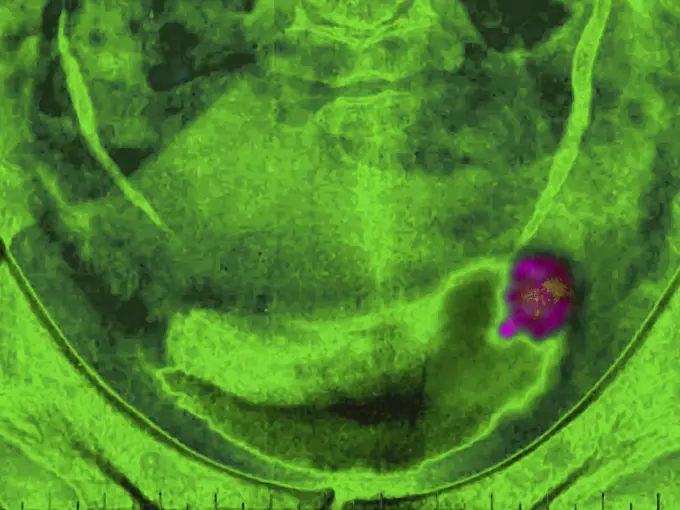
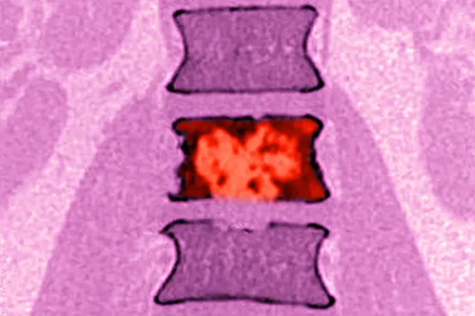
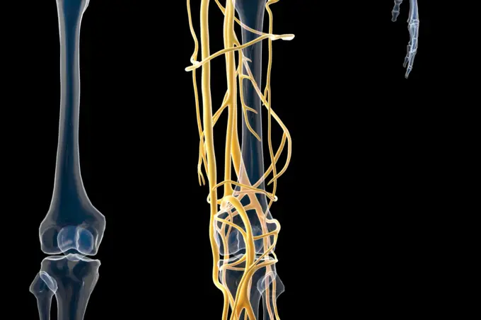
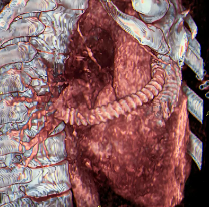
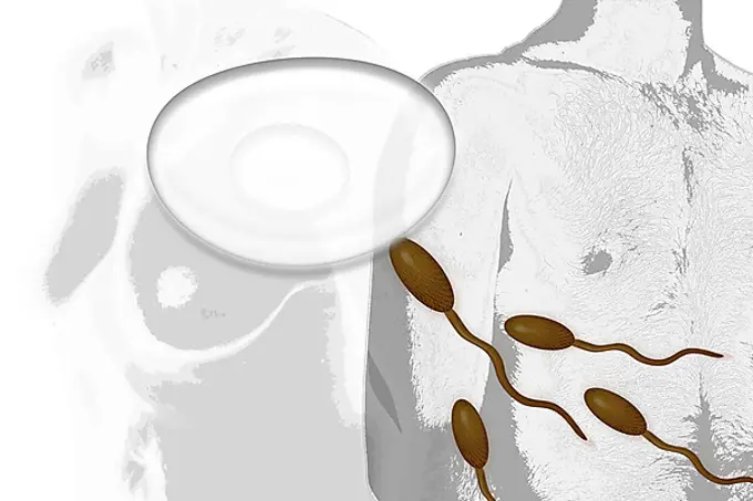
My Boards
View Pictures
  • My Boards

Medical Imaging Insights

MRI and X-ray scans depicting various medical conditions including tumors, pulmonary issues, and spinal injuries in vibrant colors and detailed cross-sections.

Tumour in the bladder (bladder papilloma), seen on a frontal cystography.
166 assets in this story
4128R-12700063
Cross section through an model of an normal upper right femur or leg bone.
6177-V53850467
4269-27765
Focused ultrasound treatment of uterine leiomyoma. ExAblateÆ 2000 system with MR guided focused ultrasound uses a combination of high-intensity ultrasound waves and Magnetic Resonance Imaging (MRI) to simultaneously locate and 3D image the uterine fibroma, treat and destroy the tissus by thermoablation. This non-invasive treatment is an alternative to surgery. Bretonneau hospital, University Hospital of Tours, France.
1525-21336589
DNA molecule
824-63206952
Lung cancer on the left pulmonary lobe, seen on a frontal x-ray of the chest.
824-63123154
Coronary angiography
4128-30418980
Colon tumour, illustration
1899-54028008
Slipped disc compression of the medulla lumbar vertebra.
824-63207420
Cancer of the liver, seen in a radial cross-section abdominal scan
6145-44950964
On March 17, 2013, NASA's Uninhabited Aerial Vehicle Synthetic Aperture Radar (UAVSAR) acquired synthetic aperture radar data over the Napo River in Ecuador and Peru.
4128R-12700068
Cross sections through models of a normal (left) and and fractured femur (right) thigh bone.
4128-19249295
Atherosclerosis, illustration.
824-63222962
Stenosis on the left popliteal artery (arteriopathy obliterating of the lower limbs). Angiography of the lower limbs.
824-63207418
Cancer of the liver, seen in a radial cross-section abdominal scan
824-63195636
Side illustration of the cervical vertebra.
1899-53511992
Retinal detachment of the left eye, visualized by radial section MRI.
1525-26227050
Colorful bright iridescent oil spill isolated on black background
824-63188624
Damaged meniscus seen on a saggital plane MRI scan.
824-63217832
Screw and plate osteosynthesis on a fracture of the fibula. Profile x-ray: tibia, fibula, malleolus and astragalus.
4128R-13817884
Kidney, CT scan. False colour 3-D computed tomography (CT) scan of a kidney in frontal view.
824-63211084
Osteoporosis with compression of L1-L2 vertebrae, seen on a sagittal plane MRI scan.
824-63217268
Pulmonary sarcoidosis seen on a frontal chest x-ray.
4128-28769218
Recurrance of cancer after surgery, PET and CT scans
824-63215953
Smoking-related lung cancer, seen on a radial section chest CT scan.
1899-53511510
Hip asymmetry, visualized by frontal pelvic x-ray.
4128R-3657
Narrowed coronary arteries, X_ray
4269-20404651
Coronary angiogram stenosed artery.
4269-39001
3D computed tomography (CT) scan of a pituitary adenoma by discovering multiple aneurysms.
4269-24998
Neurinome. 3D computed tomographic (CT) scan reconstruction of the neck showing a benign nerve tumor (neurinome) in green, localized in the vertebral canal. The acrotide arteries are seen in red, the veins in blue.
1899-53511327
Arteritis in a lower limb (progressive occlusion of the arteries supplying the limbs and organs). Angiography of a lower limb.
824-63224527
Arteritis in a lower limb (progressive occlusion of the arteries supplying the limbs and organs). Angiography of a lower limb.
1899-53511991
Retinal detachment of the left eye, visualized by radial section MRI.
824-63226741
Dislocation of the cervical vertebrae, by a spinal trauma, between the sixth and the seventh cervical vertebra visualized by MRI in sagittal section.
4128-28968800
Anatomy of the coxal bones, illustration
4128-30419354
Male bladder, illustration
1899-53510691
Stenosis on the left popliteal artery (arteriopathy obliterating of the lower limbs). Angiography of the lower limbs. (Photo by: CAVALLINI JAMES/BSIP/UIG)
4269-27130
Kyphoplasty. Color enhanced sagittal computed tomography (CT) scan of the spine showing vertebral compaction due to osteoporosis after treatment by kyphoplasty.
4128R-8004
Chromosome, artwork
4269-25250
Prostate cancer. Ultrasound scan of a cancer of the prostate, seen in green.
4128-16224160
Doll with dress, X-ray
824-57656890
Vertebral osteitis (inflammation of the bone tissue of a lumbar vertebra, caused by a bacterial infection). frontal x-ray of the lumbar vertebrae.
1525-23962832
cinema 4d rendering of The structure of human blood vessels
824-63165963
In case of stenosis, it is sometime possible to prosthetically bypass the central pulmonary arteries to revascularize the pulmonary arteries downstream of the narrowing. 3D CT scan.
4128-18929766
Spinal cord, LM
1525-24128999
science anatomy of human Blood Vessels
5507-33734454
MRI L-S spine or lumbar spine Axial T2W view with sagittal plane .
1899-53511440
Significant degenerative lumbar disc disease in an overweight 75-year-old lady. Lumbar CT scan in sagittal section.
1848-54794606
Illustration, fertilisation, a man and a woman
4297-1346
Colorized x-ray of a turtle
6145-44740192
Range 60,000 miles This image is a false-color version of a near- infrared map of lower-level clouds on the night side of Venus, obtained by the Near Infrared Mapping Spectrometer aboard Galileo. Taken at an infrared wavelength of 2.3 microns (about three times the longest wavelength visible to the human eye) the map shows the turbulent, cloudy middle atmosphere some 30-33 miles above the surface, 6-10 miles below the visible cloudtops. The image shows the radiant heat from the lower atmosphere (about 400 degrees F) shining through the sulfuric acid clouds, which appear as much as 10 times darker than the bright gaps between clouds. The colors indicate relative cloud transparency; white and red show thin cloud regions, while black and blue represent relatively this clouds. This cloud layer is at about 170 degrees F., at a pressure about 1/2 Earth's atmospheric pressure. About 2/3 of the dark hemisphere is visible, centered on longitude 350 West, with bright slivers of daylit high cloud
4409-17332809
SEMILLAS DE MANZANA - FOTOGRAFIA EN BLANCO Y NEGRO - AÑOS 60.
6145-44595838
Argyre Dust Devil Tracks
824-63207405
Prostatic adenocarcinoma seen in a transverse section ultrasound scan.
4269-27498
Epidermoid carcinoma. Colored enhanced Axial Computed Tomography (CT) scan of the calf showing a epidermoid carcinoma, a type of malignant skin  cancer. The tumor appears as the fleshy thickening at left. The section of the tibia is seen at top left, the section of the fibula, smaller, is below at right.
824-63128412
Scanner of a man pelvis and perineum. 1.Sartorius muscle (Great sartorius). 2.Right muscle of the thigh. 3.Tensor of the fascia lata. 4.Lateral great muscles of the lower limb. 5.Vastus intermedius muscle of the lower limb. 6.Obturator externus musc
824-63204740
Homogenous hepatomegaly revealed by an abdomino-pelvic scan.
4128-19246991
Gut microbiome, illustration.
4128-19490532
Gut microbiome concept
824-29057244
Pituitary adenoma visualized by MRI of the skull, in sagittal section.
1848-51501678
Plastinate, cross section through human abdomen, Body Worlds, Menschen Museum, Berlin, Germany, Europe
4128-19358525
Female breast cancer, illustration.
4269-26687
Rachis scintigram. Tomoscintigram of the rachis, showing an infectious spondylodiscitis (inflammation of the disk and vertebra) located on the lumbar vertebras L3-L4, red. Scintigraphy is a branch of diagnostic radiology that uses radioactive isotopes attached to drugs that travel to specific organs within the body. These isotopes are then imaged with a gamma camera, giving important information about an organ's function.
4128-19052438
Fatty human artery, illustration
6145-29257471
Stages of limb development
4269-25504
Breast cancer. Colored mammogram (breast X-ray) of a woman's breast showing cancerous tumor (red, left).
4269-24758
Hepatic metastasis. Axial computed Tomography (CT) scan of a liver cancer. The vertebra is visible in ocre, the ribs section in green, the kidneys (white) at bottomt. The liver (large blue mass) occupies much of the abdomen. The yellow spots are metastasis, cancerous tumors  probably associated with cancer of stomach or colon.
PREVIOUS
of 2
NEXT
2401 S. Ervay, Suite 206
Dallas, Texas 75215
United States
Get Started
Free ResearchMy BoardsMy Cart
For Creators
How To License Your ContentContributor PortalFrame of Mind
Resources
API accessPricing
Contact
+1 866 236 0087help@viewpictures.co.uk Contact form
©2026 View Pictures. All Rights Reserved. -B
This site is protected by reCAPTCHA and Google Privacy Policy and Terms of Service apply.