Medical Imaging of Abdominal Conditions

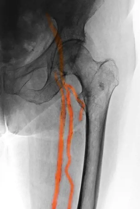
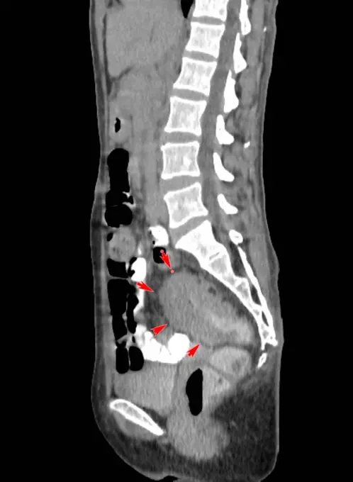
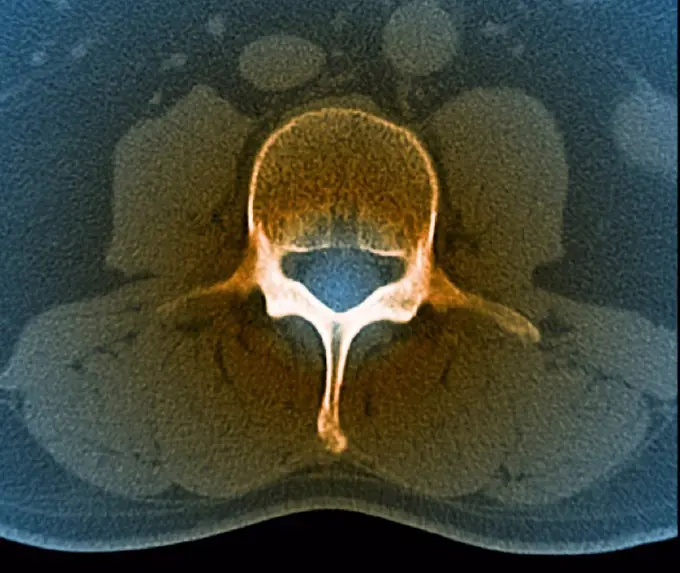
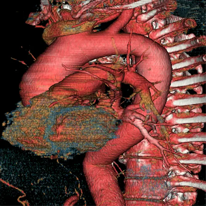
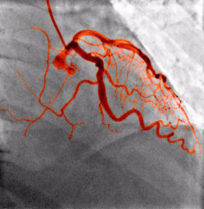
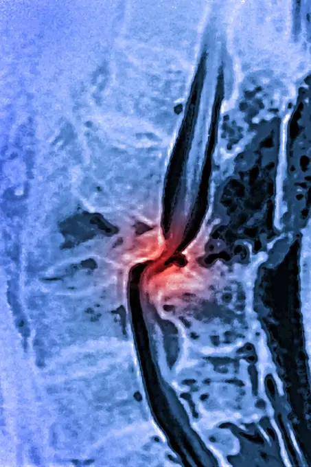
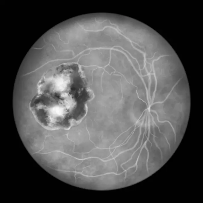
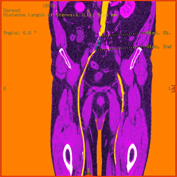
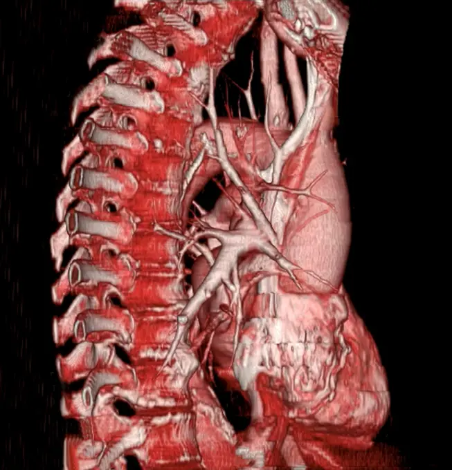
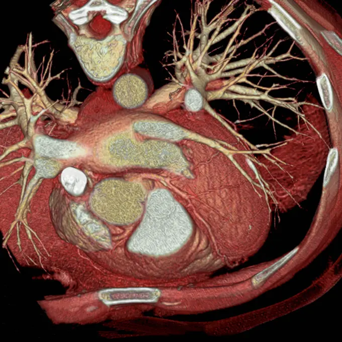
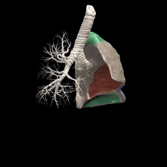
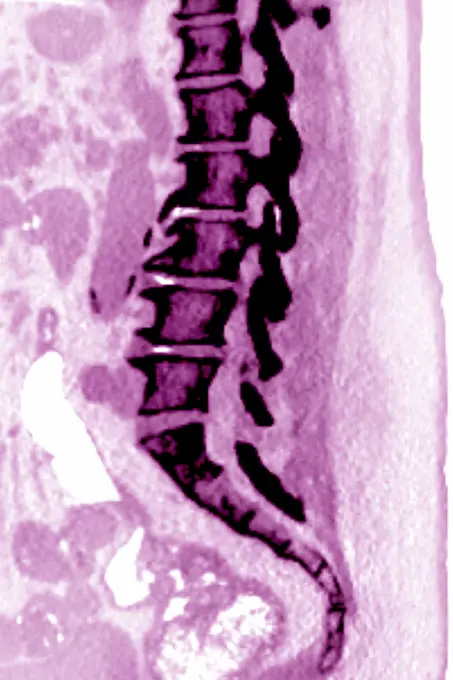
3D CT scans depicting various abdominal medical conditions, including bowel obstruction, thrombus, and hematoma, with color enhancements highlighting affected areas.

RV-PA tube (right ventricle-pulmonary artery). 20-day-old infant born without central pulmonary artery, after operation to place tube between the right ventricle and the right and left pulmonary arteries. 3D CT scan.
X-ray showing calcification of the external iliac and femoral arteries and its branchesAbdomen man, man abdomen man, man, ct recording copyright: xzoonar.com/dr.xnorberbertxlangeex 13790155Sagittal computed tomography (CT) scan of patient with rectal carcinoma showing bowel wall thickening. The patient had chronic diarrhoea and anaemia.Heart and lungs seen in a radial cross-section MRI scan.Prostate cancer (adenocarcinoma) with metastases, seen on a frontal scintiscan.Italian faience. Cup Italian faience. Cup of birth. Black and white projection plate. 1900-1930. Photograph of Jacques-Ernest Bulloz (1858-1942).Transparent Human Body with Visible Bones medical science image of human skeleton bones Copyright: xZoonar.com/StanislavxRishnyakx 14703319Image of a proton beam irradiating a brain tumor (circled in white).Coloured axial computed tomography (CT) scan of a healthy lumbar vertebra (L3) in a 33 year old patient. The front of the body is at top.A composite of several CT (computed tomography) scans shows the yellow line of a proton beam field-shaping aperture, firing at a tumor in the brain (red).Longitudinal segment through the upper part of a human femur, vintage engraved illustration. From the Universe and Humanity, 1910.Hepatic system. Three-dimensional computed tomographic (CT) scan reconstruction of the bile duct (yellow) and gall bladder (green) seen from the front. Part of the liver is seen in blue. They are comprised between the backbone in the back and the left hand side ribs in white CT scans use X-rays to take slice images through the body.A fracture of the tibia and fibula of Major William Scott-Moncrieff at the battle of Spion Kop, South Africa, April 1900. Radiograph, 1900/1904.Ascending aortic aneurysm. 3D computed tomographic (CT) scan reconstruction of aortic aneurysm. The aneurysm appears as a swollen bulge located on the ascending aorta, between the aortic arch and the heart (a part of it visible in ocre blue). An aneurysm is a bulge of a vessel due to the dilatation of its wall. A ruptured aneurysm can be fatal.Opportunity View on Sol 398 (polar)Right renal artery aneurism. Three-dimensional computed tomographic (CT) scan reconstruction of a left renal artery aneurysm.Normal blood vessels, X-rayDislocation of the lumbar vertebrae by spinal trauma, between the 3rd and 4th lumbar vertebrae, visualized by MRI in sagittal section.Retinal scar caused by toxoplasmosis, illustrationStenosis of the abdominal aorta.Aortic arch. Right side 3D computed tomographic (CT) scan reconstruction showing the heart and the aorta. The 'bended' part of the aorta is the aortic arch, from which leave, from right to left, the brachycephalic trunk, the left common carotid artery, and left sub-clavian artery. At foreground, the vasular system of the right lung.Horizontal section of a normal heart. 3D computed tomographic (CT) scan reconstruction of the upper part of the heart (seen from above). Are seen on the surface the coronary arteries, and the lung vascular system fed by the pulmonary artery (at top).X-ray of a 1 day old baby showing a normal chest, abdomen and bone structure.Kyphoplasty. Colored X-ray of the spine of a patient with vertebral compaction due to osteoporosis treated by kyphoplasty. The erosion of the bone structure of the vertebrae causes its deformation. Balloons are first inserted in the body of vertebrae and inflated in order to restaure its normal size. Surgical cement is then injected in the body (orange).Regions of the left lung IIDegenerative disk disease. Severe vertebral osteoarthritis with osteoporsis and osteophytes, seen on a side lumbar x-ray.This sagital MRI image of the lumbar spine shows a multiple myeloma, marked by bone destruction (osteolysis).Vertebral osteoarthritis with osteophytes (bone spurs) seen on a saggital plant CT-scan of the lumbar vertebrae.Released 22 June 2004 This pair of images shows part of Arsia Mons. Day/Night Infrared Pairs The image pairs presented focus on a single surface feature as seen in both the daytime and nighttime by the infrared THEMIS camera. The nighttime image (right) has been rotated 180 degrees to place north at the top. Infrared image interpretation Daytime Infrared images taken during the daytime exhibit both the morphological and thermophysical properties of the surface of Mars. Morphologic details are visible due to the effect of sun-facing slopes receiving more energy than antisun-facing slopes. This creates a warm (bright) slope and cool (dark) slope appearance that mimics the light and shadows of a visible wavelength image. Thermophysical properties are seen in that dust heats up more quickly than rocks. Thus dusty areas are bright and rocky areas are dark. Nighttime Infrared images taken during the nighttime exhibit only the thermophysical properties of the surface of Mars. The effect of suThe bones of the human fingers, wearing a finger ring. Photograph of X-ray attributed to L. Ropner, 1897.Anatomy: section of the brain showing fissures on the left cerebral hemisphere. Photograph, ca. 1900.Stomach: Kaposi's sarcomaTOPOGRAPHISCH- ANATOMISCHER ATLAS, 1872.X-Ray of Roosevelt shows bullet fired in John F. Schrank's attempted assassination of U.S. President Teddy Roosevelt in Oct. 1912.Drawings of Mars showing its 'canals' and polar ice caps from observations made at Lowell Observatory, Flagstaff, Arizona, USA 1907 July 8-12.Ultrasound scan of a baby's heartbeat.Atheromatous plaque. Transversal Doppler ultrasound scan of the femoral artery showing atheromathous plaque (yellow deposits) in the lumen. This fatty deposit narrows the vessel and restricts the blood flow (atherosclerosis)Electrocardiograph print-out.