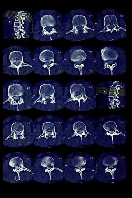
Medical Imaging of Aortic Conditions

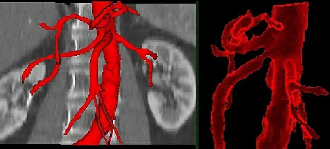
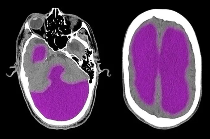
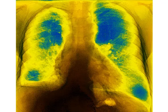
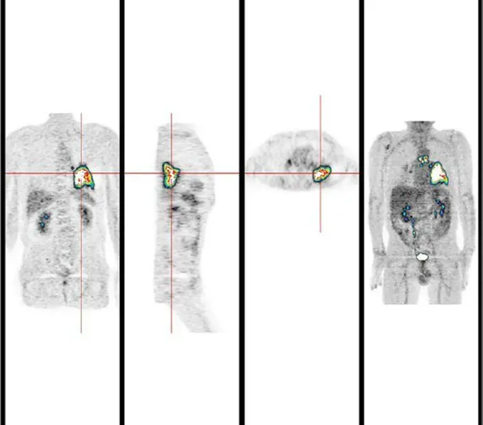
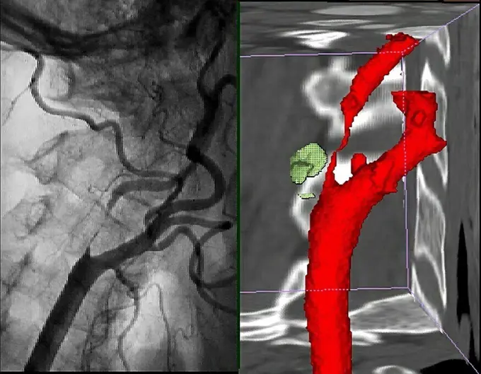
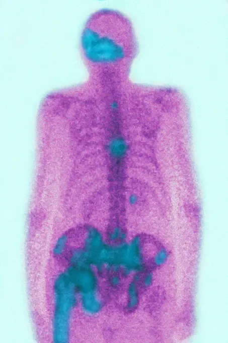
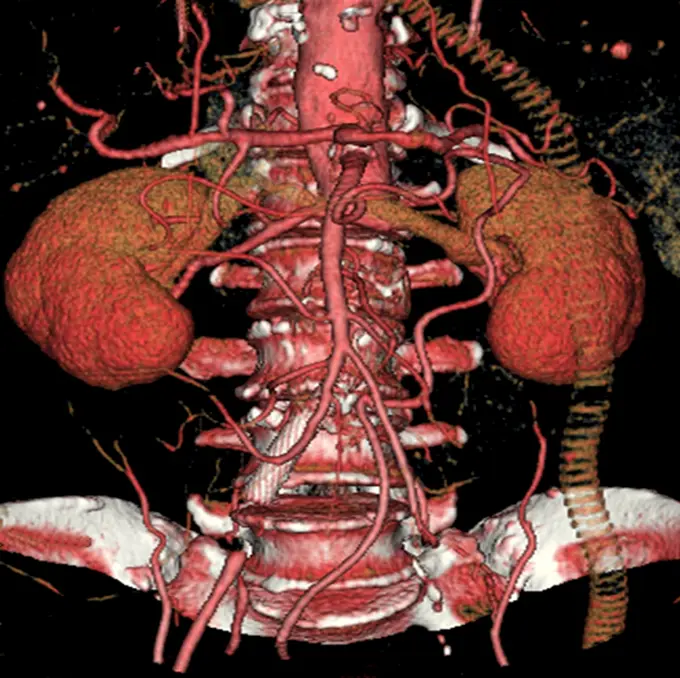
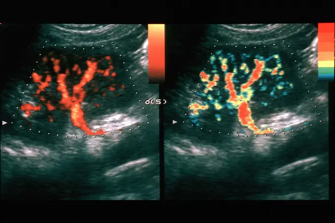
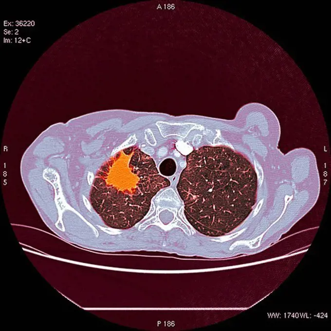
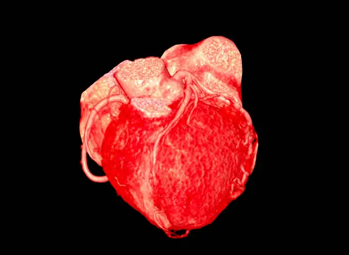
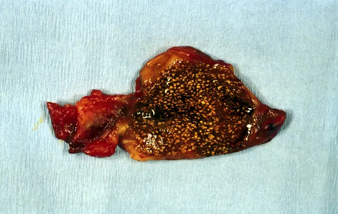
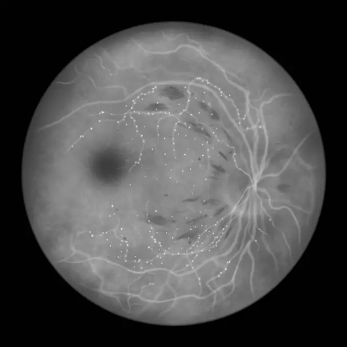
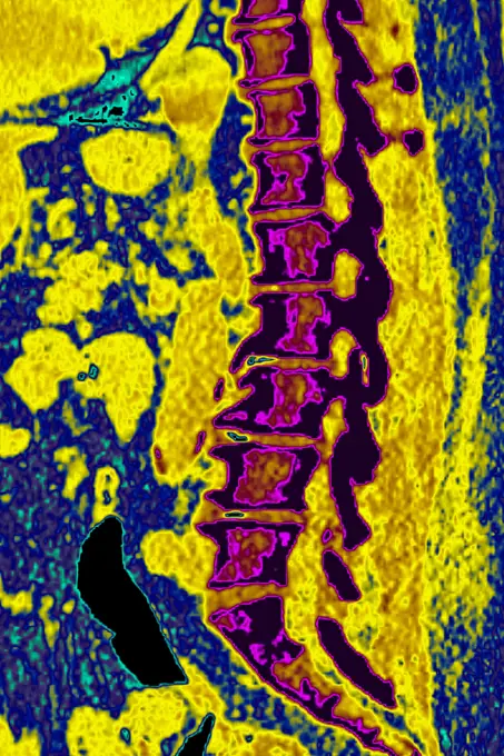
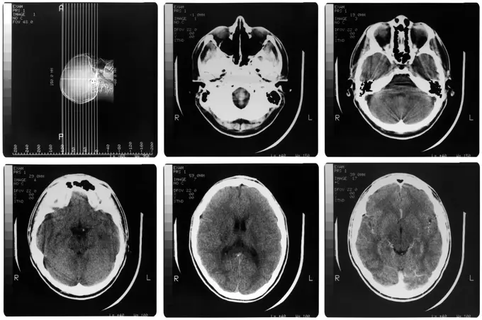
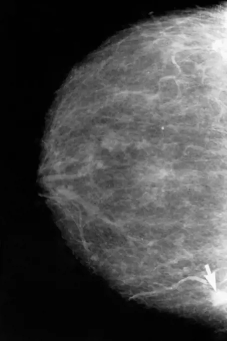
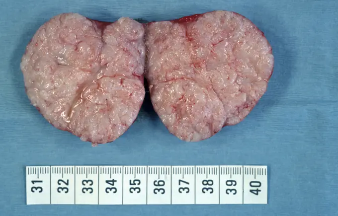
Color-enhanced scans showcasing various abdominal and thoracic medical conditions, including Takayasu's arthritis and hematome diastinum, highlighting anatomical details.

Ponytail syndrome (Herniated disc - medullary compression below the second lumbar vertebra). Cross section vertebral scanner.
Fracture of the medial cuneiform bone in the foot. Sagittal scan of the right foot.Stenosis. 3D computed tomographic (CT) scan reconstruction of an a stenosis of the superior mesenteric artery.Colorized CT scan images of a 67 year old man with severe hydrocephalusPulmonary metastases (secondary to kidney cancer), visualized by a frontal chest X-ray. (Photo by: CAVALLINI JAMES/BSIP/UIG)Pulmonary metastases (secondary to kidney cancer), visualized by a frontal chest X-ray.This frame from an animation from NASA's Cassini spacecraft shows the swirling clouds in a vortex spawned by a great northern storm on Saturn. The clouds are moving in a clockwise direction.Mucoviscidosis (accumulation of mucus in the respiratory tract), seen on a frontal x-ray of the chest.Mucoviscidosis (accumulation of mucus in the respiratory tract), seen on a frontal x-ray of the chest.Infarction in the lower left lobe following a pulmonary embolism. Frontal angiography.Calculus cholecystitis (microlithiasis), seen on an ultrasound of the gallbladder.Abdominal aorta aneurysm. hree-dimensional computed tomographic (CT) scan reconstruction of an abdominal aorta aneurysm. It appears as swollen bulge  located under the renal arteries and extending until the two comon iliac arteries. An aneurysm is a bulge of a vessel due to the dilatation of its wall. A ruptured aneurysm can be fatal.Heart. 3D computed tomographic (CT) scan reconstruction of a normal heart seen from the side. The depression seen at center is the coronary sulcus.Prostate cancer (adenocarcinoma of the prostate), visualized by ultrasound.Cross section picture of a female breast with marker point, computed tomography, magnetic resonance imaging, MRI, functional magnetic resonance imagingStent prosthesis. 3D computed tomographic (CT) scan reconstruction (side view) of an aneurysm of the abdominal aorta treated with a stent prosthesis (white).Slipped disc compression of the medulla lumbar vertebra.LUNG TUMOR. LUNG TUMOR Institute of Nuclear Medicine, University Hospital of Lille, France.   PET Scan (Positron emission tomography). Tumor in left, lower lobe.Stenosis. Angiogram at left and 3D computed tomographic (CT) scan reconstruction at right  of a stenosis of the right carotid artery.Paget's disease (bone involvement), visualized by scintigraphy.Paget's disease (bone involvement), visualized by scintigraphy.Vertebral osteoarthritis with osteophytes (bone spurs) seen on a radial CT-scan of a lumbar vertebra.Thrombosis of the cerebral venous sinuses of the dura mater, visualized by cranial MRI angiography.Brain: cytomegalovirus infectionOsteoclastoma. Front view Magnetic Resonance Image (MRI) of the knee showing a giant cell bone tumor (osteoclastoma) on the apophysis of the femur (blue and orange round shape).Slipped disc compression of the medulla lumbar vertebra.Aortic bypass. 3D computed tomographic (CT) scan reconstruction showing an occlusion of the infrarenal abdominal aorta (interruption of the aorta at the connexion with the renal arteries) treated with bypass : a prosthesis visible at right (spriral tube) maintains the blood flow from the aorta towards the iliac arteries (not seen).Hip, illustrationFrontal x-ray of a 60-year old woman's trunk.Caught in the ActTwins. Ultrasound scan of the uterus showing multiple pregnancy. The 2 embryos are visible in blue.Spinal neuroma in the 12th thoracic vertebra, seen on a sagittal section MRI scan.Kidney. Color enhanced Doppler ultrasound scan of arterial blood flow in a kidney (red).Coronal chest computed tomography (CT) scan of the lungs of a patient with lung carcinoma, showing a solid mass with an irregular border in the right ...Come Closer, CunninghamSpinal cord showing posterior sclerosis, vintage engraved illustration.Best vitelliform macular dystrophy, illustrationCancer of the liver, seen in a radial cross-section abdominal scan.LUNG CANCER, SCAN. Lungs in brownish_red with cancer in orange.LUNG CARCINOMA. LUNG CARCINOMA Institute of Nuclear Medicine, University Hospital of Lille, France.   PET Scan (Positron emission tomography). Left lung: left, upper lobectomy. epidermoid carcinoma spreading to pleura.Profile of maxillary sinuses and nose bone. 3D CT scan.Ultrasound of a 3 month old foetus.Kidney, CT scan. False colour 3-D computed tomography (CT) scan of a kidney in frontal view.Cerebral aneurysm. 3D Computed Tomography (CT) scan reconstruction of an aneurysm of the left carotid artery. An aneurysm is a bulge of a vessel due to the dilatation of its wall. A ruptured aneurysm can be fatal.NASA's Galileo orbiter looks at Jupiter's Great Red Spot, in these views from June 26, 1996. NIMS studies infrared wavelengths of light that our eye cannot see.Mammogram of the left breast of a 35-year-old patient. Slight densification in depth (Nodule) palpable here colored in yellow. The patient will have to undergo a cytological puncture.Healthy coronary arteries, CT scanSonogram    Hip bones, illustrationStrawberry gallbladder - multiple small stones in wall. Pathology specimen which has been surgically removed.Retina damage from diabetes, illustrationCoronary angiogram stenosed artery.Doppler ultrasound scan of liver. Power Doppler ultrasound scan of blood flow in a normal liver. Doppler ultrasound scanning detects moving fluids, such as blood, by the frequency change they cause when reflecting high-frequency sound waves.Cerebral aneurysm. 3D Computed Tomography (CT) scan reconstruction of a giant aneurysm of the right carotid artery. An aneurysm is a bulge of a vessel due to the dilatation of its wall. A ruptured aneurysm can be fatal.Degenerative disk disease. Severe vertebral osteoarthritis with osteoporsis and osteophytes, seen on a side lumbar x-ray.Retina damage from diabetes, illustrationProstatic adenocarcinoma seen in a transverse section ultrasound scan.A Changing Picture of ErosLumbar vertebrae, herniated disc, slice imaging, illustrationADENOCARCINOMA OF COLON. ADENOCARCINOMA OF COLON Institute of Nuclear Medicine, University Hospital of Lille, France.   PET Scan (Positron emission tomography).  Adenocarcinoma right side of colon, due to Metastasis of lung tumor (right middle lobe).This view was obtained during a distant flyby, above the moon's surface. Although NASA's Cassini will have no further close, targeted flybys of Titan, the spacecraft continues to observe the giant moon and its atmosphere from a distance.Medical Xray Medical X ray imaging of human brain skull bones Copyright: xZoonar.com/ClaudioxDiviziax 2671413A mammogram of a fatty breast with an obvious cancer, indicated by an arrow.Close-up of an x-ray image of a human breast affected with cancerFibroadenoma of breast ( benign tumour ) sectioned. Pathology specimen which has been surgically removed.