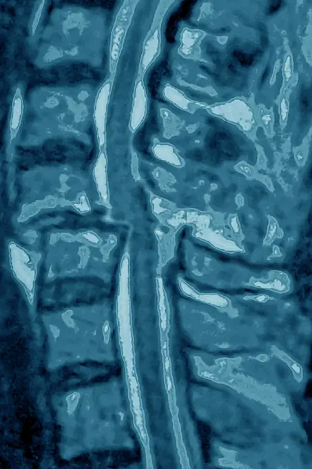
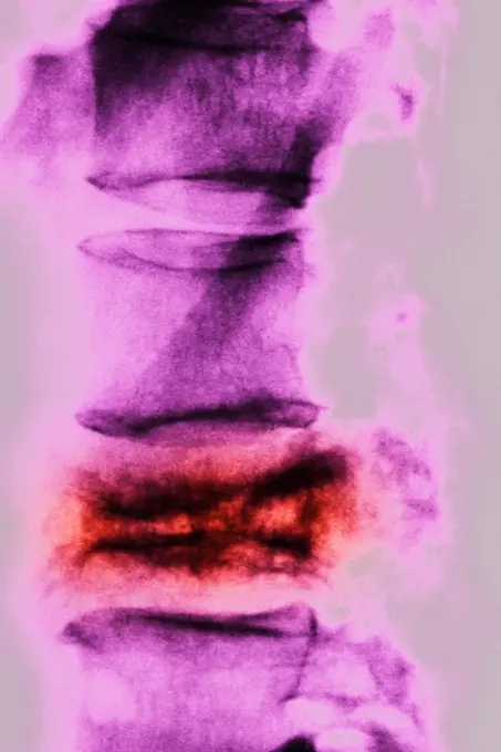
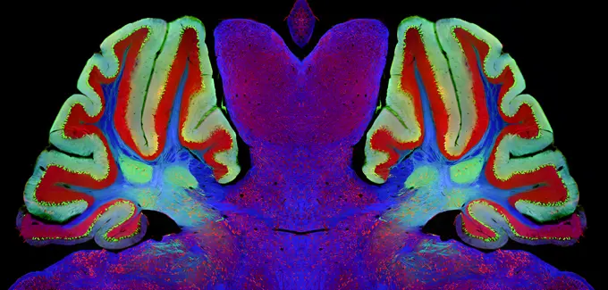
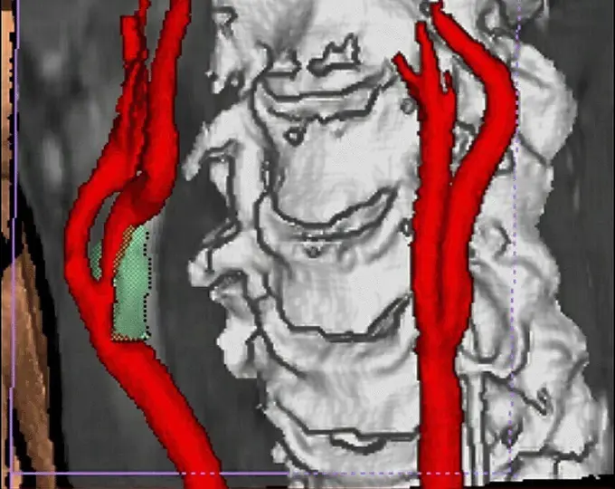
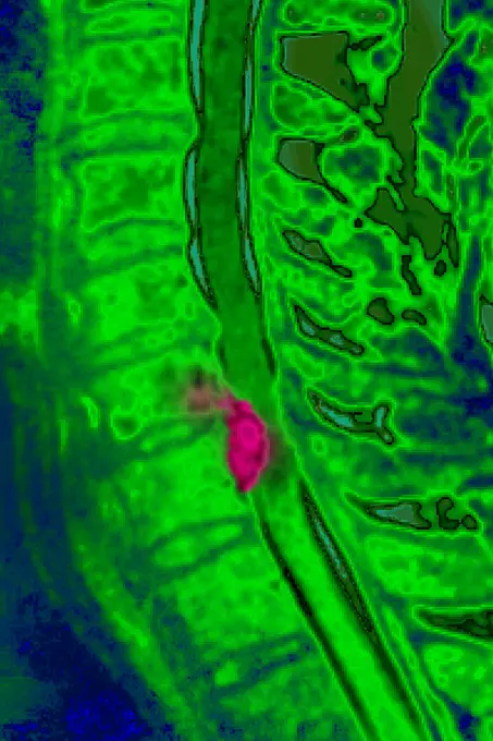
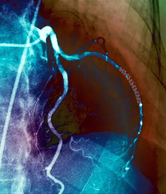
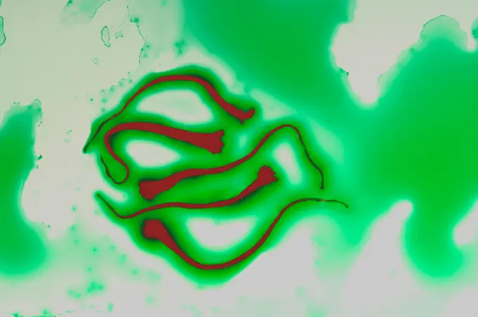
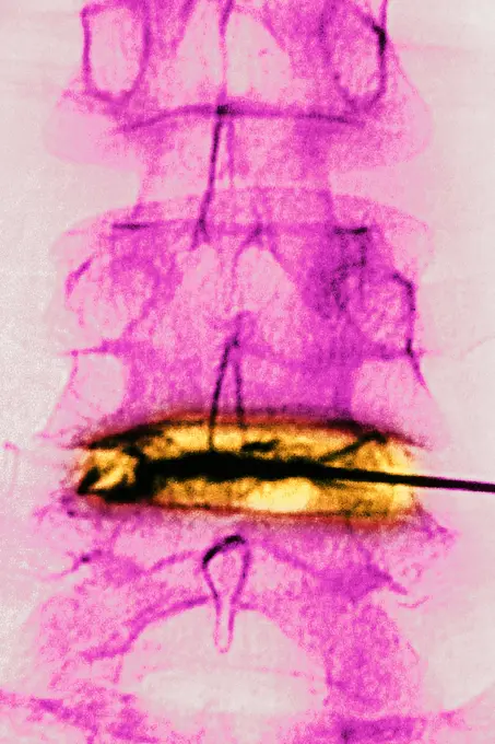
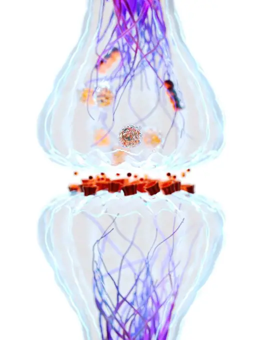
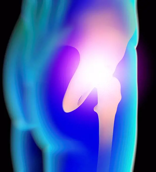
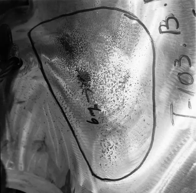
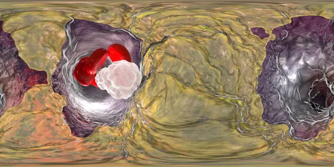
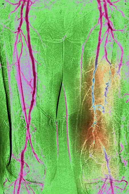
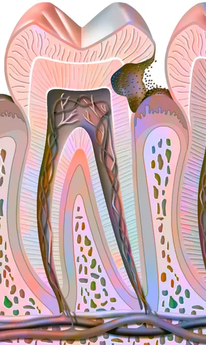
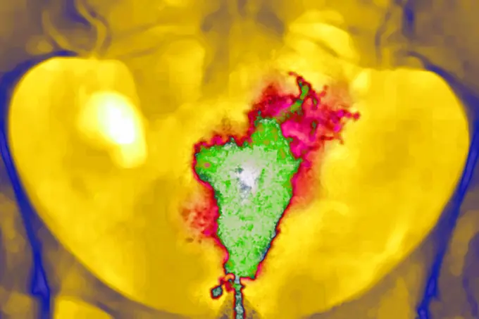
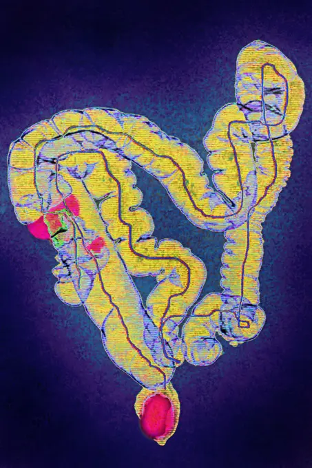
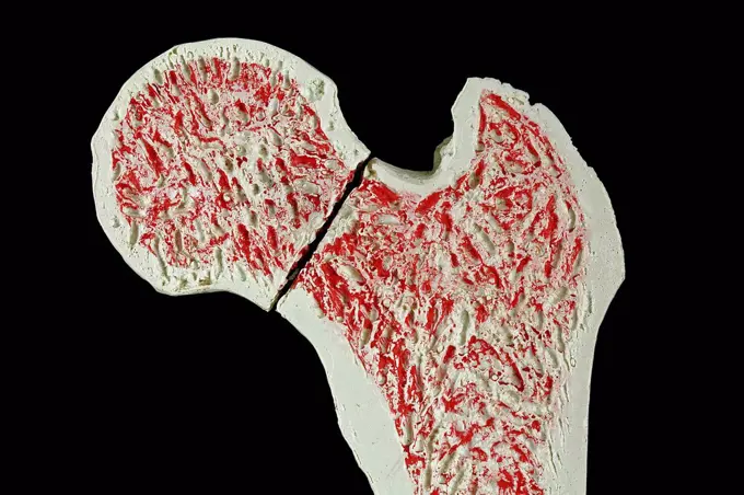
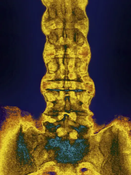
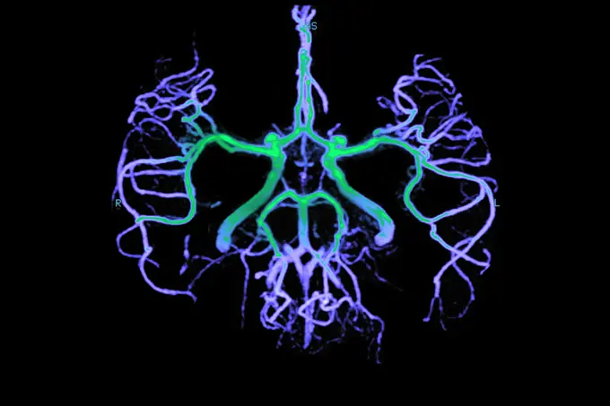
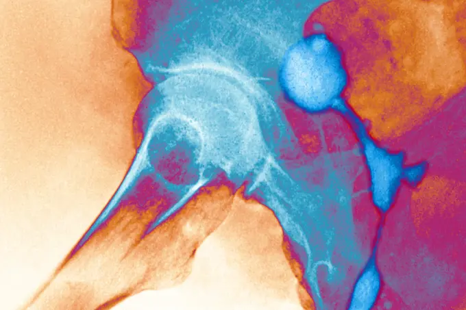
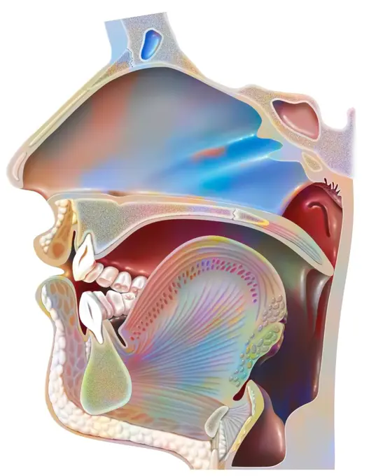
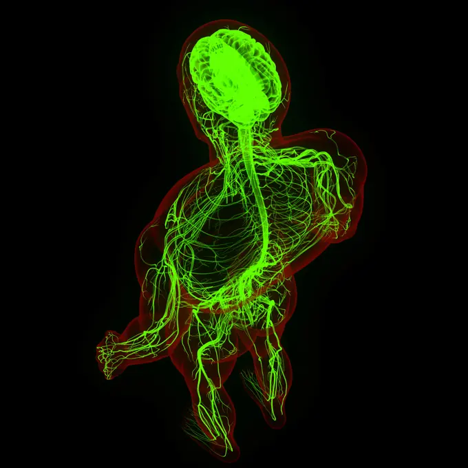
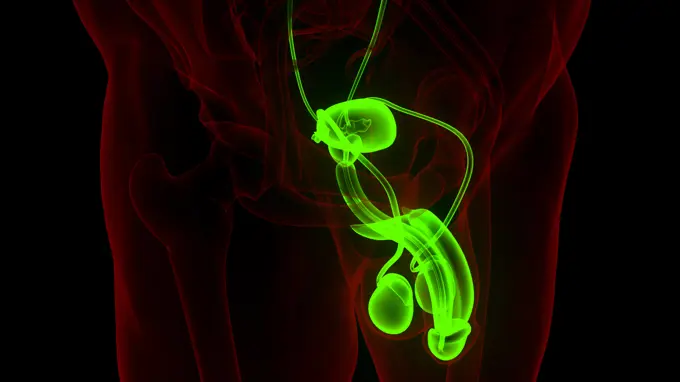
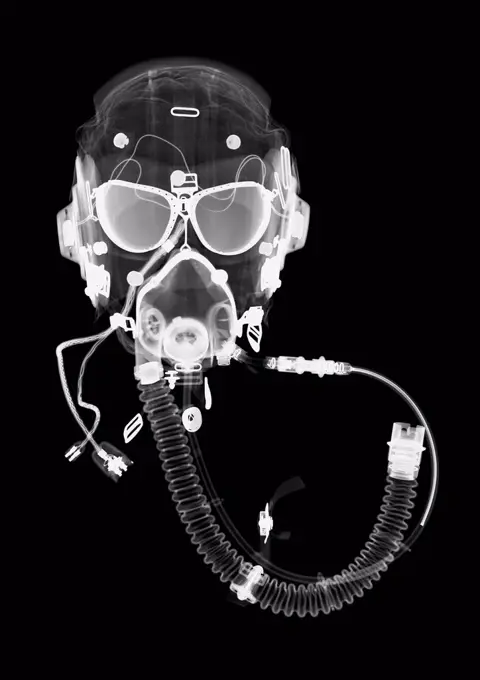
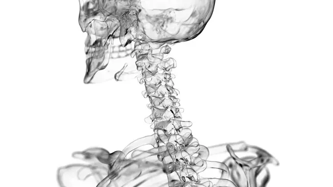
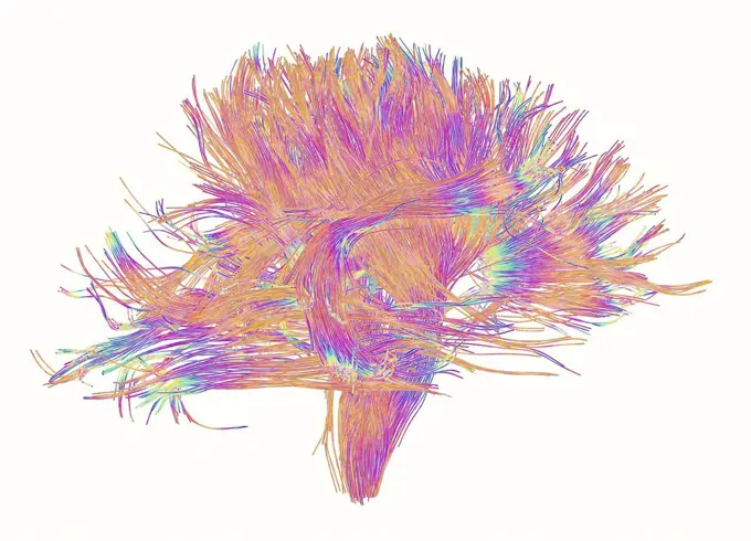
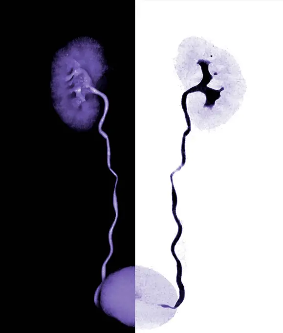
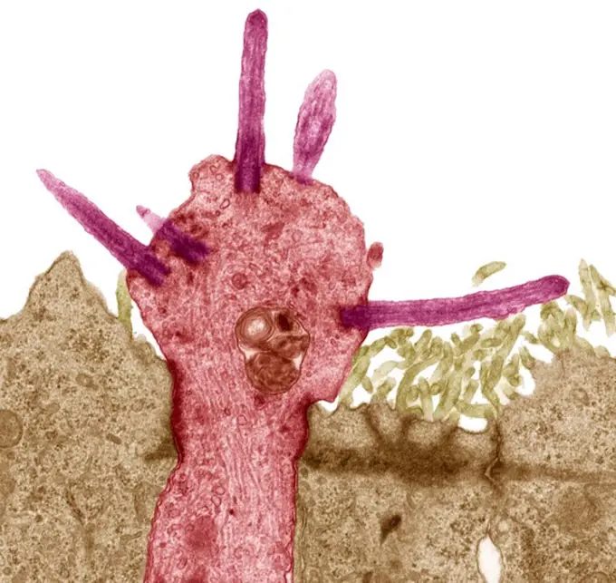
My Boards
View Pictures
  • My Boards

Medical Imaging of Spine Conditions

MRI and radiograph images highlighting cervical spine injuries and diseases, including Paget's disease and multiple sclerosis, illustrated in vivid colors.

Purple stem cell.
Purple stem cell.
172 assets in this story
824-63226740
Dislocation of the cervical vertebrae, by a spinal trauma, between the sixth and the seventh cervical vertebra visualized by MRI in sagittal section.
4378-1910
Purple stem cell.
1899-53511921
Paget's disease (bone deformity of a thoracic vertebra), revealed by a sagittal radiograph of the thorax. (Photo by: CAVALLINI JAMES/BSIP/UIG)
824-63226770
Multiple sclerosis (MS) (autoimmune disease that affects the central nervous system by dysfunction of the immune system and leads to lesions that cause motor, sensory, cognitive, visual or even sphincter disturbances). MRI of the cervical vertebrae in sagittal section.
824-63227341
Mirror image of a mouse brain with Niemann-Pick type C1 (NPC1) neurodegenerative disease. A major way to assess the impact of a therapeutic potential on the central nervous system is to assess cerebellar pathology in mice. Eunice Kennedy Shriver's National Institute of Child Health and Human Development (NICHD) researchers have found that an experimental drug can stabilize or slow the progression of NPC1.
6177-V54016779
4128-30415995
Human liver, illustration
4269-24961
Stenosis of carotid artery. 3D computed tomographic (CT) scan reconstruction of an atheromatous stenosis of the right internal carotid artery.
824-63206273
Herniated disk on the seventh cervical vertebra, seen on a sagittal section MRI scan.
4269-24953
Stenosis. 3D computed tomographic (CT) scan reconstruction of the right renal artery stenosis (yellow), responsible for arterial hypertension and renal ischemia.
1899-53512016
Normal knee visualized by MRI in sagittal section.
4128R-30966
Coronary stent. Coloured angiogram (blood vessel X-ray) of the coronary arteries of a 52 year old patient. The artery at right has had a stent placed ...
4128-V58557754
824-29057210
Lung cancer (right), visualized by an AP chest X-ray.
824-63225961
Right coronary artery visualized by frontal thoracic angiography.
824-63164485
Representation of the airways in the chimpanzee.
1848-49331301
The knee is a joint on the leg and has a kneecap
6188-63949552
Spices, saffron thread in water on green background Copyright: xHarixMahidhar/DinodiaxPhotox
1899-53511996
Retinal detachment of the left eye, visualized by radial section MRI.
824-63198906
Sinusitis in the left maxillary sinus, seen on a frontal, cross-section MRI scan.
824-63225319
Corticosteroid infiltration between the fourth and fifth lumbar vertebrae, done under radiographic control
4128-V58562683
824-63188615
Osteoarthritis of the lumbar vertebrae seen on a side x-ray.
4269-27167
Osteoclastoma. Magnetic Resonance Image (IRM) of the knee showing a giant cell bone tumor (osteoclastoma) on the apophysis of the femur vascularized by the popliteal artery and its branches.
824-63206272
Herniated disk on the seventh cervical vertebra, seen on a sagittal section MRI scan.
4128-15981229
Synapse, computer illustration.
4297-1038
Lateral view of the skull of a 43 year old woman without any teeth
4269-24984
Lung cancer. Three-dimensional computed tomography (CT) scan of upper thorax showing a Pancoast's syndrome associated with a lung cancer.
4128R-11310297
Human hip joint, computer artwork.
4269-25397
Stent prosthesis. Virtual endoscopic view of a stent prosthesis. Stent are mesh-wire like endoprosthesis (pink) inserted into a vessel during angioplasty to support tissue. It is used to treat stenosis or aneurysm of blood vessels.
4378-20013988
Basophil Cell
824-72488713
Illustration of leg pain, and more particularly venous insufficiency (here, at the level of the internal saphenous vein).
4269-27140
Bone cyst. Color enhanced axial computed tomography (CT) scan of the shoulder with aneurysmal bone cyst on the left clavicle.
1525-56190053
Nephron, the structure that actually produces urine in the process of removing waste and excess substances from blood. 3D illustration. Nephron, the structure that actually produces urine in the process of removing waste and excess substances from blood.
6145-51945804
T103 (depicted - name).T103 (depicted - name)
1899-53511747
Calcific aortic atheroma, visualized by chest echography. (Photo by: CAVALLINI JAMES/BSIP/UIG)
4128-18800611
Atherosclerosis, illustration
824-63222963
Stenosis on the left popliteal artery (arteriopathy obliterating of the lower limbs). Angiography of the lower limbs.
1848-49194846
Illustration lung man tumour
824-63163759
Second stage tooth decay (dentin damage).
1848-77403208
3D Illustration Concept of Human Digestive System Anatomy
824-63226001
Uterus cancer. Frontal pelvis x-ray.
824-63212831
Colon cancer (tumors in the caecum and rectum). Visualisation using 3D reconstruction from a CT scan (virtual coloscopy).
4128R-12700030
Cross section through an model of an fracture in the neck of the femur (thigh bone).
4269-25386
Angioplasty. 3D computed tomographic (CT) scan reconstruction showing a disjunction of a stent prosthesis placed into the aorta and the beginning of the two iliac arteries.
824-63188655
Ankylosing spondylitis in the lumbar vertebrae, seen on a frontal x-ray.
1848-49387889
Liver The organ is responsible for metabolism, illustration, natural science, biology, illustration, natural science, biology, abstract art
1525-56853468
Organs surrounding the female reproductive organ 3D rendering. Organs surrounding the female reproductive organ
6145-44625615
This anaglyph from the base of 'Burns Cliff' in the inner wall of 'Endurance Crater' combines several frames taken by Opportunity's navigation camera during the NASA rover's 280th martian day (Nov. 6, 2004). 3D glasses are necessary to view this image.
824-63128438
The circle of Willis is an arterial circle located at the base of the brain, from which depart the main arteries that irrigate the nervous tissue contained in the brain. Angio MRI. Axial view. See image n0756905 on the site.
1525-56187595
Details of Ice
824-63175219
CT scan of cholesteatoma in middle ear.
4269-24935
Dissecting aortic aneurysm. Three-dimensional computed tomographic (CT) scan reconstruction of a dissecting aneurysm of the aortic arch.
4128-15825439
Human brain, illustration.
824-63217833
Screw and plate osteosynthesis on a fracture of the fibula. Profile x-ray: tibia, fibula, malleolus and astragalus.
1525-22787236
science anatomy tomography scan of human body. Anatomy Tomography Scan of Human Body
4239-69140949
Comb Jelly in blackwater dive at night, West Palm Beach, Florida.
1848-49331319
The thyroid gland is located in the neck and has many tasks in the areas of metabolism, hormones and growth
1899-65662219
Retinal detachment occurs when the tissue at the back of the eye becomes detached from the layer of blood vessels that supply the eye with oxygen. Sagittal MRI of the eye.
824-63214275
Urethral fistulae. Frontal pelvic x-ray.
1899-53509450
Anatomy of the ear showing the eardrum, ossicles, hammer, anvil. .
4269-25719
Hip osteoarthritis. X-ray showing arthritis of the hip. At lower left and right are the thigh bones., above is the pelvis. Arthritis is a joint disease that can cause cartilage destruction (seen as a reduction in the joint space, yellow), bone erosions, and tendon inflammation.
824-63227090
Scientists have developed new image processing techniques for microscopes that can reduce post-processing time by up to several thousand times. The researchers come from the National Institutes of Health with collaborators from the University of Chicago and Zhejiang University, China.
6145-29247254
Foetal MRI scan
1899-53509449
Position of the mouth for the pronunciation of the 'U' in phonation.
6188-67688611
Zentrales Organ des menschlichen Nervensystems Anatomie des Gehirns 3D Illustration Concept of Central Organ of Human Nervous System Brain Anatomy Copyright: xZoonar.com/magicminex 23022750
6188-67688608
Anatomie des männlichen Fortpflanzungssystems 3D Illustration Concept of Male Reproductive System Anatomy Copyright: xZoonar.com/magicminex 23022742
824-63165105
Internal hemorrhoidal plexus and location of internal hemorrhoids.
5507-47241527
Human skeleton. Vector
6019-V19393528
1525-22344005
Diagrams of human organs superimposed on a human spine
824-292
X-Ray of Hands Showing Rheumatoid Arthritis
4128-16375084
Pilot helmet, X-ray
4128-19056335
Neck joint, illustration
1848-77402067
3D Illustration Concept of Central Organ of Human Nervous System Brain Anatomy
1899-54028012
Lesion of the meniscus posterior horn. Sagittal section MRI scan of the knee.
4128R-11543222
White matter fibres. Coloured 3D diffusion spectral imaging (DSI) scan of the bundles of white matter nerve fibres in the brain, depicting mirror neur...
824-63126567
Aneurysm of the brain, angiogr.
4269-25009
Left primitive iliac artery aneurysm. Three-dimensional computed tomographic (CT) scan reconstruction of left primitive iliac arteryl aneurysm located (red ballon shaped swelling).
1848-53881747
Maxima Clam or Giant Clam (Tridacna maxima), with its breathing hole open showing the gills for filtration, Queensland, Queensland, Australia, Oceania
1525-56854606
Stage II testicular cancer is characterized by the spread of cancer to nearby regional lymph nodes. 3D rendering. Stage II testicular cancer is characterized by the spread of cancer to nearby regional lymph nodes.
1525-27250465
Microscope virus cell. Pandemic bacteria pathogen medical health risk, Corona COVID-19 Alert SOS, immunology, virology, epidemiology concept. 3D rendering.
4128-30419907
Urinary system, CT scan
1525-21046587
milk splash
4128R-8073
Smell receptors, TEM
4378-2501
Wire frame anatomical model showing the bones which form the upper spine.
4128R-12580226
Male nervous system, computer illustration.
4128R-15536340
Illustration of an abstract plexus colon
4070-21285595
Bullethead parrotfish (Chlororus sordidus) tail. Red Sea, Egypt
824-63227398
In this image, the researchers used a newly developed polarized light microscope to plot the spatial orientation of neurons in a thin section of the mouse midbrain. Neurons that stretch horizontally displayed in green, while those oriented at a 45 degree angle are pinkish red and those at 225 degrees are purplish blue. These colors do not involve staining or labeling the cells with fluorescent markers: the colors are emitted strictly from light interacting with the physical orientation of each neuron.
1746-30004499
Sample of Fluorite glowing under U.V. light
4269-27604
Virtual colonoscopy. A 3D computed tomography (CT) scan of the colon, showing a small polyp.
4269-25358
Thoracoabdominal aortic aneurysm. 3D computed tomographic (CT) scan reconstruction of a thoracoabdominal aortic aneurysm. The aneurysm appears as a swollen bulge located on the descending aorta, just after the aortic arch. The upper part of the heart is visible in pink and blue. An aneurysm is a bulge of a vessel due to the dilatation of its wall. A ruptured aneurysm can be fatal
4128-16224844
Shark jaw, X-ray
4128R-23011
Male cardiovascular system, computer artwork.
824-63164195
Hepatic lobule consisting of hepatocytes, a central vein, centrilobular vein.
4269-25278
Dissecting aneurysm of the aorta. Three-dimensional computed tomographic (CT) scan reconstruction of a dissecting aneurysm of the aorta from. In this type of aneurysm, the inner wall of the aorta (intima) detaches from the outter wall (media). The blood invades the created space and generate a bulge that can break. The aneurysm is visible as a longitudinal  furrow that extends from the thoracic aorta to the common iliac arteries.
1439-57940175
Closeup image of an insect under a microscope showing intricate details
4128-15980871
Breast anatomy, computer illustration.
4378-4072
Bronchial Branches
PREVIOUS
of 2
NEXT
2401 S. Ervay, Suite 206
Dallas, Texas 75215
United States
Get Started
Free ResearchMy BoardsMy Cart
For Creators
How To License Your ContentContributor PortalFrame of Mind
Resources
API accessPricing
Contact
+1 866 236 0087help@viewpictures.co.uk Contact form
©2026 View Pictures. All Rights Reserved. -A
This site is protected by reCAPTCHA and Google Privacy Policy and Terms of Service apply.