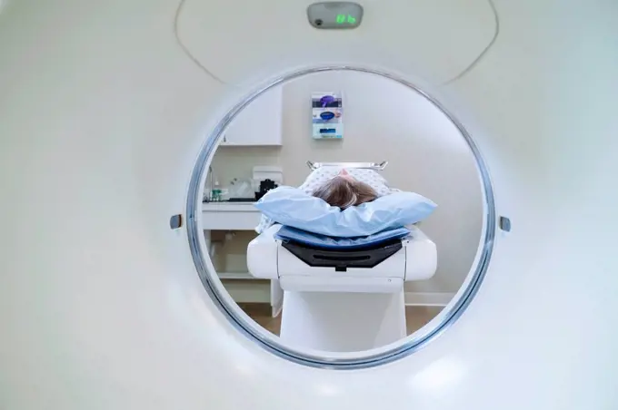
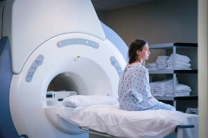
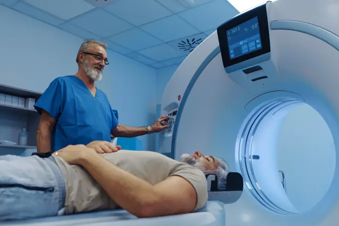
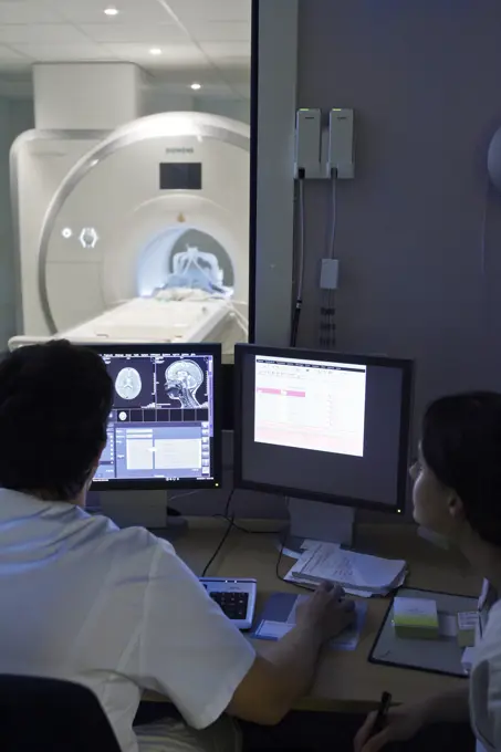
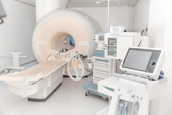
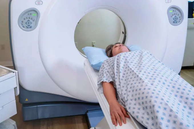
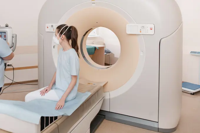
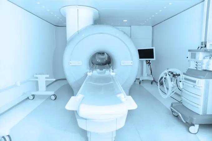
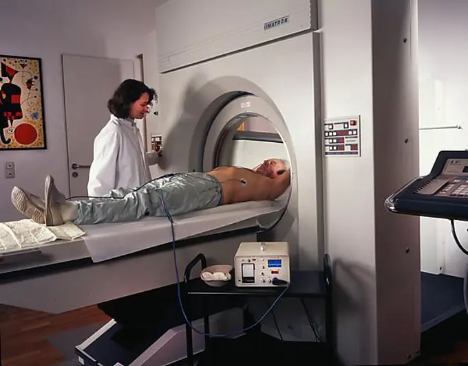
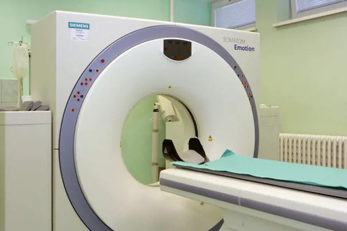
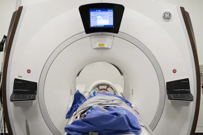
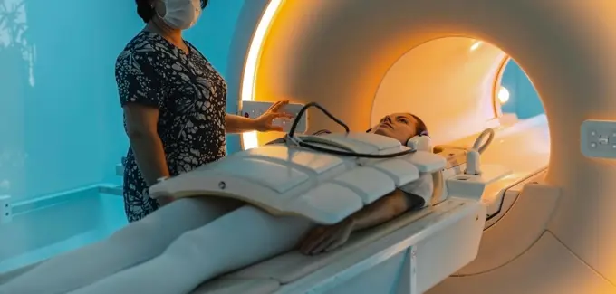
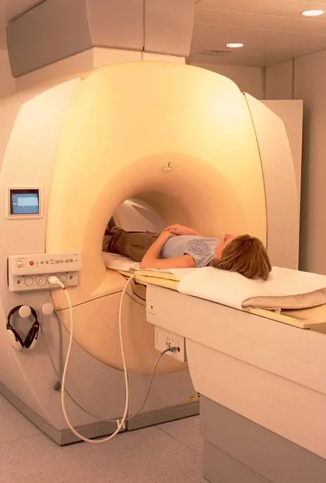
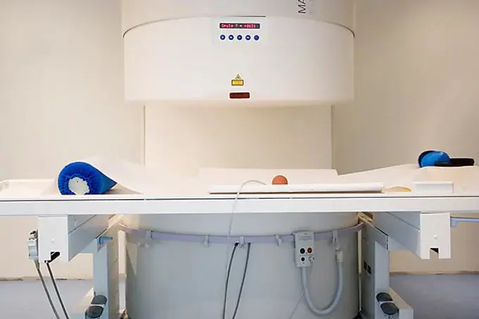
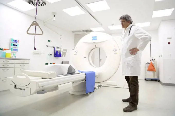
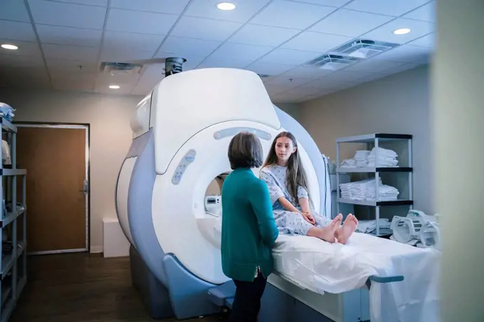
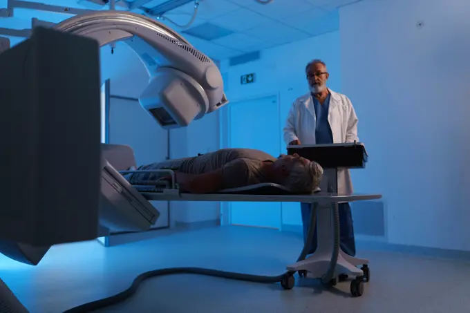
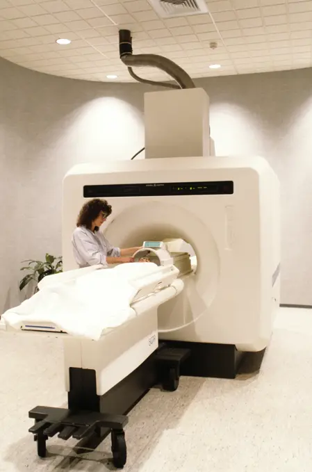
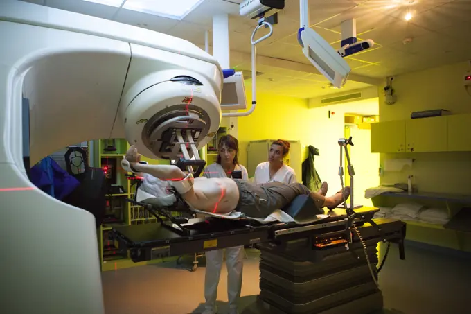
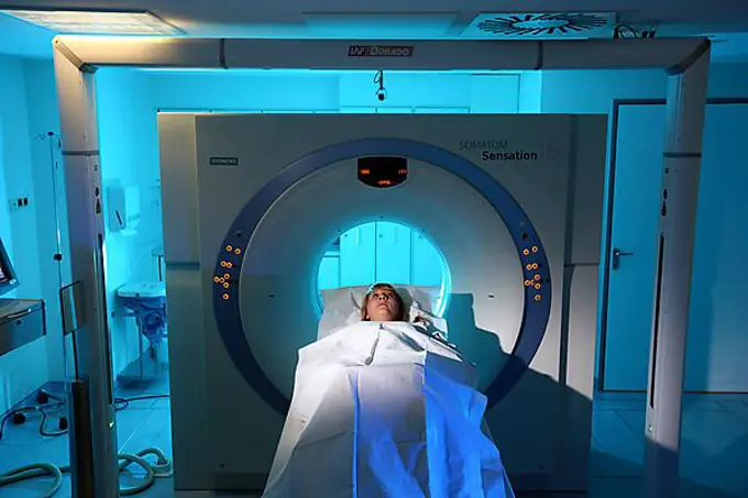
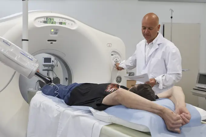
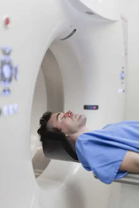
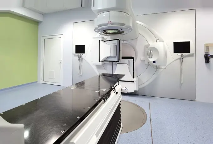
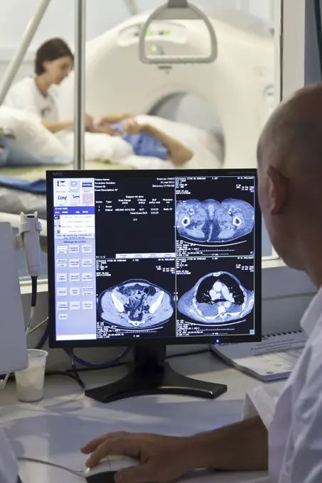
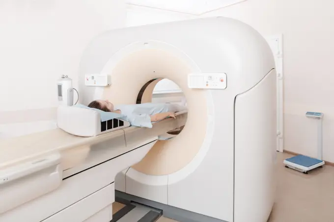
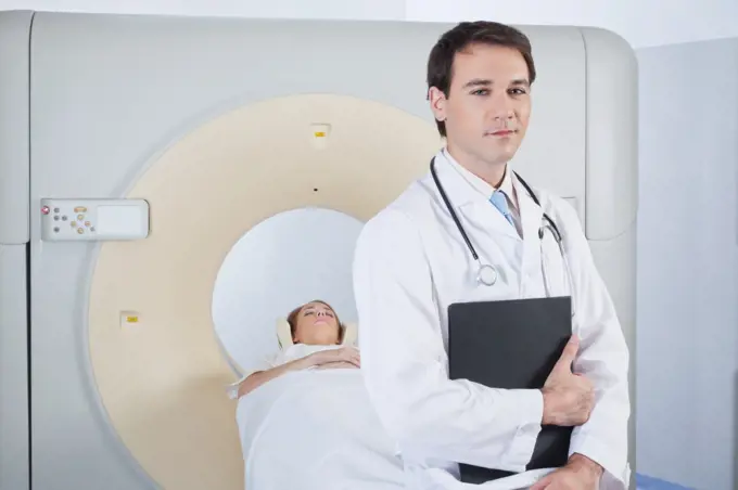
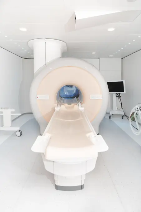
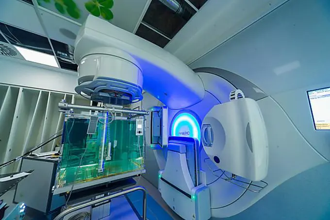
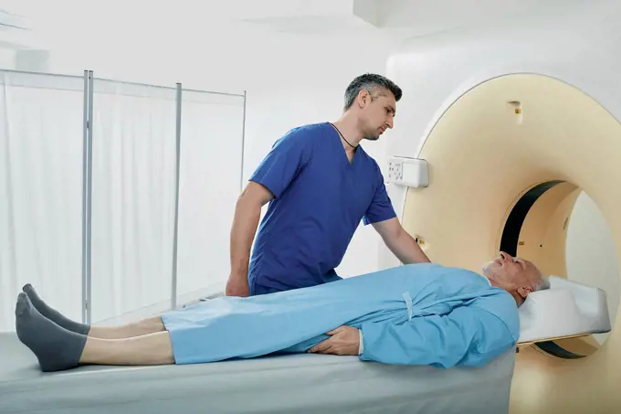
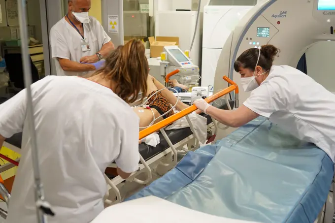
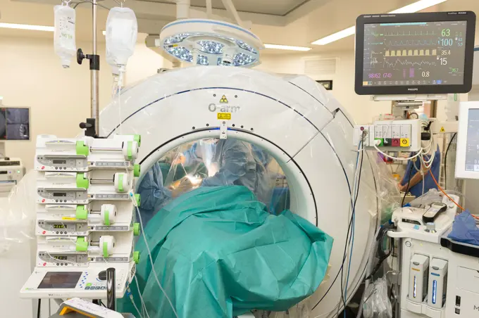
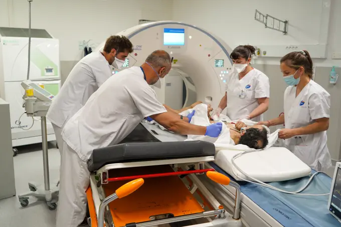
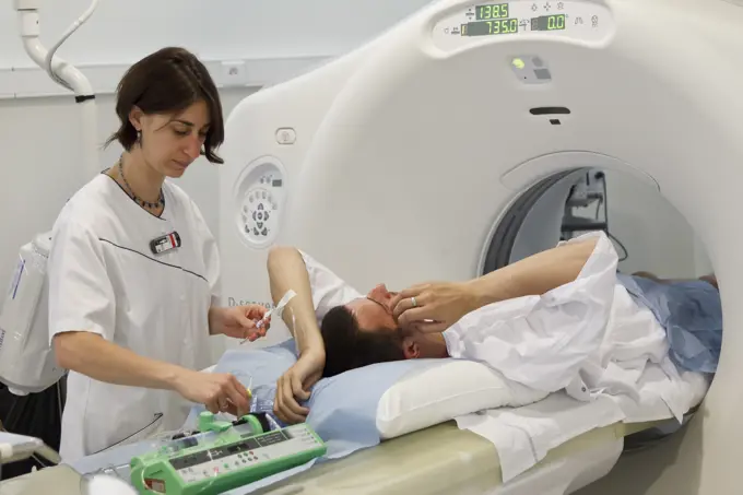
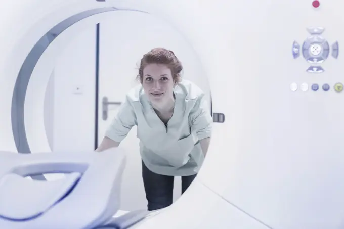
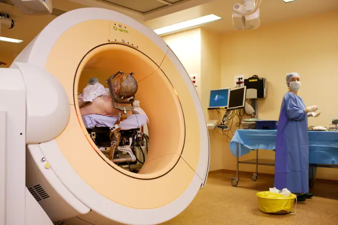
My Boards
View Pictures
  • My Boards

Medical Imaging Procedures

A series of images showcasing patients undergoing various medical imaging techniques, including MRI, CT scans, and radiotherapy in clinical settings.

Caucasian patient laying on scanner table
211 assets in this story
824-63181508
Digital medical imaging centre in Paris, France. Skull MRI.
824-63181515
Digital medical imaging centre in Paris, France. Skull MRI.
4269-3544
Department of Functional Neurosurgery and stereotaxy of the Pr Peragut, Timone hospital, Marseille, France. Treatment of a meningioma (benign brain tumor) with a Gamma knife. Here, patient at the end of treatment.
4269-33745
Radiotherapy radiographer and a female patient treated to a stomach cancer with radiotherapy. Latest generation medical particle accelerator. Radiotherapy center, pole of Oncology, Hospital of Bordeaux. Hospital Haut-Leveque.
4269-6302
Patient undergoing heart 3D CT scan, Department of Medical Imaging, Centre Cardiologique du Nord, St Denis, France. The Lightspeed VCT 64-Slice CT Scanner produces 64 slices per rotation, and reduces radiation exposure by 70%.
1589R-14623168
Caucasian patient laying on scanner table
824-63196946
Reportage in a radiation oncology service in Switzerland. The service is equipped with the latest linear accelerator, the Truebeam. A technician sets a patient up for prostate cancer treatment.
824-63211811
Report from the radiotherapy department of a hospital in Savoie. Radiotherapy session to treat brain metastases. A mask is applied to the patient to prevent movement during the treatment. The treatment is carried out with the Truebeam.
1589R-14623279
Pensive Caucasian girl sitting on scanner table
6188-68410958
Male patient undergoing X-ray examination using tilting radiographic wall stand. Male patient undergoing X-ray examination using a tilting radiographic wall stand. model released
824-63173339
Brain, mri
1660R-54942
Aerial of woman on diagnostic bed
824-63210927
Report on PET imaging. Positron Emission Tomography, or PET Scan, is used for the diagnosis and monitoring of cancer patients. This method makes it possible to detect a tumor thanks to a radioactive product which accumulates in an excessive way in the cells presenting a pathological hypermetabolism.
824-63206519
Reportage in a radiology service in a hospital in Haute-Savoie, France. MRI scan.
1848-49329147
A computer tomograph (CT) is an imaging procedure in radiology, here on 21.11.1995 in Schwerte in a radiology practice, and is used for modern diagnostics in medicine, DEU, Germany, Schwerte, Europe
6188-60030192
Medical CT or MRI Scan in the modern hospital laboratory. Interior of radiography department. Technologically advanced equipment in white room. Magnetic resonance diagnostics machine Medical CT or MRI Scan in the modern hospital laboratory. Interior of radiography department. Technologically advanced equipment in white room. Magnetic resonance diagnostics machine. ,model released, Symbolfoto Copyright: xZoonar.com/MaksimxPavlovx 17853291 ,model released, Symbolfoto ,property released
824-63196950
Reportage in a radiation oncology service in Switzerland. The service is equipped with the latest linear accelerator, the Truebeam. A technician sets a patient up for prostate cancer treatment.
824-63181510
Digital medical imaging centre in Paris, France. Skull MRI.
824-63161117
MRI or magnetic resonance imaging of a patient's hips.
1589R-14623157
Caucasian patient laying on scanner table
824-63181704
Digital medical imaging centre in Paris, France. Cranial CT scan
824-63206536
Reportage in a radiology service in a hospital in Haute-Savoie, France. MRI scan.
6188-60030237
Medical CT or MRI Scan with a patient in the modern hospital laboratory. Interior of radiography department. Technologically advanced equipment in white room. Magnetic resonance diagnostics machine Medical CT or MRI Scan with a patient in the modern hospital laboratory. Interior of radiography department. Technologically advanced equipment in white room. Magnetic resonance diagnostics machine. ,model released, Symbolfoto Copyright: xZoonar.com/MaksimxPavlovx 17856681 ,model released, Symbolfoto ,property released
824-63206544
Reportage in a radiology service in a hospital in Haute-Savoie, France. Scanner.
4269-5304
Patient undergoing a MRI scan (Magnetic Resonance Imaging).
6188-60030525
Medical CT or MRI Scan in the modern hospital laboratory. Interior of radiography department. Technologically advanced equipment in white room. Magnetic resonance diagnostics machine Medical CT or MRI Scan in the modern hospital laboratory. Interior of radiography department. Technologically advanced equipment in white room. Magnetic resonance diagnostics machine. Copyright: xZoonar.com/MariyaxLvovax 17882726
824-63206524
Reportage in a radiology service in a hospital in Haute-Savoie, France. MRI scan.
4269-6300
Patient undergoing heart 3D CT scan, Department of Medical Imaging, Centre Cardiologique du Nord, St Denis, France. The Lightspeed VCT 64-Slice CT Scanner produces 64 slices per rotation, and reduces radiation exposure by 70%.
1848-49482004
In the University Hospital of the University of Bochum, here on 19.02.1997, this open and fast CT (computer tomograph) was in use, which was supposed to prevent patients from claustrophobia during heart examinations, DEU, Germany, Europe
4286-69308
Computertomograph, CAT scan machine in hospital
824-63161112
MRI or magnetic resonance imaging of the skull.
824-63214489
Reportage in the stereotactic neurosurgery operating theatre in Pasteur 2 Hospital, Nice, France. Treating Parkinsons disease through deep brain stimulation. This technique consists of implanting electrodes in the brain, which modulate abnormal cerebral electrical activity. Carrying out the pre-op MRI scan. The scan will enable electrode targets and trajectories to be established.
4269-20408420
CT-scan.
824-63205935
Reportage at the Rinecker Proton Therapy Center in Munich, Germany. The centre has the latest equipment for proton therapy treatment. Proton therapy involves irradiating cancer cells by concentrating a proton beam on the heart of the tumour, while preserving the healthy surrounding tissue. Settings on software which allow the tumour to be targeted precisely. To treat tumours on organs that move during breathing, the patient is placed under artificial apnea.
6188-60031042
Doctor or nurse and patient with tomography CT or MRI Scan in hospital. Interior of radiography department. Technologically advanced equipment in white room. Magnetic resonance diagnostics machine Doctor or nurse and patient with tomography CT or MRI Scan in hospital. Interior of radiography department. Technologically advanced equipment in white room. Magnetic resonance diagnostics machine. ,model released, Symbolfoto Copyright: xZoonar.com/MaksimxPavlovx 18174555 ,model released, Symbolfoto ,property released
4269-40473
Female patient undergoing a CT scan, Croix Saint Simon hospital, Paris, France.
824-63221909
Emergency services of the Abbeville hospital, care in the Covid area of a patient with symptoms of infection. A nurse accompanies the patient for a scanner in compliance with health protection rules. Installation of the patient on the examination table of the scanner. The radio operator prepares the patient for the examination. Samu 80 - SMUR d'Abbeville département de la Somme (80)
1899-66322
Barium meal
4269-20589882
MRI scanner, MAGNETOM Avanto ®.
4128-28683617
Mammography
824-63214219
Reportage in the interventional scanner service in Pasteur 2 Hospital, Nice, France. Vertebroplasty- cementoplasty operation to treat a vertebral facture. The technique consists of injecting, under image guidance, a biocompatible cement mixture into the weakened vertebra to consolidate it.
824-63179549
Reportage at the Neuroimaging research centre in Pitié Salpêtrière hospital in Paris, France. Magnetoencephalography (MEG) platform. MEG detects variations in the brain's magnetic field during various types of cerebral activity. It studies normal and abnormal brain function. Recording the magnetic field produced by neuronal currents requires ultra sensitive sensors called SQUIDs (Superconducting Quantum Interference Device). The 306 sensors spread over 102 areas allow both near and far magnetic fields to be measured, and the brain's deep structures to be 'seen'. The person receives visual stimulation (faces with different expressions) from a high resolution video projector onto a projection screen. The screen is coupled with a high speed system that follows eye movement.
6177-V54158640
4269-27076
CT Scanning. Patient undergoing a liver biopsy assisted by Computed Tomography (CT) scan. This type of sampling allows to precise the diagnostic of a lesion evidenced in a previous examination.
1848-49270114
DEU, Germany: The historical slides from the times 80-90s, Iserlohn. Medicine. CT.examination.80s MR Yes!
4269-33743
Latest generation medical particle accelerator. Radiotherapy center, pole of Oncology, Hospital of Bordeaux. Hospital Haut-Leveque.
1558-14105583
Young woman / patient at the magnetic resonance
4269-25077
Radiology. Patient undergoing a MRI (Magnetic Resonance Imaging) of the brain at the HEGP (Hopital Européen Georges Pompidou), Paris, France.
1848-57215227
Magnetic Resonance Imaging MRI, magnetic resonance tomography MRT
4269-2891
Patient undergoing 3D Computed Tomography (CT) scan of the heart with a 64-Slice CT Scanner. Producing 64 slices per rotation, this advanced equipment is the first in France used only for heart diagnostic. The very high definition 3D images produced in less than 10 secondes are a usefull non invasive technique to observe a mobile organ like the heart. Heart and Lung Center, department of Pr Allal, University Hospital of Poitiers, France.
4128-20038255
Woman having CT scan
1815R-11382138
Doctor in hospital with MRT
1589R-14623257
Caucasian doctor talking to patient at scanner
6188-68410915
Male patient undergoing X-ray examination using tilting radiographic wall stand. Male patient undergoing X-ray examination using a tilting radiographic wall stand. model released
4186-58630871
1980s NUCLEAR MAGNETIC RESONANCE IMAGING SCANNER NURSE TECHNICIAN AND WOMAN PATIENT
824-63211787
Report from the radiotherapy department of a hospital in Savoie. Radiotherapy session to treat breast cancer. An electron localizer is used to provide additional surface dose. Indeed, this patient underwent surgery and some cancerous cells were able to migrate to these tissues.
824-63179564
Reportage at the Neuroimaging research centre in Pitié Salpêtrière hospital in Paris, France. Magnetoencephalography (MEG) platform. MEG detects variations in the brain's magnetic field during various types of cerebral activity. It studies normal and abnormal brain function. Recording the magnetic field produced by neuronal currents requires ultra sensitive sensors called SQUIDs (Superconducting Quantum Interference Device). The 306 sensors spread over 102 areas allow both near and far magnetic fields to be measured, and the brain's deep structures to be 'seen'. The person receives visual stimulation (faces with different expressions) from a high resolution video projector onto a projection screen. The screen is coupled with a high speed system that follows eye movement.
1848-50946607
Onco Ray
1848-54953286
Computer tomography, CT or cat-scan, computer-assisted tomography, hospital, Germany, Europe
6145-51889534
PUERTO PRINCESA, Philippines (August 4, 2022) - U.S. Navy Hospital Corpsman 1st Class Jed Rodrigues, from Los Angeles, prepares a patient for a Computerized Tomography (CT) scan aboard Military Sealift Command hospital ship USNS Mercy (T-AH 19) in support of Pacific Partnership 2022. Now in its 17th year, Pacific Partnership is the largest annual multinational humanitarian assistance and disaster relief preparedness mission conducted in the Indo-Pacific.
824-63196952
Reportage in a radiation oncology service in Switzerland. The service is equipped with the latest linear accelerator, the Truebeam. A technician sets a patient up for prostate cancer treatment.
824-63173352
Ct scanner examination
4269-7287
ExAblate® 2000 system with MR guided focused ultrasound uses a combination of high-intensity ultrasound waves and Magnetic Resonance Imaging (MRI) to simultaneously locate and 3D image the uterine fibroma, treat and destroy the tissus by thermoablation. This non-invasive treatment is an alternative to surgery. Here a patient undergoing the treatment. Bretonneau hospital, University Hospital of Tours, France.
1574R-25646
High angle view of a patient getting an MRI scan
824-63179558
Reportage at the Neuroimaging research centre in Pitié Salpêtrière hospital in Paris, France. Magnetoencephalography (MEG) platform. MEG detects variations in the brain's magnetic field during various types of cerebral activity. It studies normal and abnormal brain function. Recording the magnetic field produced by neuronal currents requires ultra sensitive sensors called SQUIDs (Superconducting Quantum Interference Device). The 306 sensors spread over 102 areas allow both near and far magnetic fields to be measured, and the brain's deep structures to be 'seen'. The person receives visual stimulation (faces with different expressions) from a high resolution video projector onto a projection screen. The screen is coupled with a high speed system that follows eye movement.
1773R-84537
Patient laying in CT scanner in hospital
4269-21551507
Patient undergoing a flash CT Scan, Angouleme hospital, France.
1525-23830918
Medical advanced linear accelerator in oncological cancer therapy in a modern hospital.
824-63173375
Ct scanner examination
1785-18811
Healthcare workers preparing patient for CAT scan
4269-7796
Medical technician monitors a patient undergoing a positron emission tomography (PET) scan on control screen.
6188-60030195
Medical CT or MRI Scan in the modern hospital laboratory. Interior of radiography department. Technologically advanced equipment in white room. Magnetic resonance diagnostics machine Medical CT or MRI Scan in the modern hospital laboratory. Interior of radiography department. Technologically advanced equipment in white room. Magnetic resonance diagnostics machine. ,model released, Symbolfoto Copyright: xZoonar.com/MaksimxPavlovx 17853295 ,model released, Symbolfoto ,property released
824-63210920
Report on PET imaging. Positron Emission Tomography, or PET Scan, is used for the diagnosis and monitoring of cancer patients. This method makes it possible to detect a tumor thanks to a radioactive product which accumulates in an excessive way in the cells presenting a pathological hypermetabolism.
4269-21307737
Bone scintigraphy, nuclear medicine department.
4269-2925
High technology digital vascular angiography unit, medical imagery center of Poitiers University Hospital, France. This scanner, combining the two highly evolved biplane and flat detector technologies, generates high quality images for diagnostic purposes and optimised therapeutic procedures. Here, patient undergoing interventional neuroradiology treatment for a cerebral aneurysm.
824-63196942
Reportage in a radiation oncology service in Switzerland. The service is equipped with the latest linear accelerator, the Truebeam. A technician sets a patient up for prostate cancer treatment.
1525-25072360
Doctor with stethoscope in front of MRI scan machine
4269-33776
Radiotherapy radiographer with a male patient treated for a prostate cancer with radiotherapy. Latest generation particle accelerator. Radiotherapy center pole of Oncology Hospital of Bordeaux. Hospital Haut-Leveque.
6145-54192685
Bridget Moran, 81st Medical Group radiation therapist, operates radiation equipment for a patient, Terry Eddy, 81st MDG medical dosimetrist, at Keesler Air Force Base, Mississippi, Aug. 30, 2022. The primary mission of the 81st MDG is to maintain medical readiness for worldwide contingencies by providing health care for more than 22,000 enrollees, including almost 4,500 active-duty members.
6188-60030270
Medical CT or MRI Scan in the modern hospital laboratory. Interior of radiography department. Technologically advanced equipment in white room. Magnetic resonance diagnostics machine Medical CT or MRI Scan in the modern hospital laboratory. Interior of radiography department. Technologically advanced equipment in white room. Magnetic resonance diagnostics machine. ,model released, Symbolfoto Copyright: xZoonar.com/MaksimxPavlovx 17857070 ,model released, Symbolfoto ,property released
1848-50946619
Medical linear accelerator in the Onko Ray
1785-18799
Radiologist holding test results next to CT scanner
4128-V58559667
824-63221908
Emergency services of the Abbeville hospital, care in the Covid area of a patient with symptoms of infection. A nurse accompanies the patient for a scanner in compliance with health protection rules. Installation of the patient on the examination table of the scanner. Samu 80 - SMUR d'Abbeville département de la Somme (80)
4294R-1485
Doctor and patient performing a mammography
4128-24794757
Patient undergoing CT scan
1899-61460297
Patient undergoing a CT scan in a university hospital
824-63211778
Report from the radiotherapy department of a hospital in Savoie. Centering under scanner before treatment. This patient is being treated for breast cancer.
1899-54027546
Orthopedic operating theatre, Pasteur 2 Hospital, Nice, France, Medullary liberation and arthrodesis, patient suffering from lumbar stenosis caused by arthritis, operation consists of freeing boney elements from the vertebral canal and consolidating the spine with an arthrodesis, carried out using the pre-op 3D imagery O-Arm system. (Photo by: BSIP/UIG via Getty Images)
1525-58426675
doctor and nurse next to mri scan
824-57657354
Patient undergoing a CT scan in a university hospital.
824-63173361
Ct scanner examination
1785-18813
Healthcare workers preparing patient for CAT scan
1899-61460299
Patient undergoing a CT scan in a university hospital
5514-28715882
Smiling nurse looking CAT scan machine
824-63181503
Digital medical imaging centre in Paris, France. MRI knee.
4269-5308
Patient undergoing a MRI scan (Magnetic Resonance Imaging).
4269-33813
Radiotherapy radiographer. Male brain cancer patient during a computed tomography (CT) scan, prepatory step to radiotherapy. Radiotherapy center, pole of Oncology, Hospital of Bordeaux. Hospital Haut-Leveque. IMG_6617 BURGER/PHANIE Radiotherapy
4269-14039
A patient with Parkinson's disease is treated with thalamic stimulation : electrods are inserted into the brain and fed with a thalamic pacemaker implanted in the thorax. This neurosurgery is performed with the O-ARM Imaging System. This motorized intra-operative multi-dimensional surgical imaging platform provides real-time high resolution fluoroscopy and 3D CT scan imaging, and allows high precision and safety surgical procedures. Here, the patient is placed into the platform. Pr Jean-Jacques
1785-18888
Doctor reviewing CT scan results with patient
PREVIOUS
of 3
NEXT
2401 S. Ervay, Suite 206
Dallas, Texas 75215
United States
Get Started
Free ResearchMy BoardsMy Cart
For Creators
How To License Your ContentContributor PortalFrame of Mind
Resources
API accessPricing
Contact
+1 866 236 0087help@viewpictures.co.uk Contact form
©2026 View Pictures. All Rights Reserved. -A
This site is protected by reCAPTCHA and Google Privacy Policy and Terms of Service apply.