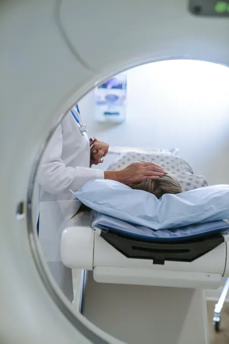
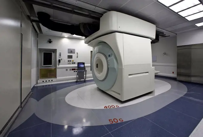
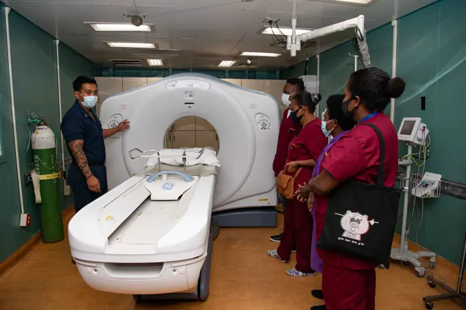
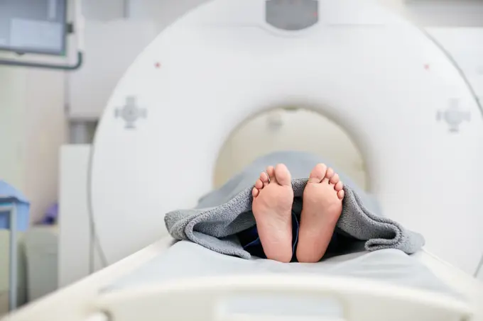
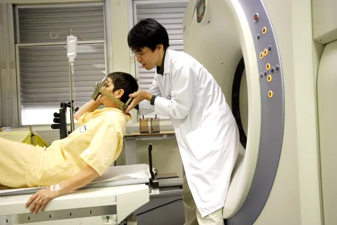
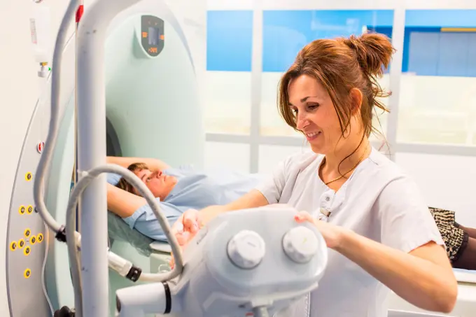
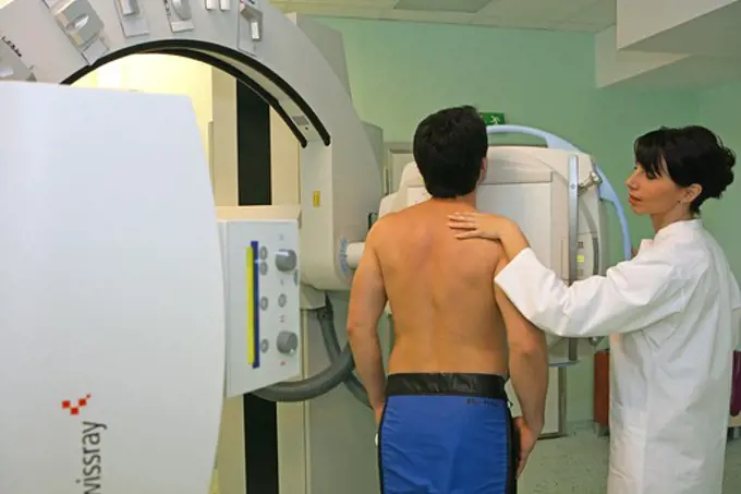
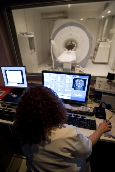
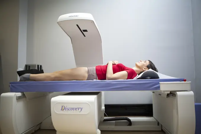
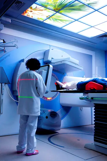
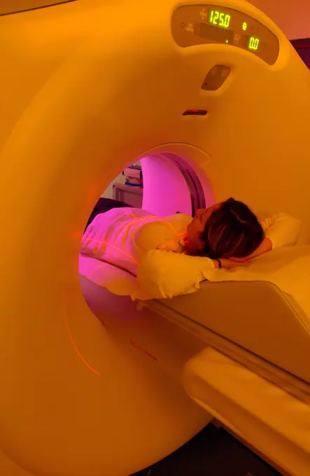
My Boards
View Pictures
  • My Boards

Medical Imaging Procedures

Images depict patients undergoing various medical imaging procedures, including CT and MRI scans, in modern hospital settings with advanced technology.

Caucasian doctor comforting patient near scanner
168 assets in this story
4362-1036
View of MRI operating room for immediate information and imaging of patients.MRI in operating room for immediate information and imaging of patients. IMRIS provides an optimized fully integrated image guided therapy environment that delivers timely information to clinicians for use during surgical or interventional procedures.
4269-6299
Patient undergoing heart 3D CT scan, Department of Medical Imaging, Centre Cardiologique du Nord, St Denis, France. The Lightspeed VCT 64-Slice CT Scanner produces 64 slices per rotation, and reduces radiation exposure by 70%.
6145-54235563
HONIARA, Solomon Islands (Sept. 1, 2022) - Hospital Corpsman 1st Class Jed Rodriguez, from Los Angeles, left, gives a presentation of a computerized tomography (CT) to medical professionals from the National Referral Hospital in Honiara aboard the Military Sealift Command hospital ship USNS Mercy (T-AH 19) during Pacific Partnership 2022. Now in its 17th year, Pacific Partnership is the largest annual multinational humanitarian assistance and disaster relief preparedness mission conducted in the Indo-Pacific. Pacific Partnership is a unifying mission that fosters enduring friendships and cooperation among many nations. The years mission in Solomon Islands will include participants from the United States, Japan and Australia.
4197-73070585
Assessment, healthcare and MRI with feet of person in machine for appointment or checkup. CT scan, diagnosis and medical equipment with patient in hospital for brain anatomy, exam or function
4128-24794707
CT scanner
6145-58911706
A radiologic technologist prepares a 3D mammography machine for use at Kimbrough Ambulatory Care Center on Fort Meade, Maryland, Feb. 12. (Defense Health Agency
6188-69110765
Doctor explaining CT scan results to patient in hospital setting Doctor explaining CT scan results to male patient in hospital setting model released
1815-69224406
Doctor encouraging patient during proton therapy session in a radiology treatment room
4269-24210
Breast cancer screening. Digital mammography.
4269-6182
Department of Medical Imaging, university hospital of Rennes, France. Patient undergoing a virtual colonoscopy. The virtual colonoscopy permits the 3D reconstruction of the inner colon from computerized coloscanner images. It is used for screening of colorectal cancer.
4286-69306
Computertomograph, CAT scan machine in hospital
824-63197786
Reportage in the National Oncology Particle Therapy Centre in Milan, Italy (The CNAO - Centro Nazionale di Adroterapia Oncologica). Particle therapy is a state-of-the-art technique used to treat cancer. It consists of using hadrons, protons (proton therapy) and cabon ions (carbon-ion treatment) to irradiate tumours. It has two therapeutic purposes, a high ballistic accuracy which allows healthy tissue surrounding the tumour to be spared, and a greater efficiency at treating certain cancers. The CNAO is the only centre to own carbon-ion treatment technology in Europe. The patient wears a mask which prevents movement during treatment.
824-63197783
Reportage in the National Oncology Particle Therapy Centre in Milan, Italy (The CNAO - Centro Nazionale di Adroterapia Oncologica). Particle therapy is a state-of-the-art technique used to treat cancer. It consists of using hadrons, protons (proton therapy) and cabon ions (carbon-ion treatment) to irradiate tumours. It has two therapeutic purposes, a high ballistic accuracy which allows healthy tissue surrounding the tumour to be spared, and a greater efficiency at treating certain cancers. The CNAO is the only centre to own carbon-ion treatment technology in Europe.
1815-69224372
Doctor and patient discussing treatment in a radiology room with advanced medical equipment
824-63173336
Brain, mri
4294R-1471
Doctor with patient and x-ray at CT scanner
4269-3556
Department of Functional Neurosurgery and stereotaxy of the Pr Peragut, Timone hospital, Marseille, France. Treatment of a meningioma (benign brain tumor) with a Gamma knife. The patient first undergoes un brain Computed Tomography (CT) scan to collect datas of the tumor (size, location etc.).
4269-39806
Female patient undergoing a CT scan, Angoulême hospital, France .
4286-69324
Radiographer taking x ray of patient
824-63197784
Reportage in the National Oncology Particle Therapy Centre in Milan, Italy (The CNAO - Centro Nazionale di Adroterapia Oncologica). Particle therapy is a state-of-the-art technique used to treat cancer. It consists of using hadrons, protons (proton therapy) and cabon ions (carbon-ion treatment) to irradiate tumours. It has two therapeutic purposes, a high ballistic accuracy which allows healthy tissue surrounding the tumour to be spared, and a greater efficiency at treating certain cancers. The CNAO is the only centre to own carbon-ion treatment technology in Europe.
824-63205924
Report at the Munich proton therapy center in Germany, the Rinecker Proton Therapy Center. Here, the PET scan room (positron emission tomography), a medical imaging method that allows 3D visualization of a tumor after injection of a radioactive product.
824-63181699
Digital medical imaging centre in Paris, France. Cranial CT scan
1848-53791496
Positioning a patient in a computed tomography, CT scanner, Germany, Europe
4269-8942
Patient undergoing a brain Magnetic Resonance Imaging scan (MRI) in a 3-tesla closed geometry MRI unit.
824-29055908
Digital medical imaging center, radiology room.
824-63181686
Digital medical imaging centre in Paris, France. Bone densitometry.
1841R-111750
4269-33747
Radiotherapy radiographer and a female patient treated to a stomach cancer with radiotherapy. Latest generation medical particle accelerator. Radiotherapy center, pole of Oncology, Hospital of Bordeaux. Hospital Haut-Leveque.
824-63179531
Reportage at the Neuroimaging research centre in Pitié Salpêtrière hospital in Paris, France. Magnetoencephalography (MEG) platform. MEG detects variations in the brain's magnetic field during various types of cerebral activity. It studies normal and abnormal brain function. Recording the magnetic field produced by neuronal currents requires ultra sensitive sensors called SQUIDs (Superconducting Quantum Interference Device). The 306 sensors spread over 102 areas allow both near and far magnetic fields to be measured, and the brain's deep structures to be 'seen'.
824-63175301
Photo essay in a aesthetic medicine office (France). Spectrum LED cold light skin therapy is a non-invasive procedure activating skin cells with low-level pulses and non-thermal light energy. The skin cells convert light energy to energy which activates collagen synthesis and skin repair. A combination of yellow and red light therapy is use for acne, scars, shingles, preventive treatment against skin cancers, haircare.
4197-64120282
Doctor, mri and woman holding hands of patient in hospital before scanning in machine. Ct scan, comfort or medical professional with senior female person in radiology test for healthcare consultation
1815-69224377
Patient undergoing proton therapy treatment in a modern medical facility
1841R-111749
1525-22527351
hospital interior.
1848-53791500
Linear particle accelerator in a radiotherapy practice, Germany, Europe
6188-64821511
Linear Accelerator at hospital Linear Accelerator at hospital Copyright: xZoonar.com/ALEXxTIHONOVx 3952180
5514-17876115
view of a neonatal incubator
824-63197463
Reportage in the Rinecker Proton Therapy Centre in Munich, Germany. The centre is equipped with the latest protontherapy treatment technology. Protontherapy consists of irradiating cancer cells by concentrating a beam of protons on the core of the tumour, while preserving healthy neighbouring tissue.
4269-33793
Radiotherapy
1848-50997679
Patient during radiation therapy, cancer treatment, brain tumor, Duesseldorf, Germany, Europe
4269-26525
PET scan. Positron Emission Tomography (PET) scan. A positon emission tomography exam allows the study of metabolic function of organs. It detects the presence of radioactive tracer molecules that have been injected into the patient's bloodstream and absorbed into the tissues.
824-63205928
Reportage at the Rinecker Proton Therapy Center in Munich, Germany. The centre has the latest equipment for proton therapy treatment. Proton therapy involves irradiating cancer cells by concentrating a proton beam on the heart of the tumour, while preserving the healthy surrounding tissue. Reportage at the Rinecker Proton Therapy Center in Munich, Germany. The centre has the latest equipment for proton therapy treatment. Proton therapy involves irradiating cancer cells by concentrating a proton beam on the heart of the tumour, while preserving the healthy surrounding tissue.
1899-53508118
Digital medical imaging center, abdominal scanner.
1848-53791508
Positioning and adjusting a patient in a linear particle accelerator for radiation treatment, Germany, Europe
6145-52245370
060816-N-3532C-039. Base: Tarakan Country: Indonesia (IDN)
6145-58888357
U.S. Navy Hospital Corpsman 3rd class Mathew Hartman, from Harve, Montana, secures a patient for a CT scan aboard the hospital ship USNS Mercy (T-AH 19) anchored off of Chuuk, Federated States of Micronesia, as part of Pacific Partnership 2024-1, Jan. 16, 2024. Pacific Partnership, now in its 19th iteration, is the largest multinational humanitarian assistance and disaster relief preparedness mission conducted in the Indo-Pacific and works to enhance regional interoperability and disaster response capabilities, increase security stability in the region, and foster new and enduring friendships.
1848-50171437
Onco Ray
6145-58888359
U.S. Navy Hospital Corpsman 3rd Class Johndenzel Macatuno, from Dededo, Guam, prepares a local patient for a CT scan aboard the hospital ship USNS Mercy (T-AH 19) anchored off of Chuuk, Federated States of Micronesia, as part of Pacific Partnership 2024-1, Jan. 16, 2024. Pacific Partnership, now in its 19th iteration, is the largest multinational humanitarian assistance and disaster relief preparedness mission conducted in the Indo-Pacific and works to enhance regional interoperability and disaster response capabilities, increase security stability in the region, and foster new and enduring friendships.
4269-25513
3D CT scan. Patient undergoing a 3D Computed Tomography (CT) scan.
1815-69224407
Doctor assisting patient in a proton therapy room with advanced medical equipment
824-63181494
Digital medical imaging centre in Paris, France. Dental x-ray.
4286-67745
magnetic resonance tomography
1848-49239390
Vertical photo of a dentist explaining the results of the scan to a patient
824-63198714
Reportage in a rehabilitation centre in Germany, specialising in neurological rehabilitation after a stroke or head injury. The centre is equipped with a number of robotic technologies which facilitate exercises. A patient who is paralysed in both legs uses the Space Curl technology. The Space Curl was developed by NASA and is the only tridimensional therapy system in the world. It improves coordination, balance and posture in patients suffering from neurological disorders.
824-63187688
Reportage in a hospital. Mammography room.
1525-21186591
Medical exam in CAT scan machine
824-63179577
Report from the Neuro Imaging Research Center located at the Pitié Salpêtrière in Paris. Magnetoencephalography (MEG) platform. MEG makes it possible to detect the variations in the magnetic field that accompanies the various cerebral activities. It is used to study normal or abnormal functioning of the brain. The recording of magnetic fields induced by the electrical activity of neurons requires the use of ultra-sensitive sensors called SQUIDs (Superconducting Quantum Interference Device). The 306 sensors are spread over 102 sites, which makes it possible to measure near and far magnetic fields, allowing a better view of the deep structures of the brain. Here the subject wears an EEG helmet (Electroencephalography) which makes it possible to combine MEG and EEG.
1899-66457
CT scan machine An image of the table or bed used
6188-56067625
Magnetic Resonance Imaging Scanner, MRI. 3D rendering Magnetic Resonance Imaging Scanner, MRI. 3D rendering isolated on white background Copyright: xZoonar.com/alexlmxx 21220377
1848-53791503
Patient lying in a linear particle accelerator for radiation treatment, Germany, Europe
1848-50171441
Onco Ray
1606-30448729
Sunbed
1899-61460777
Place infants in a phototherapy tunnel. In the first days after birth the bilirubin level of the newborn can vary. Phototherapy sessions restore the balance.Editorial Only
824-57659014
Place infants in a phototherapy tunnel. In the first days after birth the bilirubin level of the newborn can vary. Phototherapy sessions restore the balance.
1525-26515699
Young man patient standing in x-ray machine. Panoramic radiography. High quality photo.. Young man patient standing in x-ray machine. Panoramic radiography.
6145-54290284
Radiation Therapists
6145-54225255
Kailee Fetzer (pictured left) and Stephanie Hamilton are mammography technicians with Naval Medical Center Camp Lejeune's Radiology Department.The mammography section recently received reaccreditation from the American College of Radiology.
1848-50171434
Onco Ray
PREVIOUS
of 2
NEXT
2401 S. Ervay, Suite 206
Dallas, Texas 75215
United States
Get Started
Free ResearchMy BoardsMy Cart
For Creators
How To License Your ContentContributor PortalFrame of Mind
Resources
API accessPricing
Contact
+1 866 236 0087help@viewpictures.co.uk Contact form
©2026 View Pictures. All Rights Reserved. -A
This site is protected by reCAPTCHA and Google Privacy Policy and Terms of Service apply.