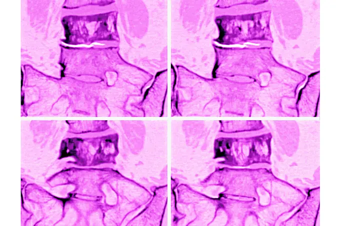
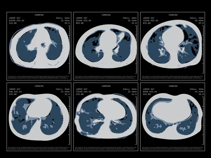
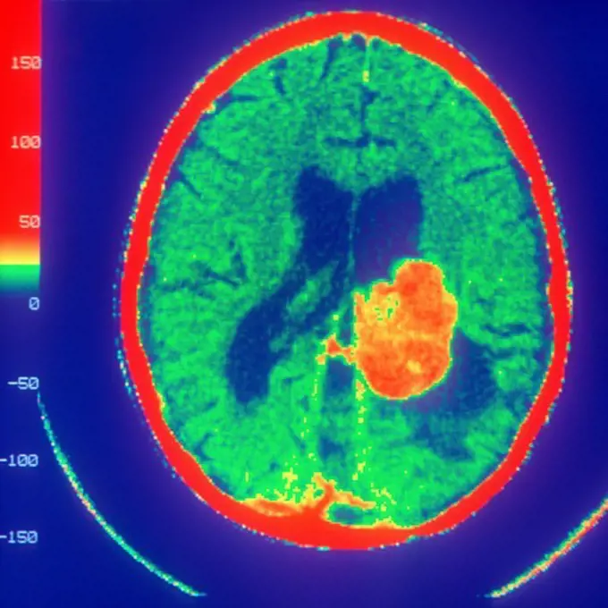
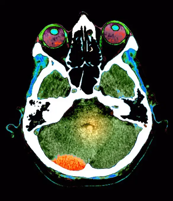
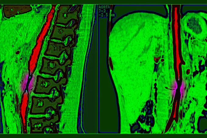
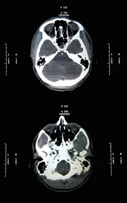
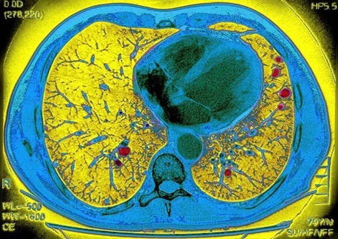
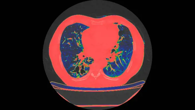
My Boards
View Pictures
  • My Boards

Medical Imaging Scan Visuals

Color-enhanced scans revealing various medical conditions within the human body, highlighting tools in diagnostics through vivid imagery.

Pagets disease, seen in a frontal MRI scan of the L4 and L5 lumbar vertebrae.
189 assets in this story
824-63184373
Fracture of the medial cuneiform bone in the foot. Frontal scan of the right foot.
824-63188650
Silicosis, seen on a transversal CT scan of the thorax.
6188-55645097
Abdomen man, man abdomen man, man, ct recording copyright: xzoonar.com/dr.xnorbertxlangeex 13790171
1899-54027148
Heart and lungs seen in a radial cross-section MRI scan.
5507-30938054
Covid-19 Computed Tomography Of The Lungs. CT Scan
1899-54027995
Normal knee seen on a frontal MRI scan.
1899-54026832
Osteoarthritis of the left fermoral head, seen on a frontal pelvic MRI scan.
1899-53511762
Lung cancer located in the upper right lobe. Frontal chest x-ray. (Photo by: CAVALLINI JAMES/BSIP/UIG)
824-63178736
Ultrasound of a 3 month old foetus.
1899-65662228
Right and left lung cancer (stage 1). Front chest x-ray.
1899-53508442
3D CT scan of thorax: lungs, aorta, and pulmonary arteries are visible.
4391-302
A magnetic resonance image (MRI) of a Ewing sacrcoma, the white area on the right.
824-63224769
Uterine cancer visualized by frontal MRI.
6188-55645102
Abdomen man, man abdomen man, man, ct recording copyright: xzoonar.com/Dr.xnorberbertxlangeex 13790176
824-63226758
Pulmonary metastases (secondary to kidney cancer), visualized by a frontal chest X-ray.
4269-25377
Dissecting aneurism of the aorta. Three-dimensional computed tomographic (CT) scan reconstruction of a dissecting aneurysm of the aorta. In this type of aneurysm, the inner wall of the aorta (intima) detaches from the outter wall (media). The blood invades the created space and generate a bulge that can break. The aneurysm is visible as a longitudinal  furrow that extends from the thoracic aorta (hidden behind the upper part of the heart) to the common iliac arteries.
824-63224757
Uterine cancer visualized by sagittal MRI.
4269-27486
Shoulder chondrosarcoma. Sagittal MRI of the shoulder showing a chondrosarcoma of the shoulder blade. The tumor is evidenced by the presence of a heterogenous lytic area with bone calcifications. The black round shape at right is the head of the humerus inserted into the shoulder blade.
824-63217236
Pulmonary infarction (pulmonary embolism) seen on a radial cross-section chest CT scan.
824-63217800
Normal knee seen on a sagittal section MRI scan.
1899-53508418
Ectopic spleen, the spleen is tipped downwards, 3D CT scan. This anomalous position is not pathological.
4377-278
CAT Scan of the Brain Shows Tumor
4128R-3942
Extradural haematoma, CT scan
1899-54026881
Takayasu"s arthritis, seen by 2 adominal scanners (sagittal section on the left and frontal on the right), at the descending aorta.
824-63206991
Takayasus arthritis, seen by 2 adominal scanners (sagittal section on the left and frontal on the right), at the descending aorta.
824-63205982
Gallstone seen on an ultrasound scan of the abdomen.
4269-27560
Fractured head of the humerus. 3D Computed Tomographic (CT) reconstruction scan of the shoulder showing a fragmented fracture of the head of the humerus (bone of the arm).
1899-54028011
Slipped disc compression of the medulla lumbar vertebra.
1815R-96131
Ct scan report of head and skull, close up
4269-25330
Angioplasty. 3D computed tomographic (CT) scan reconstruction of a stenosis of the abdominal aorta and common iliac arteries treated by placing a stent.
824-66802
LUNG METASTASIS, SCAN. Pulmonary metastases secondary cancer, carcinoma in a 79_year_old man. Thoracic scan in axial section.
4128-V58573820
6188-55645101
Abdomen man, man abdomen man, man, ct recording copyright: xzoonar.com/dr.xnorberbertxlangeex 13790175
1899-54026862
Intercranial neurinoma, seen on a radial section MRI scan.
4128-20041871
Bronchiectasis, CT scan
4269-20404653
Coronary angiogram stenosed artery.
824-63225313
Stress fractures on the shins, visualized by scintigraphy.
1899-53511984
Lung cancer (right), visualized by an AP chest X-ray.
4269-25335
Stenosis. 3D computed tomographic (CT) scan reconstruction of a stenosis of the right external iliac artery.
6145-44685379
Forty Meters from Entry to Victoria Crater (Polar)
1439-57939915
Xray image revealing the intricate details of human lungs and ribs.
824-63203270
3mm renal lithiasis (microlithiasis) in the left kidney, seen in an abdominal ultrasound.
4128R-3191
Pleural effusion, X_ray
824-63198907
Sinusitis in the left maxillary sinus, seen on a frontal, cross-section MRI scan.
4269-25366
Dissecting aneurism of the abdominal aorta. Three-dimensional computed tomographic CT) scan reconstruction of a dissecting aneurism of the sub-renal abdominal aorta. In this type of aneurysm, the inner wall of the aorta (intima) detaches from the outter wall (media). The blood invades the created space and generate a bulge that can break. The aneurysm is visible as a longitudinal  furrow that extends from the renal arteries to the common iliac arteries.
824-63211590
Acute pneumonia, seen on a cross-section thoracic CT-scan.
4269-24999
Neurinome. Three-dimensional computed tomographic (CT) scan reconstruction of the neck showing a benign nerve tumor (neurinome) in green, localized in the vertebral canal.
824-52936
LUNG CANCER, SCAN. Lungs in brown with cancer in pink.
824-63218252
Osteosarcoma of the knee (malignant bone tumor). Frontal scintigraphy of the entire body.
4269-27288
Cerebral aneurysm. 3D Computed Tomography (CT) scan reconstruction of an aneurysm of the right carotid artery. An aneurysm is a bulge of a vessel due to the dilatation of its wall. A ruptured aneurysm can be fatal.
4269-25346
Stenosis. Virtual endoscopic view of the left internal carotid artery showing a stenosis (narrowing) and atheromatous plaques in pink.
4128-48285498
Bone metastasis, CT scan
6145-44601402
Dune and Dust Devil Streaks
4269-3383
3D Computed Tomographic (CT) reconstruction scan, external view, internal organs and skeleton of the trunc of person with multiple traumas : fracture of the 11th lower rib and pelvis bone, after a car accident.
4269-27875
Kidney cancer. Three-dimensional computed tomographic reconstruction scan of of a kidney with massive tumor (red).
4269-25816
Knee arthrosis. X-ray of knees affected by arthosis (green), also called osteoarthritis or gonarthrosis. It is a joint disease that can cause cartilage destruction (seen as a reduction in the joint space), bone erosions, and tendon inflammation.
4269-25352
Abdominal aorta aneurysm. hree-dimensional computed tomographic (CT) scan reconstruction of an abdominal aorta aneurysm. It appears as swollen bulge  located under the renal arteries and extending until the two comon iliac arteries. An aneurysm is a bulge of a vessel due to the dilatation of its wall. A ruptured aneurysm can be fatal.
4269-25687
Back. 3D computed tomographic (CT) scan reconstruction (longitudinal side-section) of the trunk showing the bonny structures in blue (skull and backbone) and the internal organs in grey. The nasal cavity (sinus) and the trachea appear in white.
4269-24982
Lung cancer. Three-dimensional computed tomography (CT) scan of upper thorax showing a Pancoast's syndrome associated with a lung cancer. In purple, the subclavian vein and branches, in red the sub-clavian artery. The tumor of the superior lobe of the left lung (blue) pushes strognly the lungs (yellow).
824-63203306
Calculus cholecystitis (microlithiasis), seen on two ultrasounds (longitudinal and transversal) of the gallbladder.
824-40562
RECTAL CARCINOMA. RECTAL CARCINOMA Institute of Nuclear Medicine, University Hospital of Lille, France.   PET Scan (Positron emission tomography).  Colorectal carcinoma. Several liver and lung abnormalities which contra-indicate surgical intervention.
824-29057246
Pituitary adenoma visualized by MRI of the skull, in sagittal section.
6145-44755483
Opportunity's Surroundings After Sol 1820 Drive (Polar)
4128-30420968
Autosomal recessive bestrophinopathy, illustration
4269-20408356
Ultrasound scan of the prostate.
824-63226756
Pulmonary metastases (secondary to kidney cancer), visualized by chest CT scan in axial section.
824-63211057
Fractured thumb at the metacarpal, seen on a hand x-ray.
4269-27286
Cerebral aneurysm. 3D Computed Tomography (CT) scan reconstruction of a giant aneurysm of the right carotid artery. An aneurysm is a bulge of a vessel due to the dilatation of its wall. A ruptured aneurysm can be fatal.
4269-27544
Renal artery ruptured. 3D Computed Tomographic (CT) reconstruction scan of the kidneys showing a partial rupture of the right kidney (at left). An hemorrhage of the renal artery is also evidenced by a S shaped bleeding behing the ureter (white). Consequence of a chock (read accident).
5513-51516033
Mouse Floats In Space -- Here are photos from movie film that recorded actions of two mice during 40-mile high rocket flight. Top-Mouse at left floats in air in front half of two-section drum; other mouse in rear has shelf to which he clings, thus controlling his movement. Photo made after nose was separated from rocket and during so-called zero gravity period before -parachute opened. Bottom-picture are normal Air Force says these tests emphasize belief man will be able to withstand unusual forces expected in rocket flight to outer atmosphere. September 26, 1952. (Photo by Dept. of Defence Photo).
6145-52890687
A stereo photograph by Romanus Schmehlik showing a water flea at a scale of 30:1 on silver gelatin paper in 1912. The image documents microcrustacean anatomy and behavior.
4128-19249284
Peripheral artery disease, illustration.
824-63207427
Breast cancer (adenocarcinoma), seen in two mammograms.
6145-29604475
Collection Dutch Heidematschappij. Damage, senior plants, mushroom, yellow lupine, root milk. undated
1899-53511361
Right coronary artery. Angiography with injection of contrast product.
4269-27873
Lung cancer. Colored Computed Tomography (CT) scan of the abdomen showing early stage lung cancer. The tumor is seen as a small red spot.
4269-24951
Stenosis. 3D computed tomographic (CT) scan reconstruction of a stenose of the right internal carotid artery. In blue, the carotid veins, in yellow, the respiratory tracts.
824-63217822
Lesion of the meniscus posterior horn. Sagittal section MRI scan of the knee.
824-40561
RECTAL CARCINOMA. RECTAL CARCINOMA Institute of Nuclear Medicine, University Hospital of Lille, France.   PET Scan (Positron emission tomography).  Colorectal carcinoma. Several liver and lung abnormalities which contra-indicate surgical intervention.
4269-27143
Bone cyst. Color enhanced axial 3D computed tomography (CT) scan of the shoulder with aneurysmal bone cyst on the left clavicle.
4409-17232668
Haekel's hypothesis. The phrase "ontogeny recapitulates phylogeny".
1899-53511492
Lumpy tumor of the thyroid gland (benign tumor), thyroid ultrasound.
824-63204739
Homogenous hepatomegaly revealed by an abdomino-pelvic scan.
6145-29290761
Anatomy: a dissection of the lumbar region showing the anterior relations of the kidneys. Photograph, ca. 1900.
824-29057245
Pituitary adenoma visualized by MRI of the skull, in sagittal section.
4128-19357506
Ultrasound scan of a 16-week old foetus showing the gestational sac.
4269-26919
Aneurysm of the abdominal aorta. Color Doppler ultrasound scan of an aneurysm of the abdominal aorta showing circulating lumen of the artery (orange), the rest of the bulge is occupied by a thrombus (clot)  seen in dark grey. Doppler ultrasound or echodoppler scanning detects moving fluids. Color is used to illustrate moving fluid, not flow direction.
824-63206262
Doppler ultrasound scan of a foetus in the fifth month of pregnancy.
1525-20359834
Radiology or Radiologist Medical Field Specialty As Art
PREVIOUS
of 2
NEXT
2401 S. Ervay, Suite 206
Dallas, Texas 75215
United States
Get Started
Free ResearchMy BoardsMy Cart
For Creators
How To License Your ContentContributor PortalFrame of Mind
Resources
API accessPricing
Contact
+1 866 236 0087help@viewpictures.co.uk Contact form
©2026 View Pictures. All Rights Reserved. -A
This site is protected by reCAPTCHA and Google Privacy Policy and Terms of Service apply.