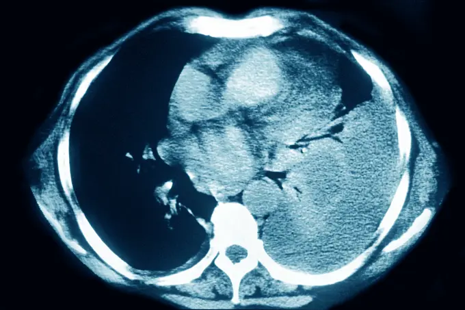
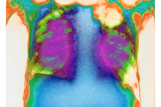
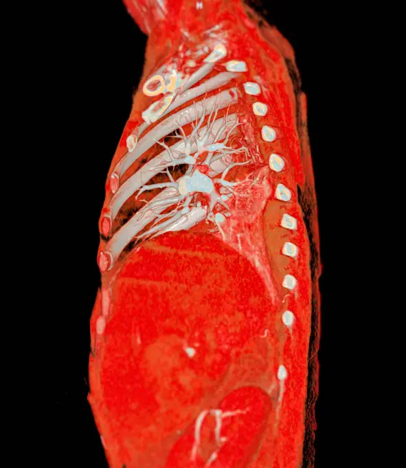
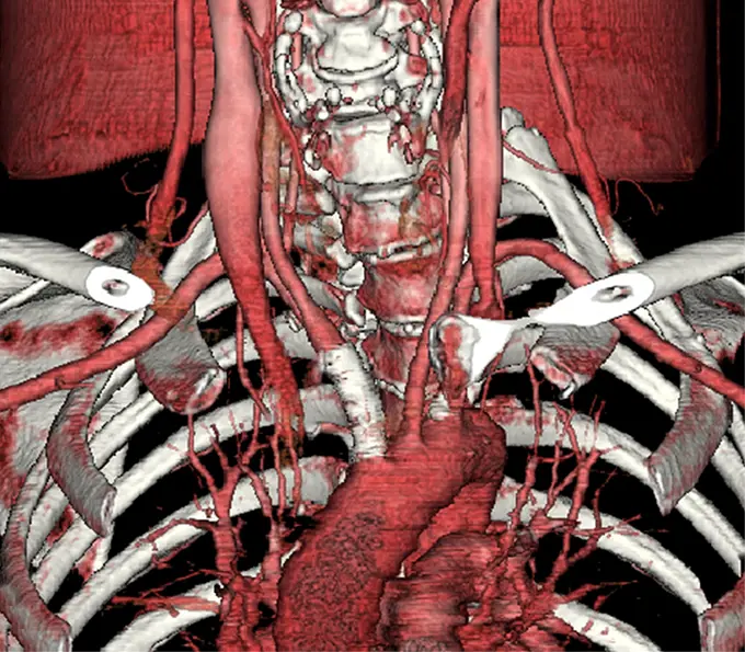
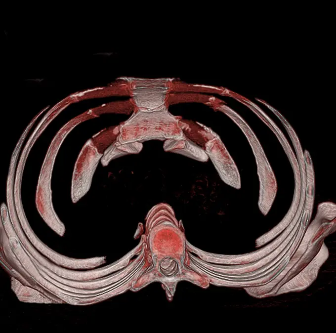
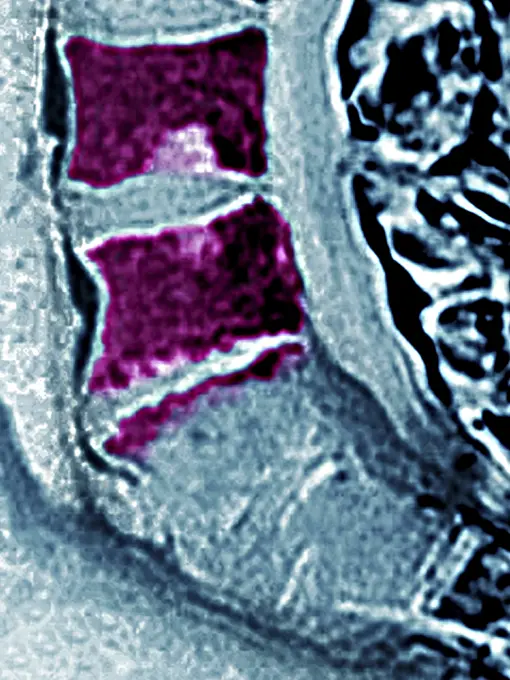
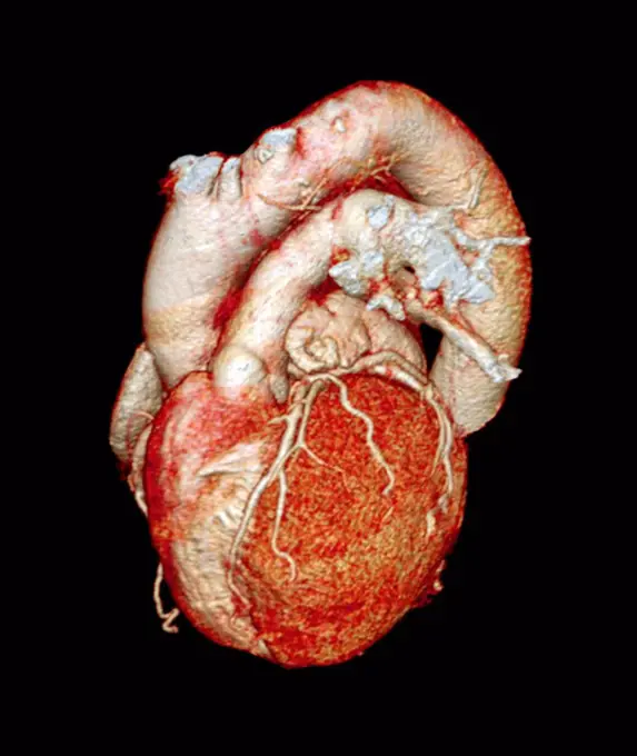
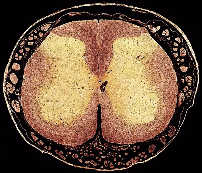
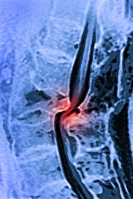
My Boards
View Pictures
  • My Boards

Medical Imaging Scans

Detailed medical imaging scans highlighting various abdominal conditions, showcasing cross-sectional views of organs and tissues.

Pleurisy (left lung), seen on a radial section chest scan.
135 assets in this story
4269-27282
Cerebral aneurysm. 3D Computed Tomography (CT) scan reconstruction of a small aneurysm of the left carotid artery. An aneurysm is a bulge of a vessel due to the dilatation of its wall. A ruptured aneurysm can be fatal.
4128-30419914
Stent in aortic aneurysm, CT scan
4269-27564
False aneurysm of the aorta. Sagittal Computed Tomography (CT) scan of false aneurysm of the aorta seen as a swelling on the upper aorta (pink), below the aortic arch. This phenomenon usually occurs after trauma to an artery, such as surgery.
4128R-14008
Abdominal aorta and spine. Coloured three_dimensional computed tomography CT scan of a side view of a healthy abdomen. The spine is at left and part o...
4297-1082
Chest x-ray of a 71 year old smoker showing lung cancer in the left lung
4269-25004
Right femoral infectious aneurysm. Three-dimensional computed tomographic (CT) scan reconstruction of right femoral artery infectious aneurysm.
824-63218244
Dislocated cervical vertebrae (traumatic lesion of cervical vertebrae C1-C2). X-ray in profile.
824-63215947
Allergic bronchopulmonary aspergillosis (ABPA), seen on a frontal chest x-ray.
4269-24719
Pulmonary embolism. Angiography of the lungs showing a pulmonary embolism. This occurs when one of the pulmonary arteries is blocked.
4269-25381
Prosthetic cardiac valves. 3D computed tomographic (CT) scan reconstruction of prosthetic mitral valve below (situated between the left atrium and the left ventricle), and an prosthetic aortic valve above (between the left ventricle and the aorta).
4128-30419939
Pulmonary artery, CT scan
4269-27280
Cerebral aneurysm. 3D Computed Tomography (CT) scan reconstruction of an aneurysm of the left carotid artery (side view). An aneurysm is a bulge of a vessel due to the dilatation of its wall. A ruptured aneurysm can be fatal.
4269-25329
Angioplasty. 3D computed tomographic (CT) scan reconstruction of a stenosis of the brachiocephalic trunk, treated with a stent prosthesis (visible in white, above the aortic arch).
1899-54027716
Pulmonary sarcoidosis seen on a frontal chest x-ray.
4269-26729
Asbestosis. Computer tomography (CT) scan of a longitudinal slice through the chest of a patient suffering from asbestosis, a kind of pneumoconiosis caused by the inhalation of asbestos particules from the air that have damaged the lung tissus and can lead to breathing problems and heart failure. The disease generated pleural plaques (yellow) in the most affected parts.
4128-28514437
Heart, CT scan
4269-25418
Thoracic cage. 3D computed tomographic (CT) scan reconstruction of the thoracic cage seen from above showing the sternum (at top), the costal cartilages and the ribs, and the rachis.
824-63188635
Osteitis on the L4-L5 lumbar vertebra, seen on a saggital plane MRI scan.
6188-60976253
Human Respiratory System Diaphragm Anatomy 3D Illustration Concept of Human Respiratory System Diaphragm Anatomy Copyright: xZoonar.com/magicminex 21748048
4269-24936
Aortic aneurysm. Three-dimensional computed tomographic (CT) scan reconstruction of aorto-iliac aneurysm.
824-63193294
An image produced by a new, noninvasive way to diagnose liver fibrosis using a new technique called MR Elastography which uses sound waves to determine tissue firmness. Doctors are able to see inside the liver without biopsies. The red indicates diseased tissue while the blue indicates healthy tissue. Source: Richard Ehman, Mayo Clinic
4128-48286064
Lung cancer tumour and light micrograph, illustration
4128R-12687
Normal heart. 3D computed tomography CT scan of a healthy heart from a 58 year old male.
4128R-13447185
Spinal cord. Light micrograph (LM) of a cross-section through the human spinal cordin the lumbar region. The spinal cord consists of a butterfly-shape...
4409-17312847
ECOGRAFIA DE NIÑA 20 DIAS ANTES DEL NACIMIENTO (ROCIO).
1899-53511901
Dislocation of the lumbar vertebrae by spinal trauma, between the 3rd and 4th lumbar vertebrae, visualized by MRI in sagittal section. (Photo by: CAVALLINI JAMES/BSIP/UIG)
4297-1145
CT scan images showing an abdominal aortic aneurysm which developed between the renal arteries and the common iliac arteries
6145-51832446
Portrait of Don Tadeo Bravo de Rivero Francisco de Goya y Lucientes (Spanish, 1746-1828). Portrait of Don Tadeo Bravo de Rivero, 1806. Oil on canvas, 81 1/2 x 45 11/16in. (207 x 116cm).  In their formal full-length portraits, Old and New World elites proudly defined themselves through their things. The colonial Peruvian nobleman Tadeo Bravo de Rivero sat for Francisco de Goya y Lucientes wearing the brilliant scarlet uniform of a cavalry officer. The medal of the chivalric Order of Santiago displayed on his lapel points to one of the ways that Creoles (American-born Spaniards) elevated their rank within the empires social and racial hierarchy. At the officers feet is a dog, the traditional symbol of fidelity, suggesting the subjects devotion to his king. European Art 1806
824-63203304
Calculus cholecystitis (microlithiasis), seen on two ultrasounds (longitudinal and transversal) of the gallbladder.
824-63188632
Vertebral edema seen on a saggital plane MRI scan of the lumbar vertebrae.
1525-25029923
Medical Xray. Medical X ray imaging of human brain skull bones
6145-50449195
X-Ray of Roosevelt shows bullet
4269-25935
Heart scintigraphy. Gamma scan of a heart affected by ischemia during physical activity (lack of blood).
824-63178740
Ultrasound of an 8 month old foetus. Head circumference measurement.
4128-20040498
Pituitary adenoma cancer, illustration
PREVIOUS
of 2
NEXT
2401 S. Ervay, Suite 206
Dallas, Texas 75215
United States
Get Started
Free ResearchMy BoardsMy Cart
For Creators
How To License Your ContentContributor PortalFrame of Mind
Resources
API accessPricing
Contact
+1 866 236 0087help@viewpictures.co.uk Contact form
©2026 View Pictures. All Rights Reserved. -A
This site is protected by reCAPTCHA and Google Privacy Policy and Terms of Service apply.