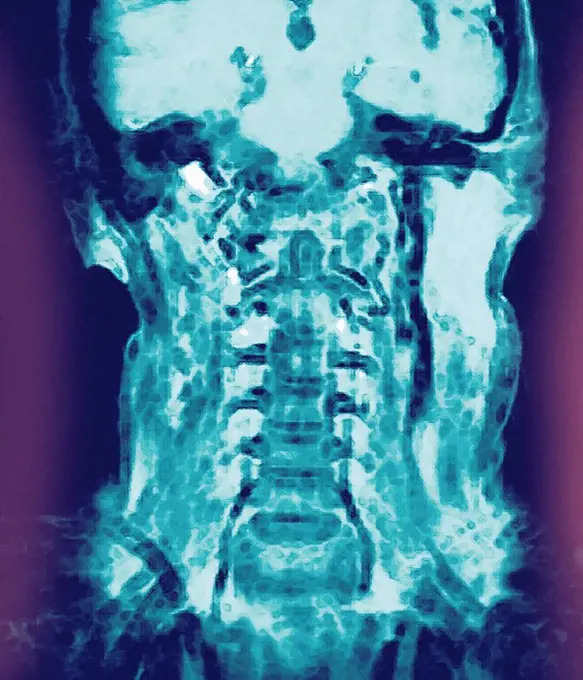
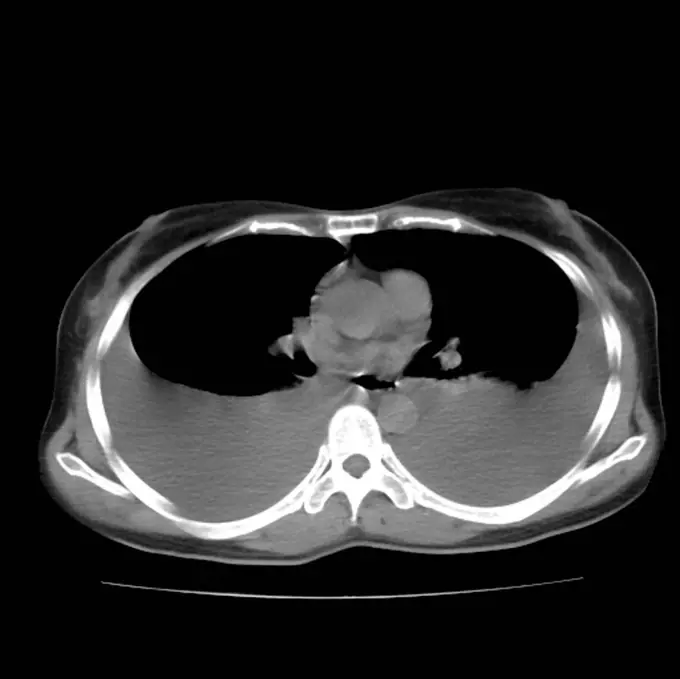
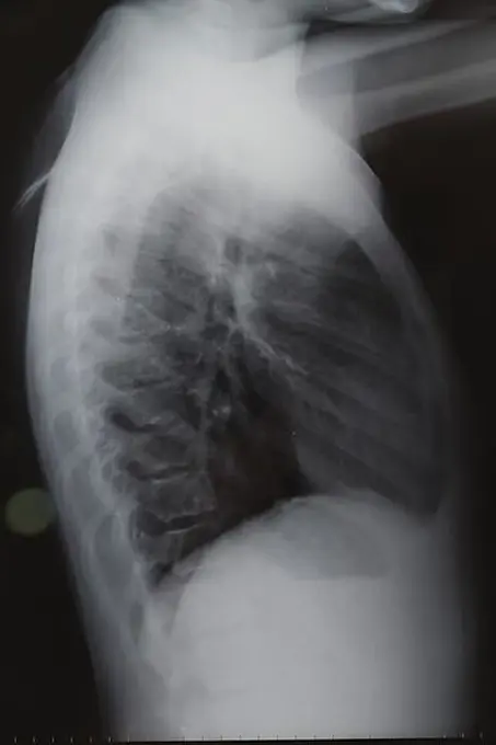
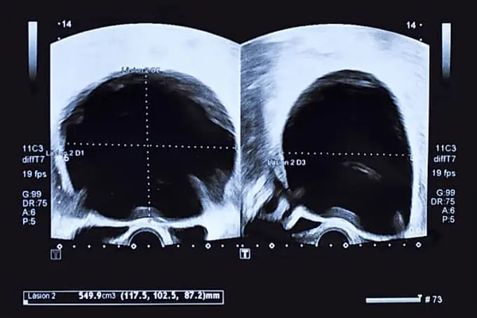
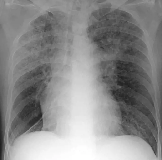
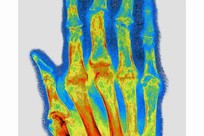
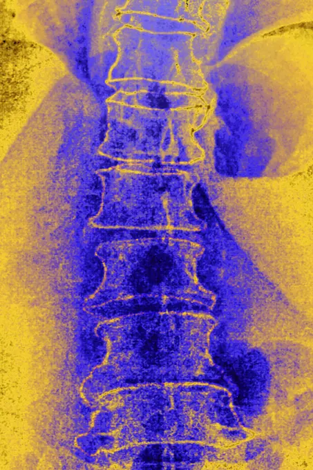
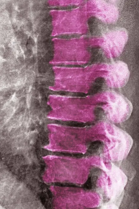
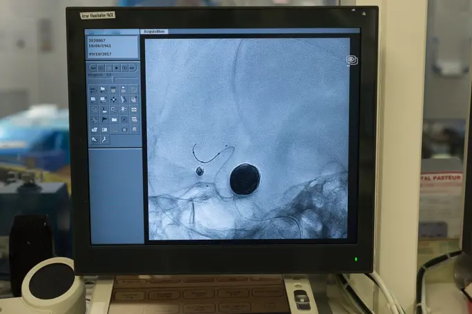
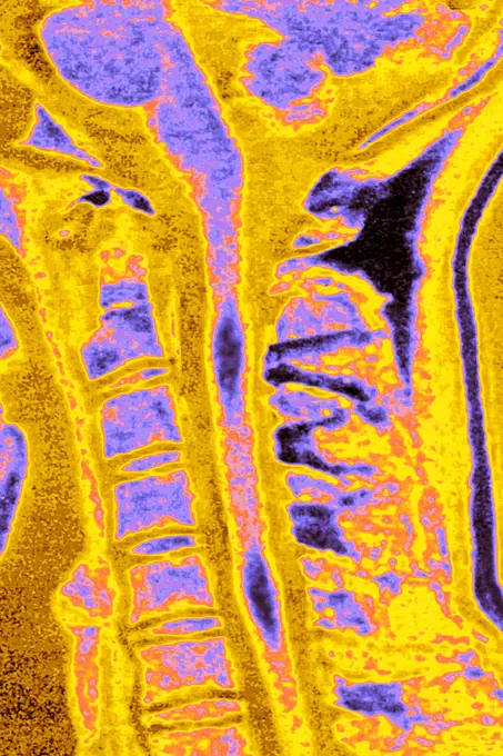
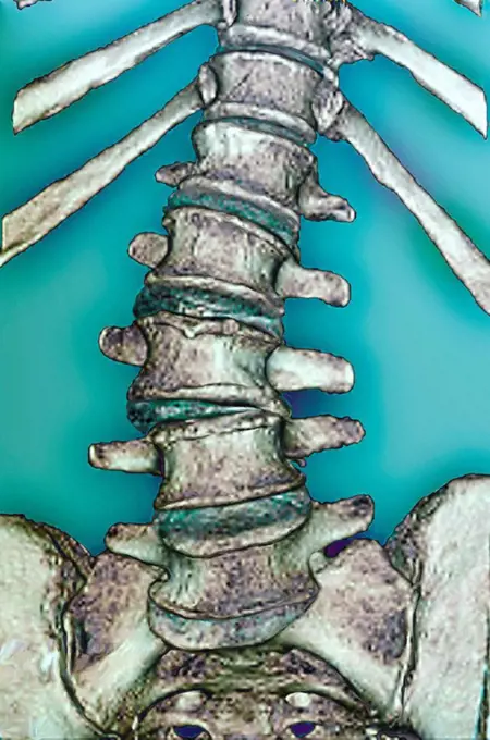
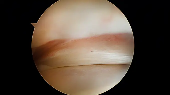
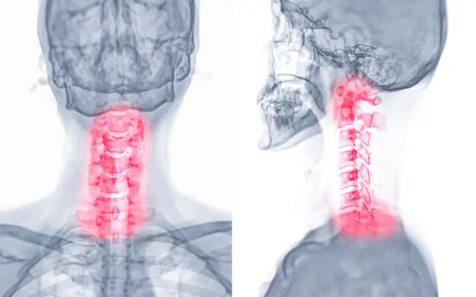
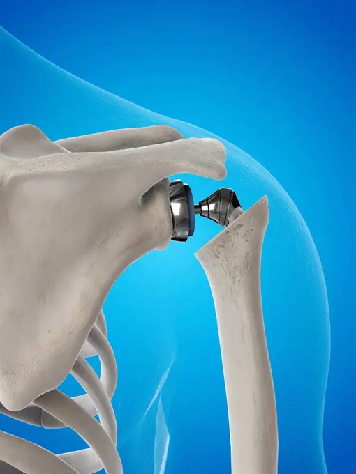
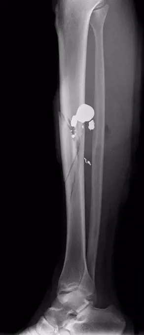
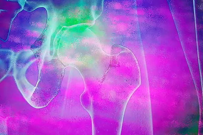
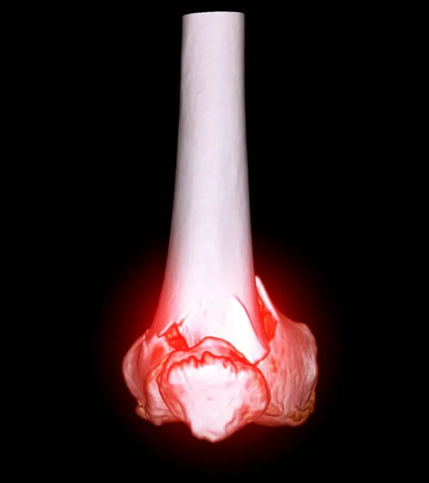
My Boards
View Pictures
  • My Boards

Medical Imaging Techniques

X-ray and MRI scans displaying various conditions such as spinal issues and joint placements. Highlights diagnostic imaging in healthcare.

Normal head and neck, MRI scan
291 assets in this story
4128-20239775
Pleural effusion, CT scan
4269-27571
Rachis fracture. Sagittal Computed Tomography (CT) scan of fractured backbone with a total rupture of the vertebral canal (road accident).
1525-22179998
MRI
824-63194051
Healthy dental panoramic x-ray in a 29-year old woman.
1848-49258513
X ray scan back
1848-60337065
Ultrasound image of large round cyst in lower abdomen of woman printed on a piece of paper
1899-61460136
Knee fracture (caused by a violent shock, causing trauma with serious lesions to the tibial plateau and to the lower end of the femur. X-ray of the left knee from the front
1899-53511916
Fixation by screws and plates on the sixth, seventh cervical vertebrae and the first thoracic vertebra, after vertebral dislocation, visualized by radiography in sagittal section. (Photo by: CAVALLINI JAMES/BSIP/UIG)
824-63224949
Dental panoramic of a 70 year old man with an implant and many crowns, the crosses visualizing the teeth that will need care. Paris, Ile-de-France, France.
4128-19358740
Computed tomography (CT) scan of the brain of a 32 year old male a patient with severe traumatic brain injury after being involved in a road traffic a...
4128-19358039
Chest X-ray of 64 year old female patient showing heart enlargement (cardiomegaly). The cardio-thorax ratio should not be greater than 50% in people o...
255-28641409
Vintage Photograph. X Ray images of a foot, Anterior Posterior view and Oblique view
824-63178573
Sinus radiography of a young child. No anomaly detected
4128-19358057
Pneumothorax, X-ray. Clear space in the left hemithorax suggestive of a left pneumothorax.
4297-1199
Colorized x-ray of the hand of a 16 year old boy who dislocated his thumb at the metacarpal phalangeal joint
1525-22988764
Panoramic radiograph is a scanning dental X-ray of the upper jaw maxilla and lower jawbone mandible. The photo shows a young man aged thirty seven 37 years. Panoramic dental x-ray of young man
4128-28970850
Pulmonary giant bulla, CT scan
824-63206994
Right and left ureter, seen on two frontal abdominal x-rays.
4128-V58573827
1525-26235798
Swollen and inflamed knuckles, a condition that causes joint inflammation from a joint infection.3D illustration. Swollen and inflamed finger joints
4128-19357515
Axial, mediastinum window, chest computed tomography (CT) scan of a 50-year-old male with cough showing right loculated pleural effusion with pleural ...
4128-19358211
X-ray of a proximal ulnar fracture with radial head dislocation, known as a Monteggia fracture.
824-63206974
Pacemaker implanted, visualized by an AP chest X-ray.
824-63178579
Nose-forehead x-ray of the sinuses of a 42-year-old woman. Normal transparency of all the sinuses of the face. Bilateral hypertrophy of the inferior turbinates.
1899-65505
Radiology.
4128-28970851
Pulmonary giant bulla, CT scan
824-63211562
Vertebral osteoarthritis with osteophytes (bone spurs) seen on a frontal CT-scan of the lumbar vertebrae.
1899-65500
Radiology.
6188-55645079
Abdomen Mensch, Mann Abdomen Mensch, Mann, CT Aufnahme Copyright: xZoonar.com/Dr.xNorbertxLangex 13790128
824-63123162
In purple, wisdom teeth.
824-63178658
Trauma-contusion of the right thumb after a shock. No fracture. Bone tearing
4128-19357512
Coronal mediastinum window chest computed tomography (CT) scan of a 50-year-old male with cough, showing a right loculated pleural effusion with pleur...
824-63215938
Mediastinal adenopathy caused by sarcoidosis. Frontal chest x-ray.
4128-19358062
X-ray of a patient with miliary nodules. Miliary refers to innumerable small 1-4 millimetre pulmonary nodules throughout the lungs. On a chest X-ray, ...
824-63216820
Digital x-ray of the right foot.
824-63207457
Rheumatoid arthritis seen on an x-ray of the hand.
1899-53511506
Significant degenerative lumbar disc disease in a 75-year-old woman, frontal x-ray.
4128R-12576676
X-rays of hands.
1899-53511503
Significant degenerative lumbar disc disease in a 75-year-old woman, lateral x-ray.
1525-22232567
Mid-adult Caucasian male doctor pointing at an x-ray.
1525-28468528
X ray film for human hands and legs
1525-23996544
x ray scan. Resolution and high quality beautiful photo. x ray scan. High quality and resolution beautiful photo concept
824-63214349
Reportage in the interventional neuroradiology service in Pasteur 2 Hospital, Nice, France. Treating a cerebral aneurysm through embolization. This endovascular treatment consists of introducing a catheter into the brain and filling the aneurysm with platinum coils in order to clot it and prevent it splitting. The embolization is carried out under fluoroscopic guidance. On the screen, x-ray showing the freshly treated aneurysm (small) and another previously treated aneurysm (larger).
1899-53511927
Multiple sclerosis (MS) (autoimmune disease that affects the central nervous system by dysfunction of the immune system and leads to lesions that cause motor, sensory, cognitive, visual or even sphincter disturbances). MRI of the cervical vertebrae in sagittal section. (Photo by: CAVALLINI JAMES/BSIP/UIG)
824-63226772
Multiple sclerosis (MS) (autoimmune disease that affects the central nervous system by dysfunction of the immune system and leads to lesions that cause motor, sensory, cognitive, visual or even sphincter disturbances). MRI of the cervical vertebrae in sagittal section.
1848-55284666
X-ray after osterosynthesis, metal implant for correction of joint position
4128-V58573803
4128R-15047349
Lower leg X-rays
4128R-6980
Arthritis of the spine, CT scan
1525-19878959
X-ray photo of human knee joint isolated on white background
824-63191309
An arthroscopy of the hip. Normal labrum in the middle, fermoral head at the bottom.
4128-30419978
Fractured knee, CT scan
1525-19778486
Panoramic x-ray image scan of humans teeth and dental mirror
824-72488764
Osteoporosis of the femoral neck and location of fracture sites.
6145-46761669
AGFA autochrome. Medical photographs.A-blood cell B-serum, high DRP. Satisfy immersion.
4297-1243
Chest x-ray of a 76 year old man with COPD, chronic obstructive pulmonary disease, Chest x-ray of a 76 year old man with a history of smoking and COPD
6177-V53821086
4128-30420008
Neck pain, conceptual X-ray
824-63224565
Fracture of the foot on the wedge bone. Foot scanner
824-63178487
Knee, x-ray
4128R-15536607
Illustration of a shoulder replacement
4128R-11287607
Dislocated shoulder. Coloured X-ray of a 77 year old female patient with a dislocated shoulder.
4128-30420927
Best vitelliform macular dystrophy, illustration
1525R-232820
MRI
1570R-132785
CAT Scan of the spine
1899-65296
Two surgeons examining x-ray of hand before surgery
4269-18998
This 2D and 3D digital X-ray imaging equipment is dedicated to the orthopedic practice and permits a static study of the musculoskeletal system. It takes by low dose X-ray scanning two simultaneous, perpendicular planar views in the standing position. A reconstruction provides a 2 and 3D image of the osteoarticular structures of the patient. The university hospital of Bordeaux is the first hospital to use this diagnostic technique in France.
4297-1051
Hand x-ray showing a dislocation of the middle finger at the proximal interphalangeal joint
4128-30420963
Retinal scar caused by toxoplasmosis, illustration
4128-18929578
Gun shot in leg, X-ray
1848-49273070
The hip joint is a so-called ball and socket joint because it is round and, above all, more than 50 per cent surrounded by the acetabulum. The upper end of our thigh bone forms the femoral head
1525-24588620
Backbone or spine icon, human skeleton anatomy isolated sketch. Spinal cord, vertebrae sketch. Spine bone sketch icon, human skeleton
4128-30419972
Fractured knee, CT scan
824-63203276
Osseous edema on the knee. Sub-cortical lesion of the condyle, seen on a frontal section MRI.
4269-27065
Micro-herniaectomy. The micro-herniaectomy consists in removing a slipped disk with an aspiration tube introduced through a 2 mm incision under local anesthesia.
824-63209864
Reportage in the Rossetti Centre in Nice, France, during Professor Griffets monthly consultations. He is an orthopedic surgeon from Grenoble Hospital, and founder of this centre. X-ray of a teenager with scoliosis, wearing a back brace. The x-ray uses the EOS low-emission radiology system. It enables simultaneous frontal and profile images to be produced from head to foot on a standing patient, and enables the 3D reconstruction of all the osteo-articular areas, with a considerable reduction in x-ray levels.
1899-53511369
Fracture of the foot on the wedge bone. Foot scanner.
1841R-111787
1838-17347851
Dental X-Ray of Teen Girl
1899-65662215
Retinal detachment occurs when the tissue at the back of the eye becomes detached from the layer of blood vessels that supply the eye with oxygen. Radial MRI of the eye.
6188-63925416
Physiotherapy center specializing in hand, elbow and shoulder pathologies before or after operation. Hand skeleton. Physiotherapy practice 016809_016
1899-53511491
Lumpy tumor of the thyroid gland (benign tumor), thyroid ultrasound.
824-63193978
Thyroid ultrasound scan on a 32-year old patient, showing a disturbance in the biological check-up with collapsed levels of TSH. The scan shows an uneven hypoechoic thyroid gland (25.8cc), suggesting thyroiditis.
4391-150
A mammogram of normal breast tissue.
6188-55643169
Bolar tooth with tooth root molar with tooth root
6177-V53806538
4128-28575111
Normal mammogram
1899-65662235
Ischemic mitral insufficiency (MI) - Pathology of the left ventricle (mitral ring and subvalvular apparatus). transthoracic echocardiography (TTE).
824-63162854
Optical coherence tomography (OCT) showing the optic nerve of a patient at risk for glaucoma.
4443-21167971
excision of metacarpo-phalangeal joint, medical equipment, surgical instrument, history of medicine
824-63193980
Thyroid ultrasound scan on a 32-year old patient, showing a disturbance in the biological check-up with collapsed levels of TSH. The scan shows an uneven hypoechoic thyroid gland (25.8cc), suggesting thyroiditis.
PREVIOUS
of 3
NEXT
2401 S. Ervay, Suite 206
Dallas, Texas 75215
United States
Get Started
Free ResearchMy BoardsMy Cart
For Creators
How To License Your ContentContributor PortalFrame of Mind
Resources
API accessPricing
Contact
+1 866 236 0087help@viewpictures.co.uk Contact form
©2026 View Pictures. All Rights Reserved. -A
This site is protected by reCAPTCHA and Google Privacy Policy and Terms of Service apply.