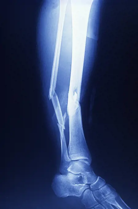
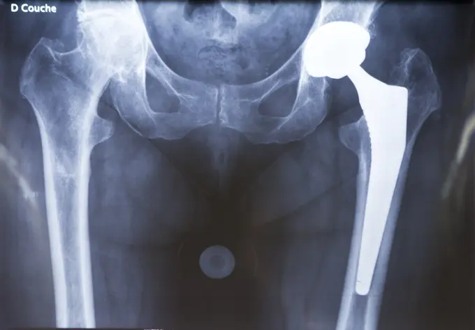
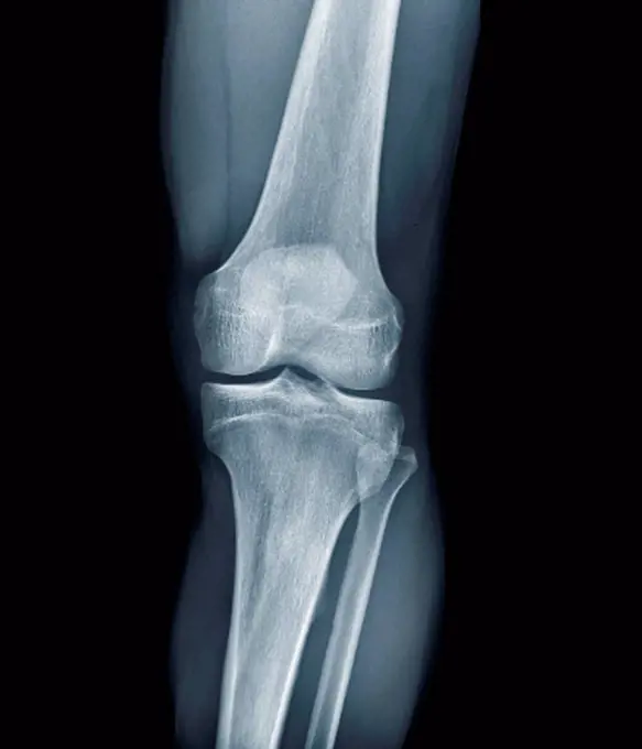
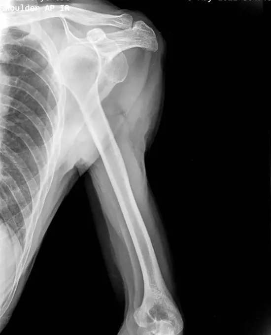
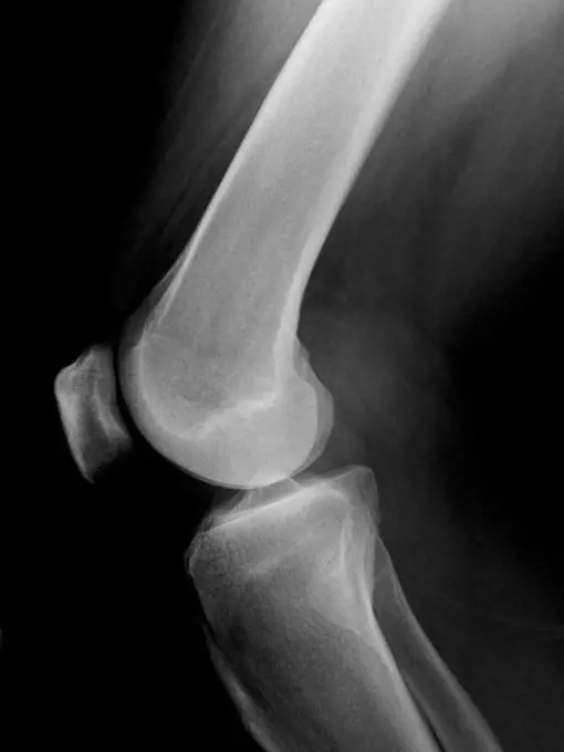
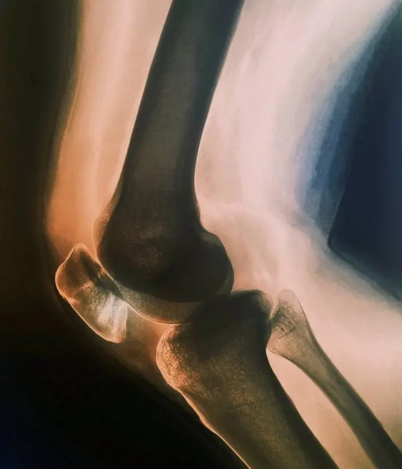
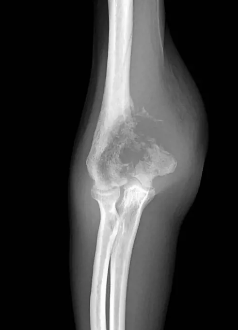
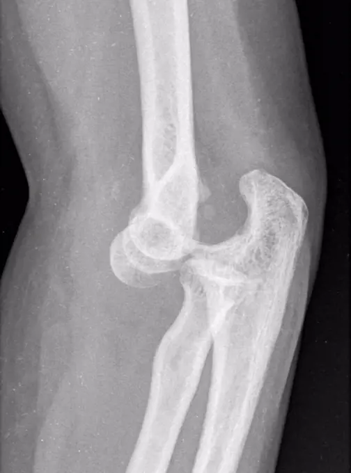
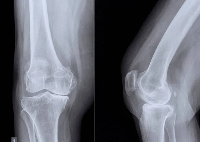
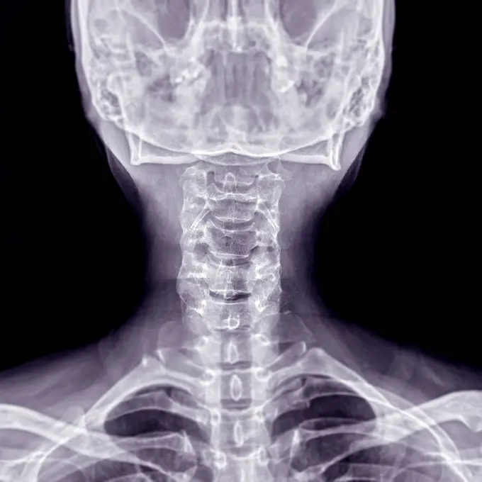
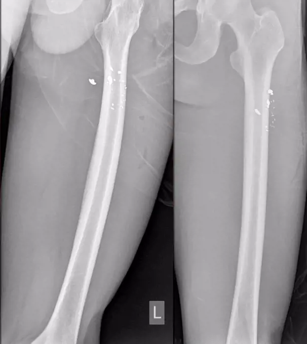
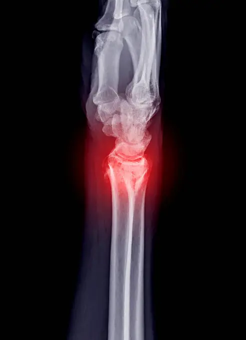
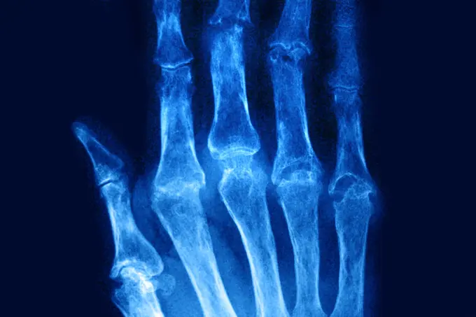
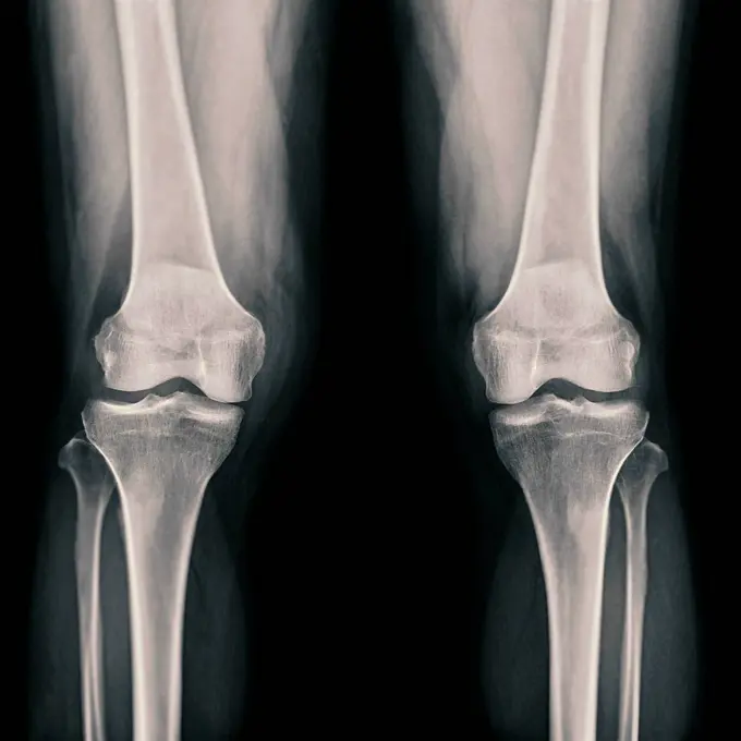
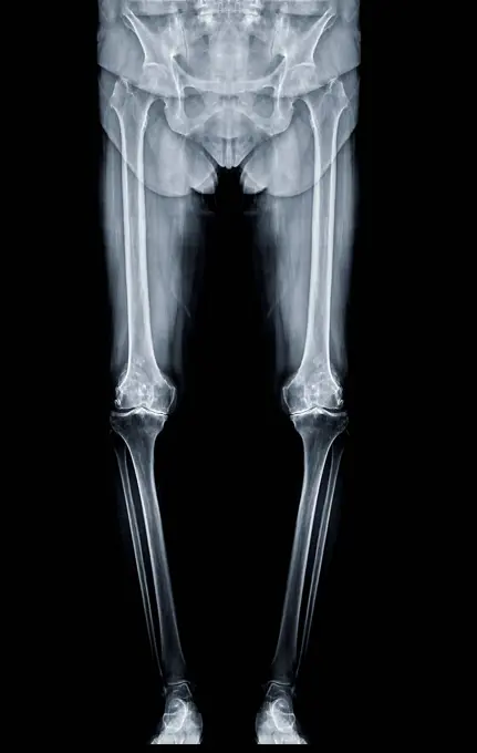
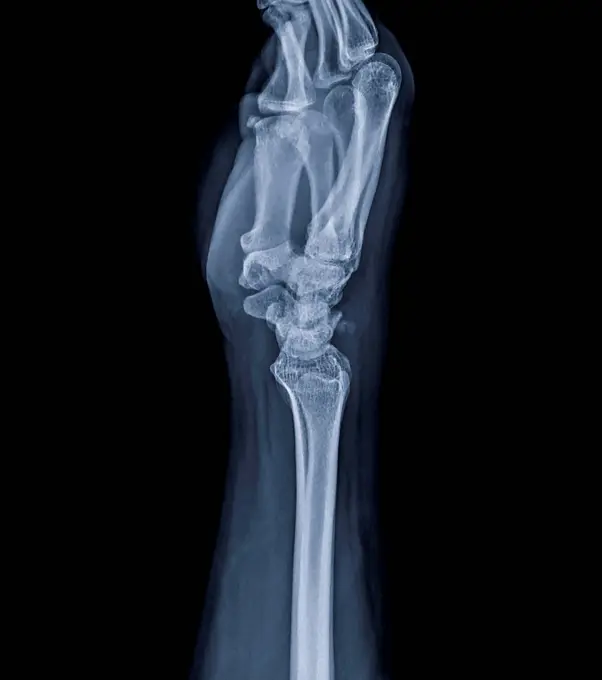
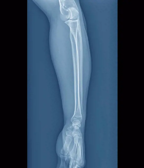
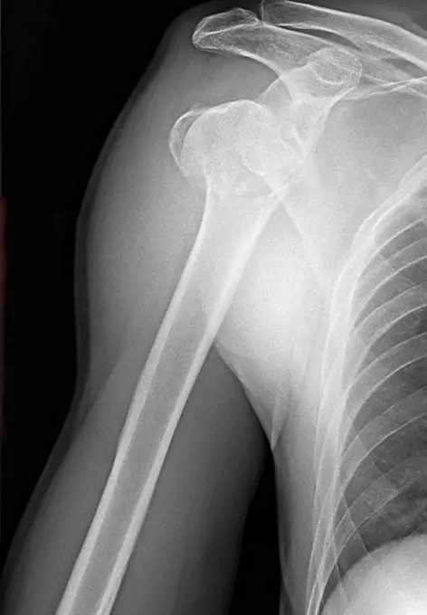
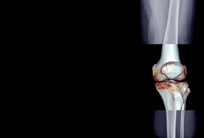
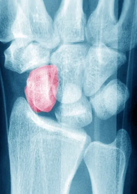
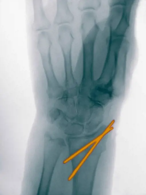
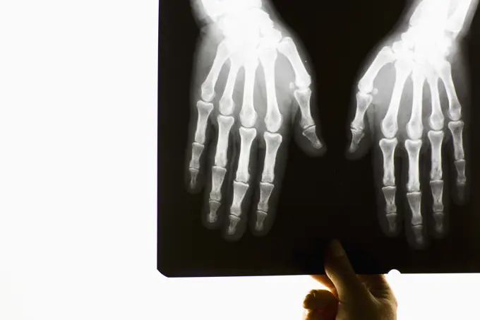
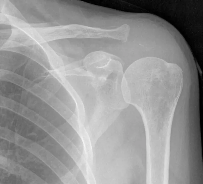
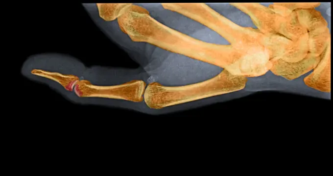
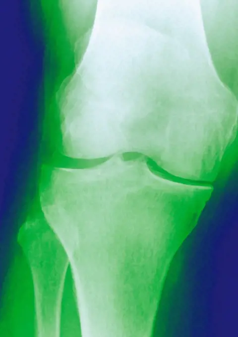
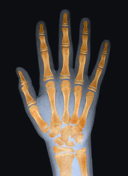
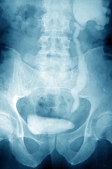
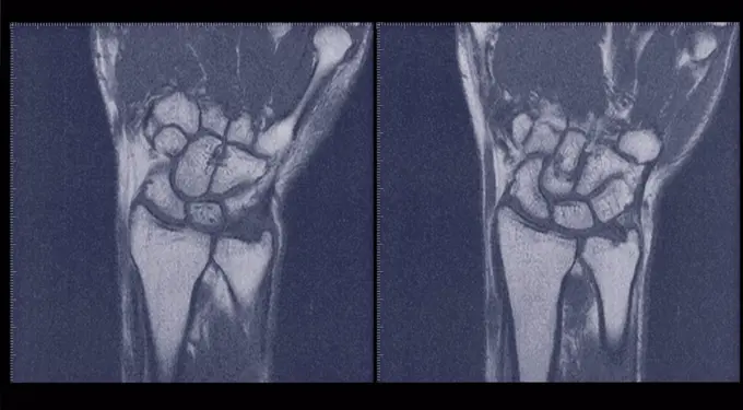
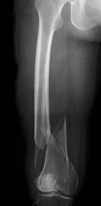
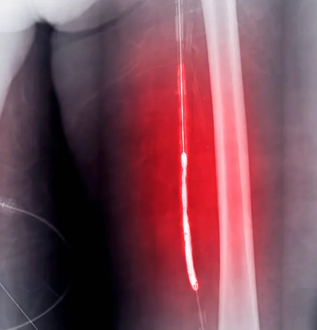
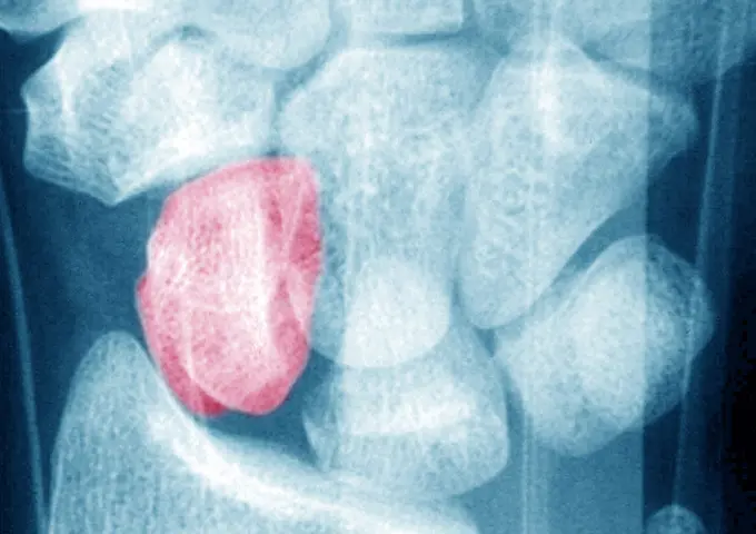
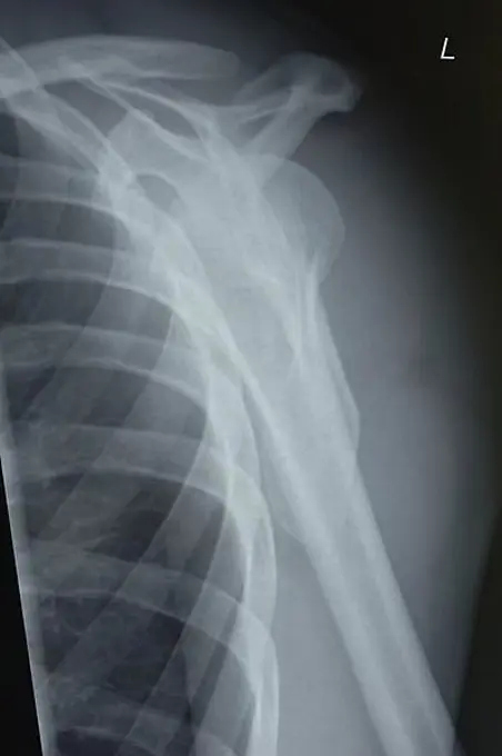
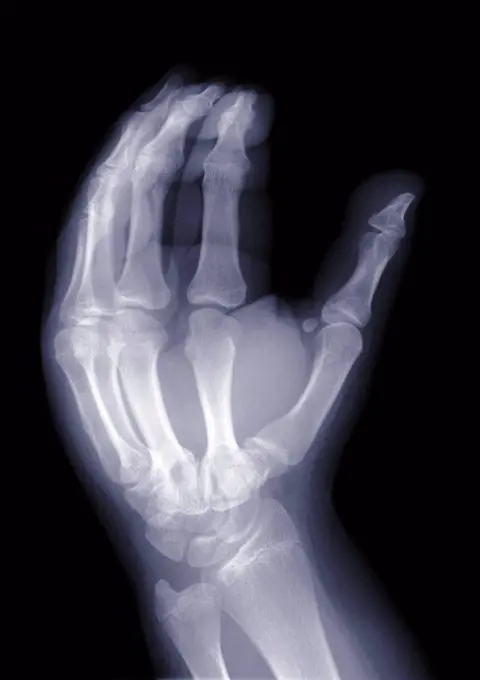
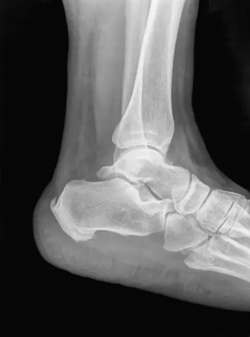
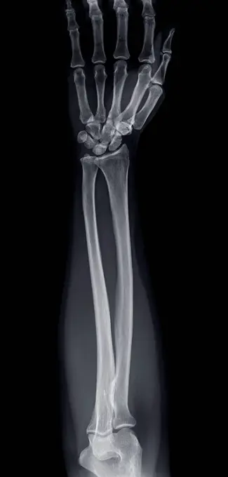
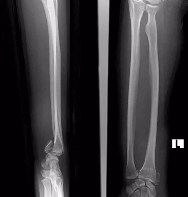
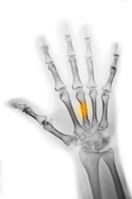
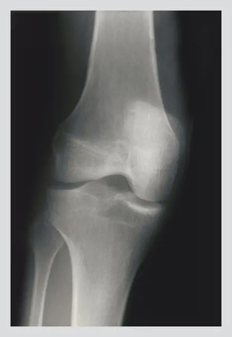
My Boards
View Pictures
  • My Boards

Medical X-Rays and Imaging

Various X-ray images showcasing conditions like osteonecrosis, arthritis, and fractures. Clinical, informative style with a focus on skeletal health.

Fractured Fibula and Tibia
211 assets in this story
4269-24813
Hip. Femoral head osteonecrosis X-ray
824-63170783
Post-traumatic wrist arthritis. X-ray of left hand.
824-63170798
Post-traumatic wrist arthritis. X-ray of left hand.
1815R-97530
X ray of knee, close up
824-777
Fractured Fibula and Tibia
824-63176441
Reportage in the orthopaedic surgery unit in the Diaconesses Croix Saint Simon hospital in Paris. Placing a second hip prosthesis. X-ray of the patient before the operation.
4128R-12577964
Normal knee. Frontal X-ray of the healthy left knee of a 28 year old.
824-63178632
Calculation of the bone age with a radiography. 12 years old child elbow radiography. Normal
4128-24795306
Dislocated shoulder, X-ray
6145-29252334
A deformed foot: X-ray. Photograph.
4297-1069
Normal x-ray of the knee of a 51 year old man
1841R-111782
1899-86152
Knee X Ray
824-63224566
Fracture of the foot on the wedge bone. Foot scanner
4128R-15731
Broken knee. Coloured X_ray of the knee of a 38 year old patient with a fractured patella kneecap.
824-63178421
Normal X-ray of the knee of a young child
1841R-111781
4128-19357561
X-ray showing humerus osteosarcoma with an ill-divined lytic lesion with wide zone transition and a Codman triangle: a type of periosteal reaction. A ...
1848-61113908
Cervical spine
4128-18929524
Dislocated elbow, X-ray
4128-20043294
Knee osteoarthritis, X-ray scans
4128-19489793
Tibial plateau fracture, X-ray
4128-28769196
Cervical spine, X-ray
4128-28766912
Fractured shoulder, X-ray
4128-18929587
Gun shot in thigh, X-ray
4128R-6956
Normal shoulder and elbow joints, X_ray
4128-28769204
Fractured ulna, X-ray
4269-27139
Bone cyst. Colored X-ray of aneurysmal bone cyst on the left clavicle (yellow).
824-63207455
Rheumatoid arthritis seen on an x-ray of the hand.
4128-28769209
Healthy spine, X-ray
4408-1009
Foot, Medical - Xray / Radiology
824-66065934
X-ray of severe scoliosis in an adult.
4128-19489756
Supracondylar fracture, X-ray
4408-1014
Fixed Tibia, Medical - Xray / Radiology
4128-20043159
Ureteral mass, X-ray
4128-28766904
Healthy knees, X-ray
4128-19489661
Humerus fracture, X-ray
1899-53511770
Osteosynthesis of the wrist by bone metal plate and screw, following a fracture of the radius. sagittal x-ray of the wrist. (Photo by: CAVALLINI JAMES/BSIP/UIG)
4128-28766911
Healthy lower body, X-ray
4128-28769206
Healthy wrist, X-ray
4129-133
shoulder and arm x-ray
4128R-6458
Normal elbow and wrist joints, X_ray
4128-19489697
Humerus fracture, X-ray
5507-34876639
X-ray of the heart
4128-28766905
Healthy knee joint, MRI scan and X-ray
824-63170797
Fracture of the carpal scaphoid. X-ray of a left wrist.
4297-1092
X-ray of a radius fracture stabilized with two pins
824-63194045
Knee x-ray on a 73-year old woman. The exam shows widespread bone demineralisation, a discrete pinching in the internal femorotibial joint space and an osteosynthesis of the left tibial plateau (screw).
1570-30032482
Hand of healthcare professional holding x-ray of a pair hands
1899-53511595
Osteosynthesis of the hip by a nail-plate prosthesis, following a fracture of the neck of the femoral head. X-ray of the left hip.
4128-20239758
Missing acromion, X-ray
4128-19489709
Humerus fracture, X-ray
824-63226002
Osteosynthesis of the wrist by bone metal plate and screw, following a fracture of the radius. sagittal x-ray of the wrist.
4128-19489635
Broken ankle, X-ray
4128R-11287858
Fractured collarbone. Coloured X-ray of a 16 year old male with a fractured clavicle (collar bone).
1899-54027169
Rheumatoid arthritis seen on an x-ray of the hand.
824-63178677
X ray of the left foot of a 29 years old male patient. Normal
824-63178660
Trauma-contusion of the right thumb after a shock. No fracture. Bone tearing
824-63178674
Bone calcification of the big toe on a 29 years old foot patient. X ray
4128R-6420
Arthrosis of the knee, X_ray
1525R-76797
X-ray of a hip replacement
1841R-111796
824-63178664
X-ray of the right hand and fourth finger of a 10-year-old boy. No visible recent bone lesion.
1899-54027399
Nephritic colic caused by a stenosis on the left ureter. Frontal abdominal-pelvic x-ray. (Photo by: BSIP/UIG via Getty Images)
824-63178575
Normal X-ray of a 30-year-old patient.
4128-16073304
Scans of wrists, MRI
4269-2538
Colored X-ray showing an implanted insulin pump.
4128-18680906
Kneecap fracture, X-ray
4128-19499658
Comminuted femur fracture, X-ray
4128-19357513
X-ray showing the lower (lumbosacral) spine taken from a patient with ankylosis spondylitis.
4297-1050
Normal hand x-ray of a 56 year old man, who dislocated his the middle finger at the interphalangeal joint, and had the dislocation reduced in the emergency room
824-63225317
Corticosteroid infiltration between the fourth and fifth lumbar vertebrae, done under radiographic control
4128-30419894
Treatment for blocked arteries, angiogram
4128-28769187
Fractured shoulder, X-ray
4297-1385
Normal x-ray of the shoulder of a 30 year old man
1525-28468529
X ray film for human hands and legs
4128-28766906
Healthy knees, X-ray
1525R-76795
Three x-rays of a wrist
1899-61460134
Knee fracture (caused by a violent shock, causing trauma with serious lesions to the tibial plateau and to the lower end of the femur. X-ray of the left knee from the front
4128-19357539
Abdominal X-ray of a patient with prostate carcinoma. The X-ray shows am osteoblastic lesion on the pelvis bone and vertebral lumbalis indicating bone...
1848-49597916
X-ray of a patient with a distal radial extension fracture on the right with PSU avulsion, stabilised by plate osteosynthesis with angular stable plate
4128-30419925
Fractured heel, CT scan
1773-97598
wrist X-ray of comminuted fracture of radius
824-57656875
Knee fracture (caused by a violent shock, causing trauma with serious lesions to the tibial plateau and to the lower end of the femur. X-ray of the left knee from the front.
824-63170807
Fracture of the carpal scaphoid. X-ray of a left wrist.
1525-25247097
hand holds radiograph  isolated on a white background
1848-61113906
Shoulder joint, X-ray
4128-16072253
Hand, X-ray
4269-1060
Side viewed three-dimensional computed tomographic reconstruction of the lombar vertebras and the intervetebral spaces. The sacrum is at the bottom
1439-57940614
Xray image of a human ankle showing bones
4128-28766902
Healthy elbow joint, X-ray
1899-53511368
Fracture of the foot on the wedge bone. Foot scanner.
1525-24886763
close up of xray hands with black background backlight
1899-53511596
Osteosynthesis of the hip by a nail-plate prosthesis, following a fracture of the neck of the femoral head. X-ray of the left hip.
4128-18929604
Smith fracture, X-ray
1899-53511924
Multiple sclerosis (MS) (autoimmune disease that affects the central nervous system by dysfunction of the immune system and leads to lesions that cause motor, sensory, cognitive, visual or even sphincter disturbances). MRI of the cervical vertebrae in sagittal section. (Photo by: CAVALLINI JAMES/BSIP/UIG)
4297-1515
Colorized hand x-ray showing a fracture of the middle metacarpal. A heavy object fell on this man's hand
4128-28766914
Fractured shoulder, X-ray
4408-1007
Knee, Medical - Xray / Radiology
4128-18929455
Fractured hand in cast, X-ray
PREVIOUS
of 3
NEXT
2401 S. Ervay, Suite 206
Dallas, Texas 75215
United States
Get Started
Free ResearchMy BoardsMy Cart
For Creators
How To License Your ContentContributor PortalFrame of Mind
Resources
API accessPricing
Contact
+1 866 236 0087help@viewpictures.co.uk Contact form
©2026 View Pictures. All Rights Reserved. -B
This site is protected by reCAPTCHA and Google Privacy Policy and Terms of Service apply.