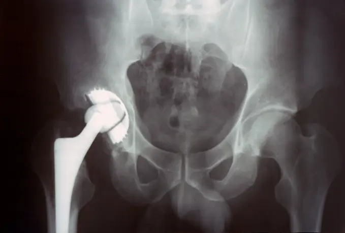
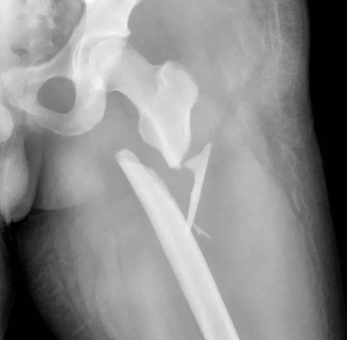
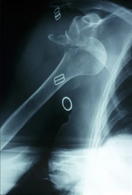
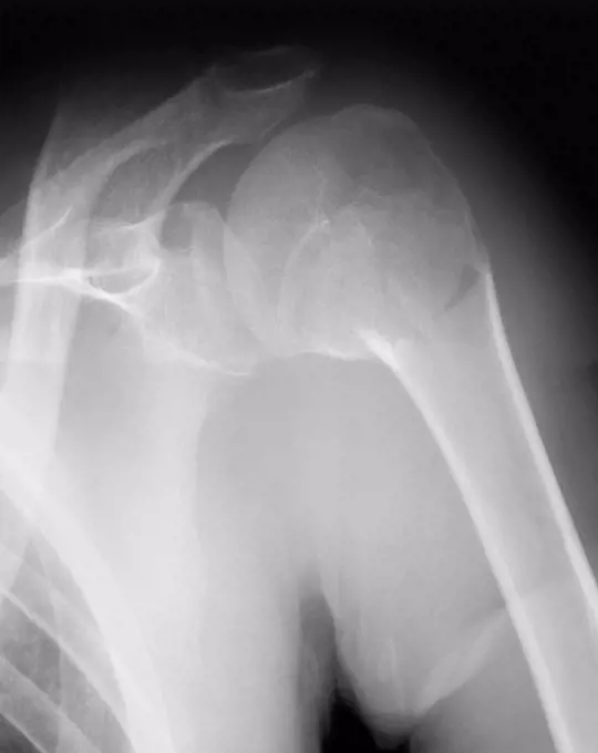
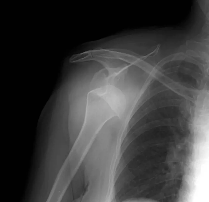
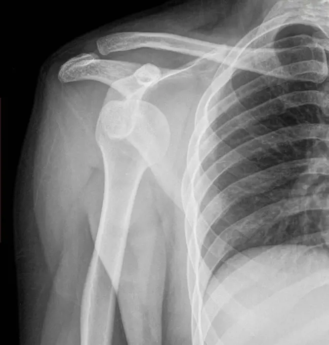
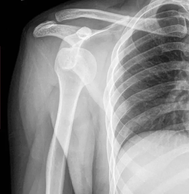
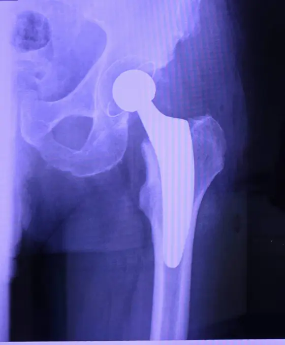
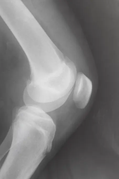
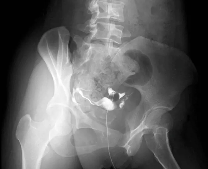
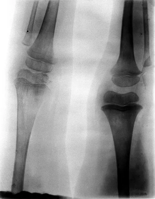
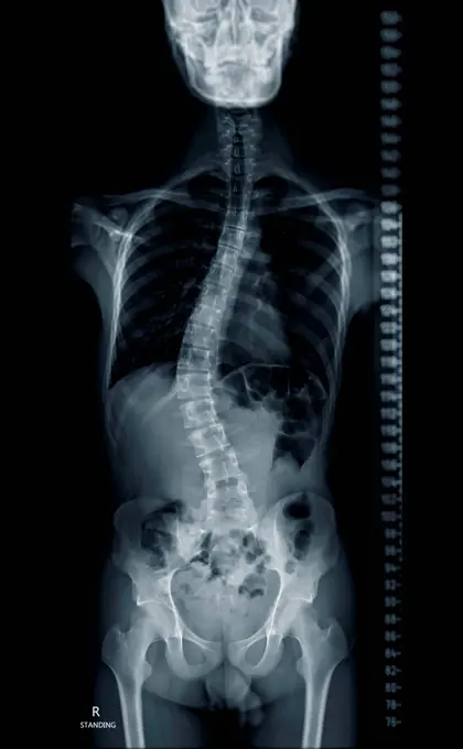
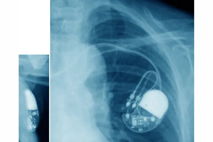
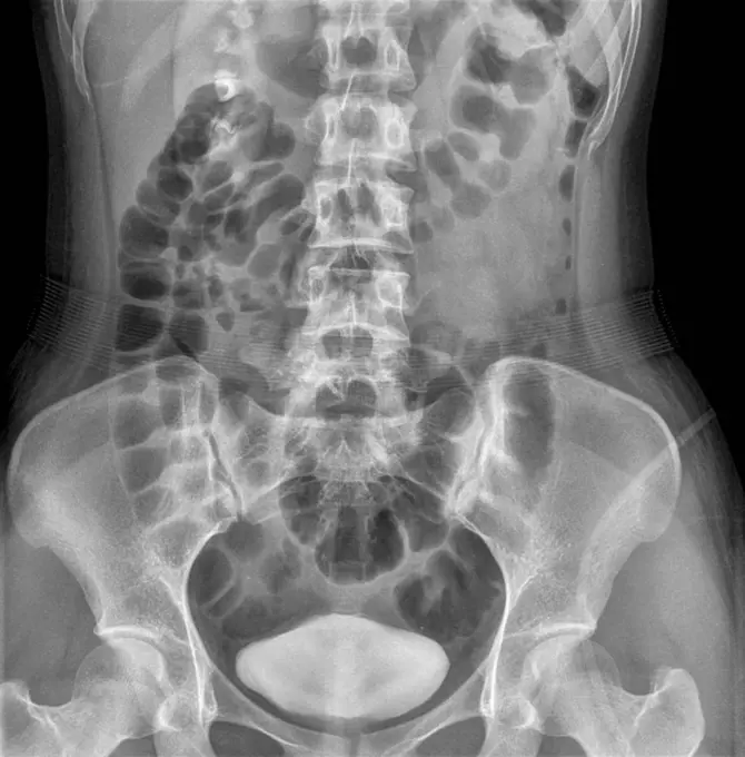
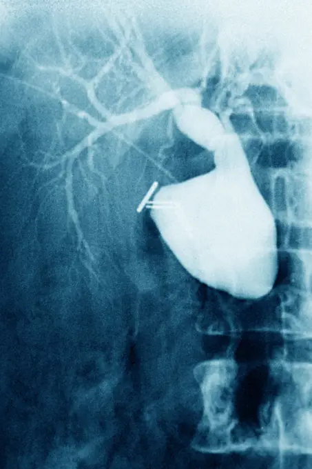
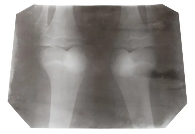
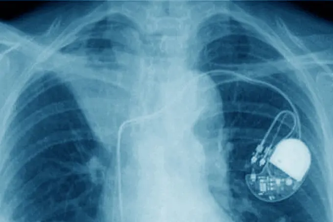
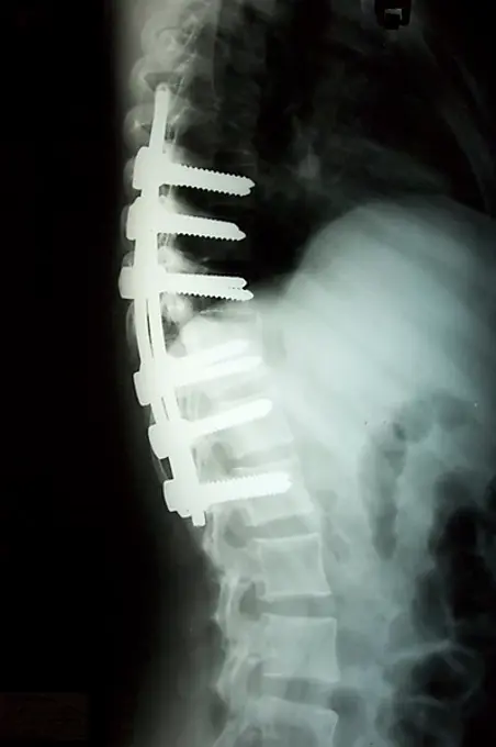
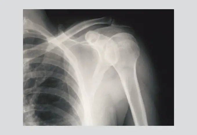
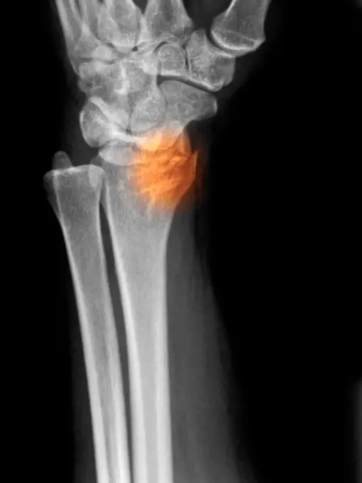
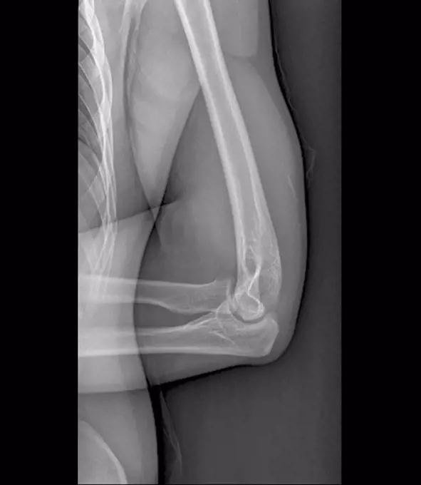
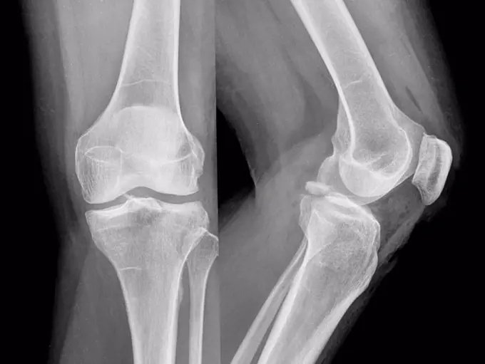
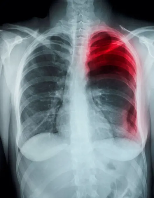
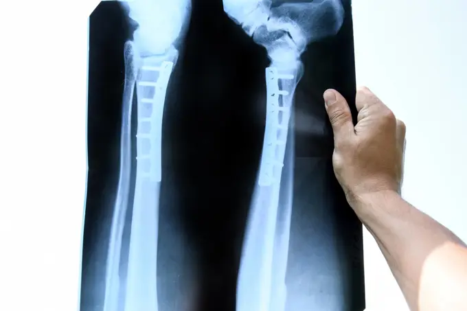
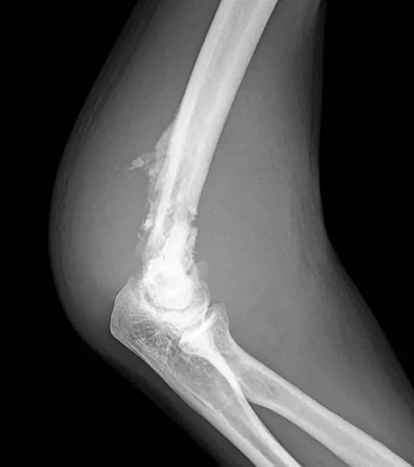
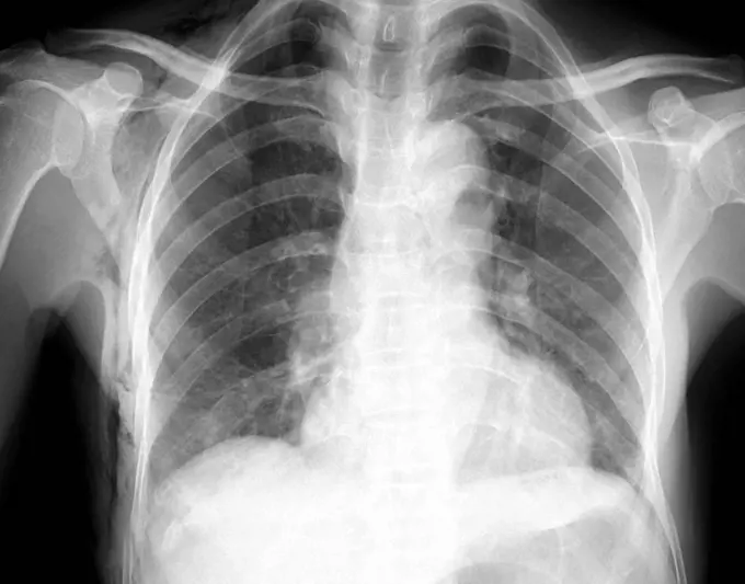
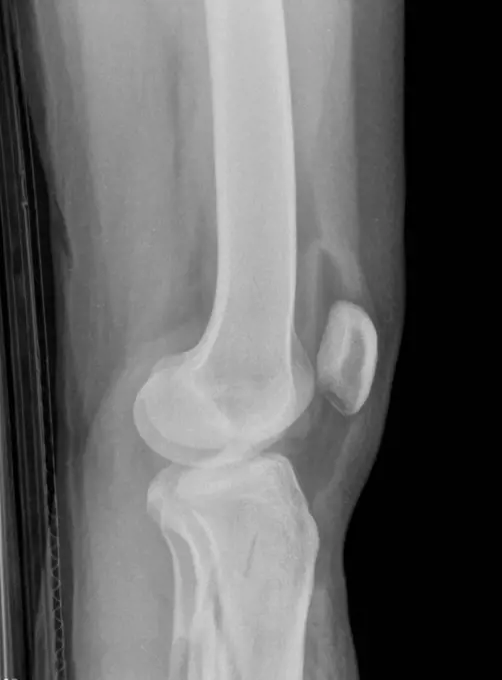
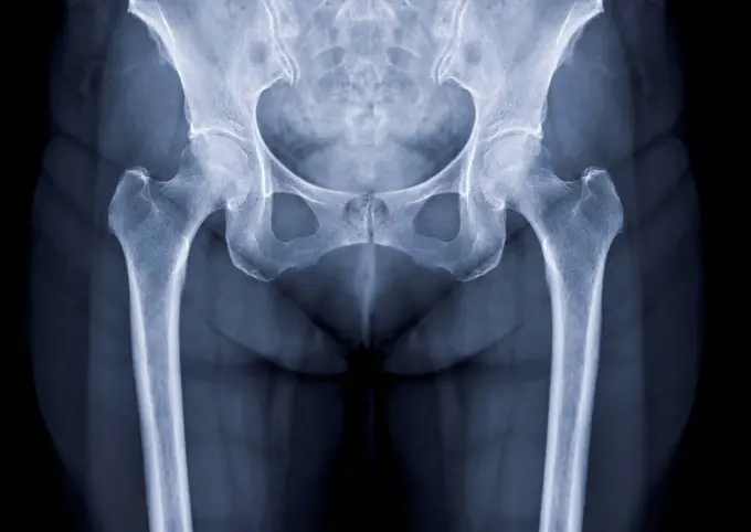
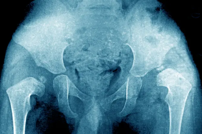
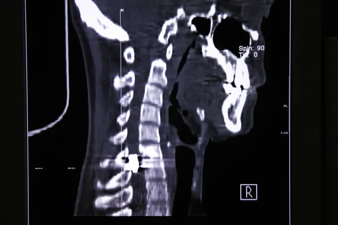
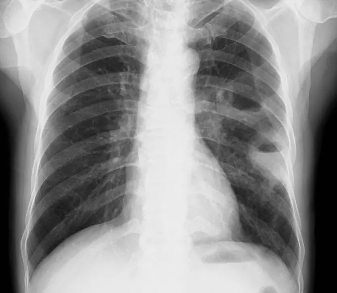
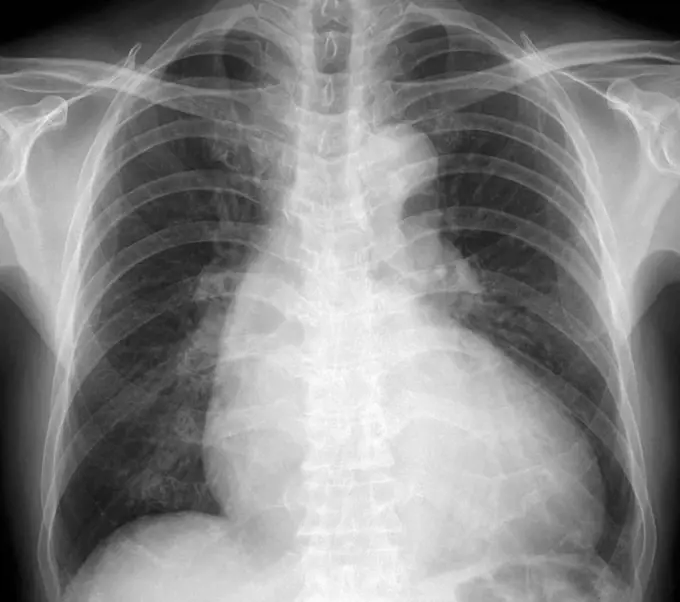
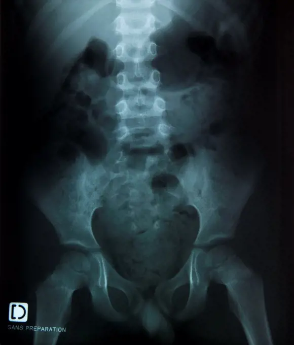
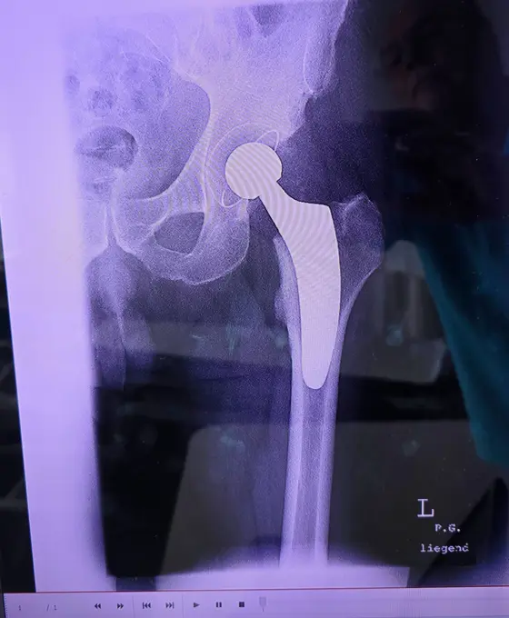
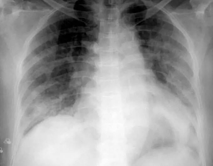
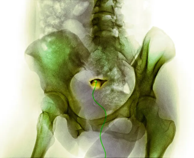
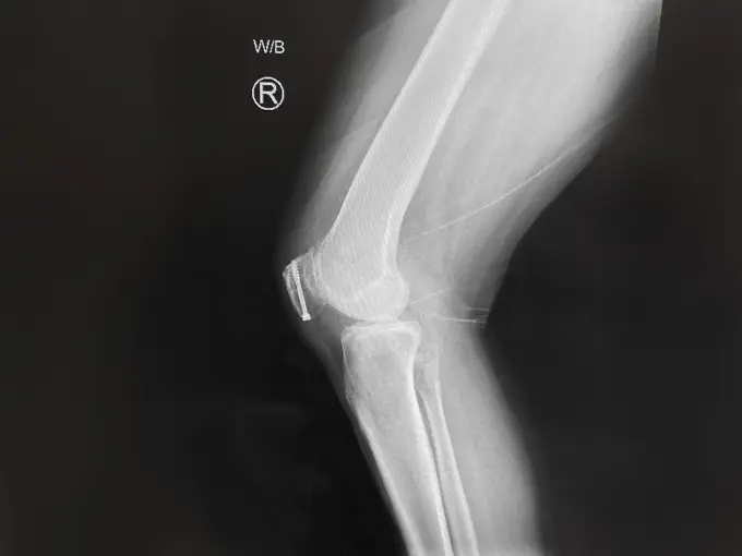
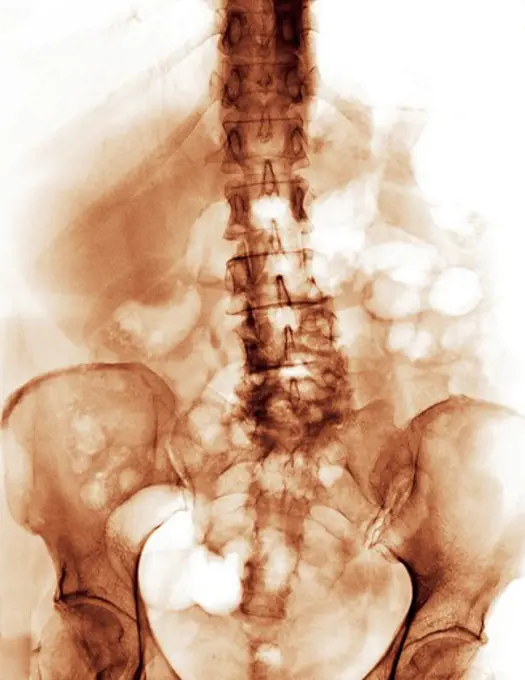
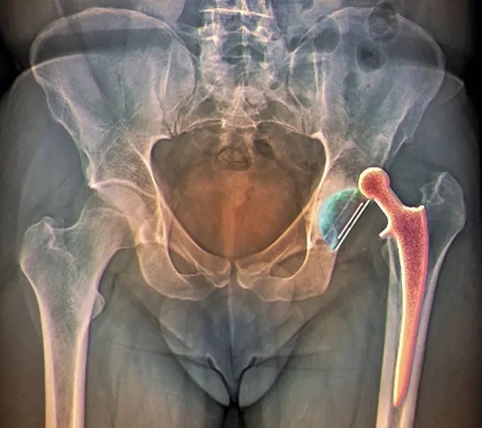
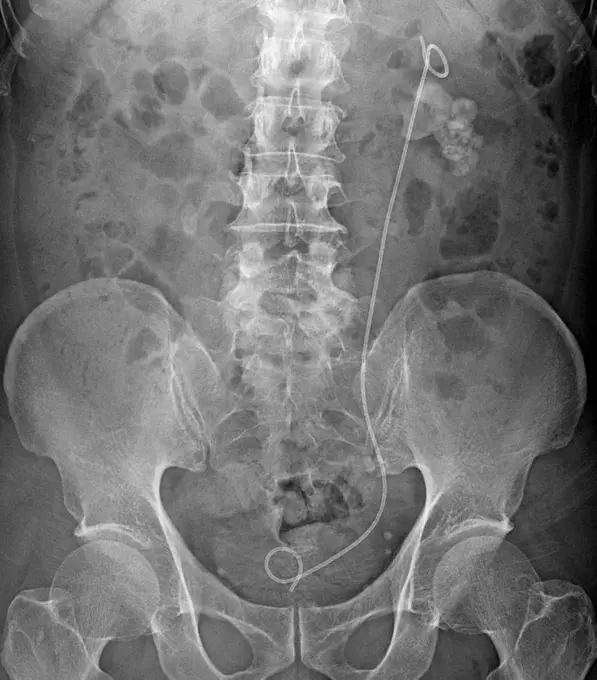
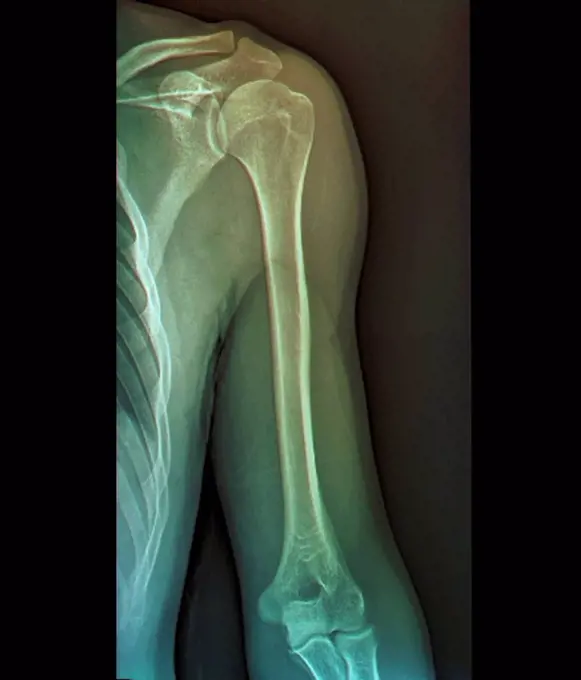
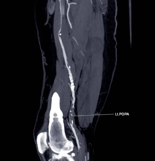
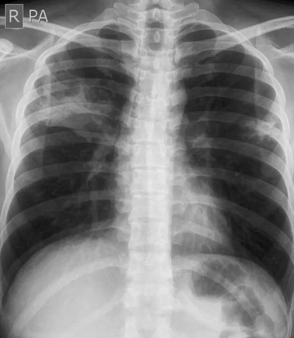
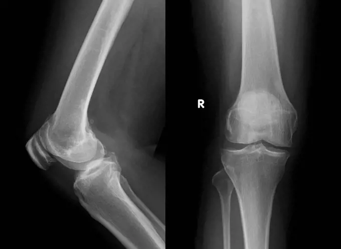
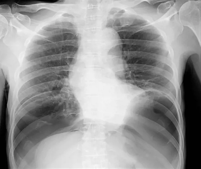
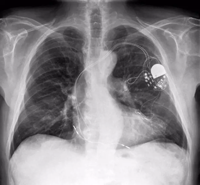
My Boards
View Pictures
  • My Boards

Medical X-Rays of Injuries

X-ray images representing various fractures and dislocations, focusing on shoulder and femur injuries, highlighting medical diagnostics.

An x-ray of a total hip replacement.
147 assets in this story
4128-19357551
X-ray of communitive left femur fracture.
1841R-111776
6114-50976661
An x-ray of a dislocation of shoulder.
4128R-15198
Broken shoulder. X_ray of the shoulder of a patient with a fracture in the neck of the humerus upper arm bone.
4128-24795637
Dislocated shoulder, X-ray
4128-19358077
X-ray showing a dislocated shoulder, where the head of the humerus is detached from the shoulder joint.
4128-24795641
Dislocated shoulder, X-ray
1848-55997741
X-ray image of broken hip in a dog
6114-50976670
An x-ray of a total hip replacement.
6188-69050246
Röntgen Aufnahme mit Hüft Prothese Röntgen Aufnahme mit Hüft Prothese LicenseRF Copyright: xZoonar.com/HGVorndranx 23243032
6188-55478602
human knee-joint with kneecap on X-ray image side view of human knee-joint with kneecap on X-ray image Copyright: xZoonar.com/ValeryxVoennyyx 13808390
4128-28769186
Fractured shoulder, X-ray
4128-19358050
Left oblique hysterosalpingography (X-ray) of a 35 year old women with infertility for 3 years showing bilateral patent tuba fallopian and bilateral c...
4128-19489655
Humerus fracture, X-ray
4128-18680257
Miliary tuberculosis, X-ray
6188-69050248
Röntgen Aufnahme Schulter links Röntgen Aufnahme Schulter links LicenseRF Copyright: xZoonar.com/HGVorndranx 23243063
824-63193989
A healthy lung x-ray on an 18-year old patient.
6145-29295404
The left and right knee-joints of Frank Burgess, probably a soldier in the South African War, after excision of the left knee. Radiograph, 1900/1904.
4128-19489710
Humerus fracture, X-ray
4128-28769213
Scoliosis of the spine, X-ray
824-29055706
X-ray of the hip before fitting a hip prosthesis.
1899-54027689
Pacemaker implanted in the chest. Frontal and side chest x-ray.
824-63194030
X-ray of the collar bone and the left sternoclavicular joint. The sternoclavicular joint space seems a little pinched with a small subchondral osteosclerosis, degenerative in appearance. The exam shows a probable degenerative arthropathy of the sternoclavicular joint.
4128-19358051
Intravenous urography of a 30 year old women with right abdominal pain, showing right hydroureteronephrosis. Hydroureteronephrosis is defined as a dil...
824-63195617
Choledochal cyst. A choledochal cyst is a congenital dilation of the main biliary tract.
1525-19878755
X-ray picture of two human knee-joints isolated on white background
1841R-111801
4297-1102
Normal shoulder x-ray of a 69 year old woman
824-63217239
Pacemaker (cardiac pacemaker) implanted in the thorax. Frontal chest X-ray.
4128-19357521
X-ray of a patient who had a femur (thigh bone) fracture with internal fixation. The X-ray shows the patient underwent trauma again and shows a broken...
1848-54974060
X-ray image of a lung with pulmonary tuberculosis, St. Thomas Home, Shibpur district, Howrah, Kolkata, West Bengal, India, Asia
1848-55375179
X-ray of a spinal column after an operation relating to spinal tuberculosis, damaged vertebrae have been bridged with steel, without this paralysis may occur, operations are financed by aid organisations, Howrah, Hooghly, West Bengal, India, Asia
824-63222237
Pneumonia with radiological appearance in frosted glass, it is due to partial filling of the alveolar lumens and / or thickening of the alveolar partitions, during pneumonia with infection in an immunocompromised patient (ex COVID 19), or a demed, or hemorrhage, or lung inflammation.
4408-1006
Shoulder, Medical - Xray / Radiology
4128-19357541
X-ray showing the dislocated elbow of a patient who was in a traffic accident.
824-63205983
Nephritic colic seen on a frontal x-ray of the abdomen.
1525-28468525
X ray film for human chest
824-224
X-Ray of Prostate Gland Tumor
6188-55478598
film with X-ray image of front view of human knees film with X-ray image of front view of two human knees isolated on white background Copyright: xZoonar.com/ValeryxVoennyyx 13808346
4128R-3006
Pancreas examination, X_ray
4297-1091
X-ray showing a distal radius fracture in a 52 year old woman
4128-20243404
Fractured femur, X-ray
4128-18929633
Fractured leg, X-ray
4128R-6534
Normal elbow joint, X_ray
1841R-111803
4128-19489606
Broken ankle, X-ray
824-63178607
X ray of the hip of a 6monts old girl. Normal
4128-19489774
Supracondylar fracture, X-ray
1841R-111790
4128-18680907
Posterior cruciate ligament avulsion fracture, X-ray
4128-19357546
Chest X-ray of a patient with prostate carcinoma with bone metastases. The X-ray shows an osteoblastic lesion on the rib, vertebral and proximal humer...
1773-97664
Chest X-ray, spontaneous pneumothorax
6188-54821817
steel plate holding together the tibia of the leg. x-ray of the leg. A steel plate holding together the tibia of the leg. Copyright: xZoonar.com/LeonidxEremeychukx 14368828
4128-19357548
X-ray showing osteosarcoma of the distal humerus showing an ill-divined lytic lesion with wide zone transition and a Codman triangle, a type of perios...
4128-20239853
Subcutaneous emphysema, X-ray
4128-20043110
Lipohemarthrosis, X-ray
4409-17333418
RADIOGRAFIA DE TIBIA CON ROTURA Y TORNILLOS EN EL TOBILLO.
4128-28766896
Healthy hip joints, X-ray
4128R-3761
Normal shoulder and elbow joints, X_ray
4128-20043114
Pneumonia, X-ray
824-63206956
Congential dislocation of the right hip in a 2 and a half-year old child, seen on a frontal pelvic x-ray.
824-63226723
Prostate cancer seen by frontal pelvic x-ray.
4297-1665
Hip x-ray of an 85 year old woman with breast cancer showing a metastatic bone lesion in the femur that has resulted in a fracture below her hip replacement
6188-64531328
Röntgenbild Steckschuss Röntgenbild Steckschuss im Hals. X-ray picture showing gun shot wound in neck. Copyright: xZoonar.com/AndreasxWolfx 306292
4128-19489727
Hydropneumothorax, X-ray
4128-48285514
Lung abscesses, X-ray
4128R-15463
Normal shoulder. Coloured X_ray of the healthy right shoulder of a 28 year old patient.
4128-19358202
Chest X-ray showing heart enlargement (cardiomegaly). The cardio-thorax ratio should not be greater than 50% in people of normal size.
824-63214252
Prostate caverns and diverticulum in the bladder. Frontal pelvic x-ray.
1899-85962
Retrograde Cystography
4128-20043103
Lipohemarthrosis, X-ray
6188-69050250
Röntgen Aufnahme mit Hüft Prothese Röntgen Aufnahme mit Hüft Prothese LicenseRF Copyright: xZoonar.com/HGVorndranx 23243079
4128-19358045
X-ray showing the chest of a Covid-19 pneumonia patient showing consolidation (opacities) in both lungs.
4128-30420610
Blocked fallopian tube, X-ray
1838-48084399
Medical x-ray of mid-adult woman's repaired patella
4128-19358056
15 minutes Intravenous urography of 30 year old women with right abdominal pain, showing right hydroureteronephrosis. Hydroureteronephrosis is defined...
4297-1079
Lumbar spine x-ray of a 64 year old male with degenerative arthritic changes in the lower back
4128R-30957
Dislocated hip replacement. Coloured frontal X-ray of the pelvis of a 35 year old patient with an artificial left hip (right) that has become dislocat...
4128-28970847
Urinary system stent, X-ray
4128R-3159
Normal shoulder joint, X_ray
4128-28767065
Popliteal artery, CT scan
4128-20043155
Lung abscess, X-ray
1841R-111775
4128-19489796
Tibial plateau fracture, X-ray
4128-30419884
Treatment for blocked arteries, angiogram
4297-1178
Chest x-ray of a 2 year old boy showing right middle lobe pneumonia
4128-20043630
Lung abscess, X-ray
4128-19358214
Lateral left decubitus (LLD) abdominal X-ray of a patient with abdominal pain showing a crescent shape radiolucent lesion above the liver suggestive o...
1525-22326474
Close-up of a dental X-Ray
1841R-111820
824-63211086
Ruptured Achilles tendon, seen on a sagittal section MRI scan of the ankle.
1525-26807273
Dental x ray film for dental care concept
4269-20408381
A knee x-ray of a patient with ACDC, a rare calcification disorder, reveals calcification in the main artery supplying blood to the lower leg.
1439-57940620
Chest Xray showing heart and lungs with a visible pacemaker
4128-20043131
Knee osteoarthritis, X-ray
824-63178603
Sinus X-ray of a 35-year-old woman. No anomaly noted
4128-20041843
Pneumoperitoneum, X-ray
824-63217267
Pulmonary sarcoidosis seen on a frontal chest x-ray.
4128R-7208
Heart pacemaker, X_ray
4408-1021
Lower Spine, Electronics - Radio & Wireless
PREVIOUS
of 2
NEXT
2401 S. Ervay, Suite 206
Dallas, Texas 75215
United States
Get Started
Free ResearchMy BoardsMy Cart
For Creators
How To License Your ContentContributor PortalFrame of Mind
Resources
API accessPricing
Contact
+1 866 236 0087help@viewpictures.co.uk Contact form
©2026 View Pictures. All Rights Reserved. -A
This site is protected by reCAPTCHA and Google Privacy Policy and Terms of Service apply.