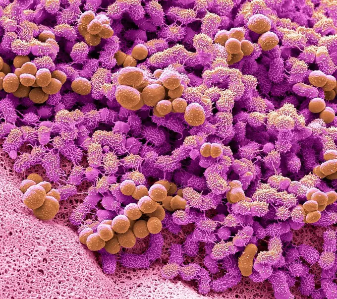
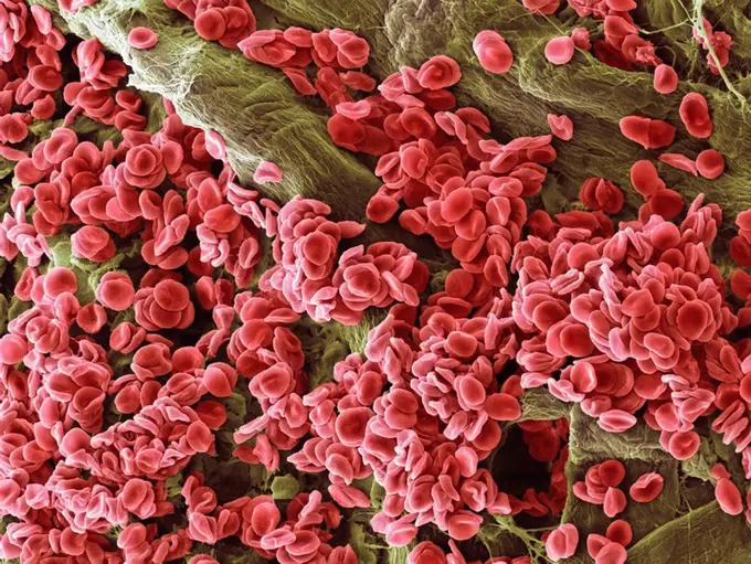
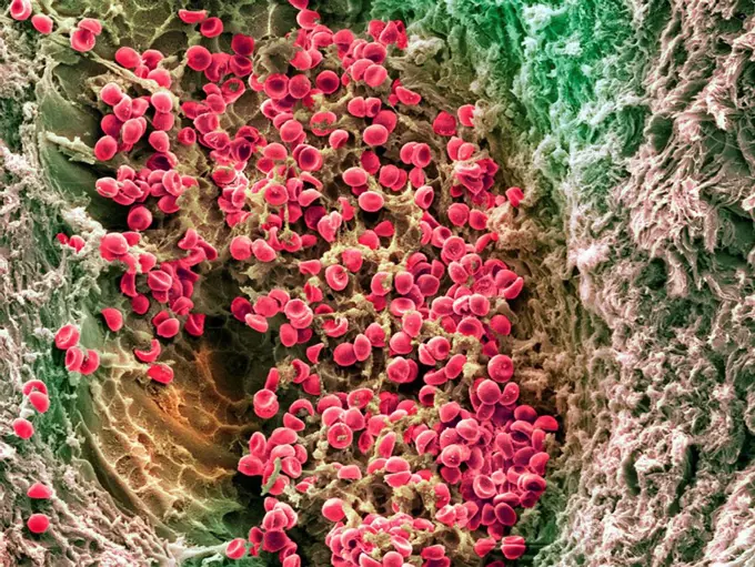
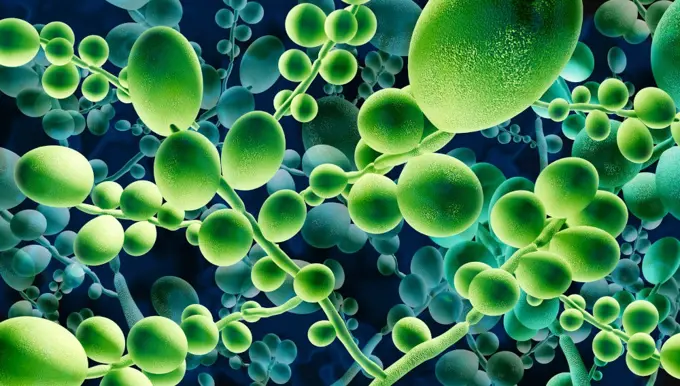
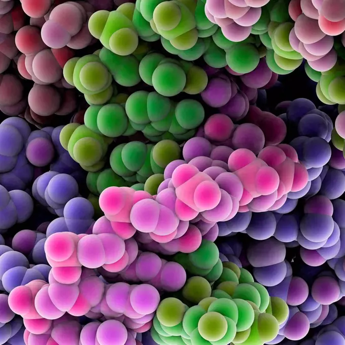
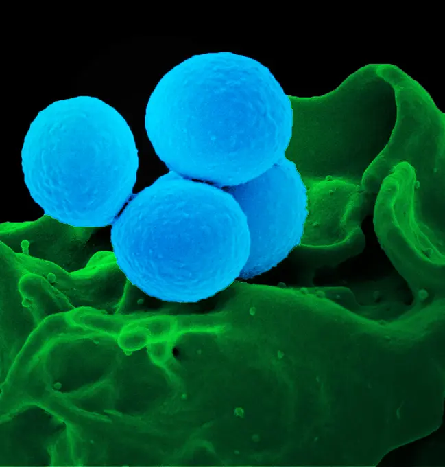
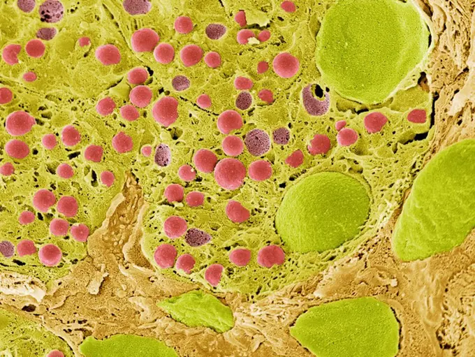
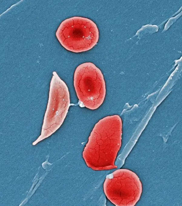
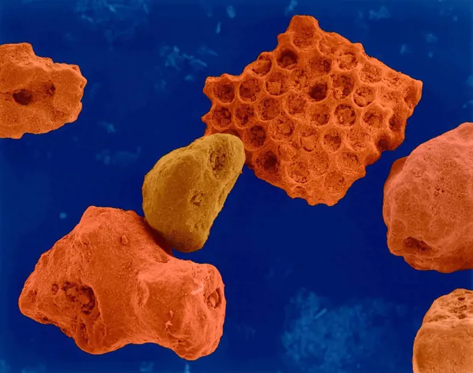
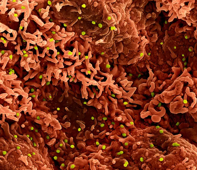
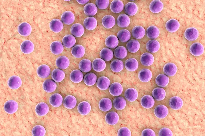
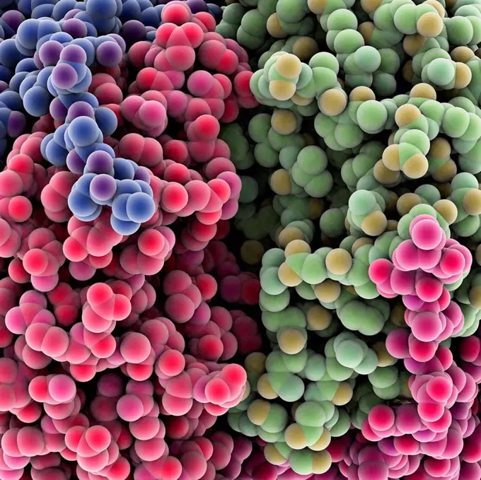
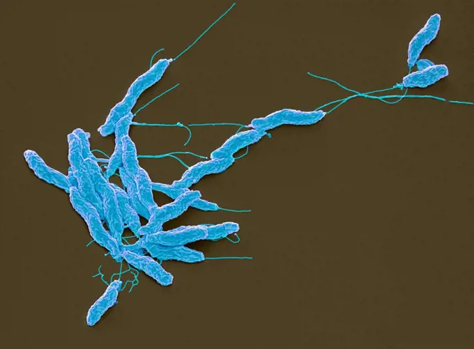
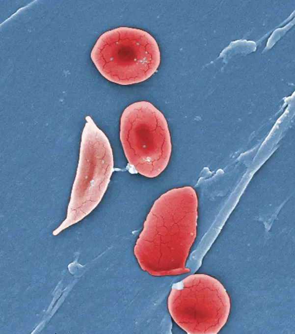
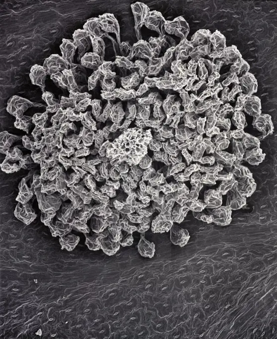
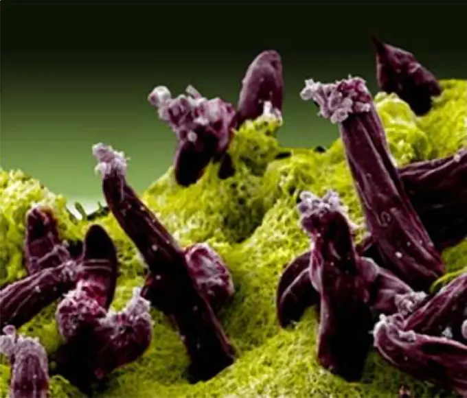
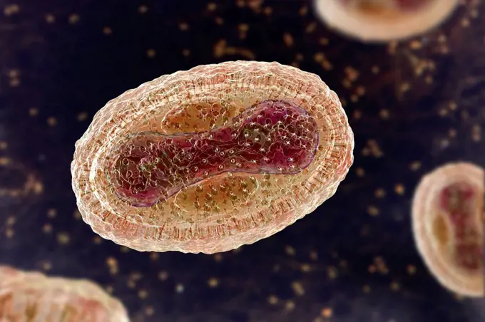
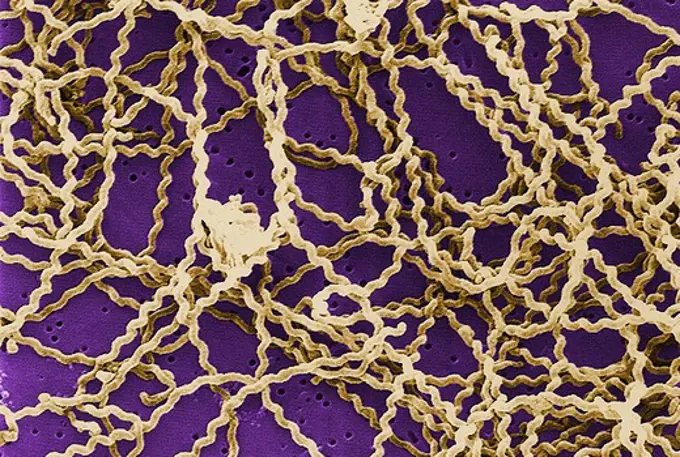
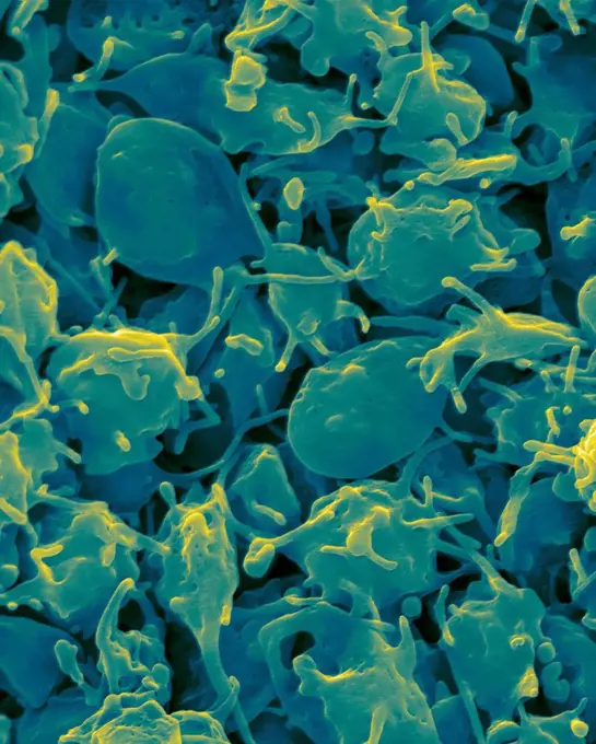
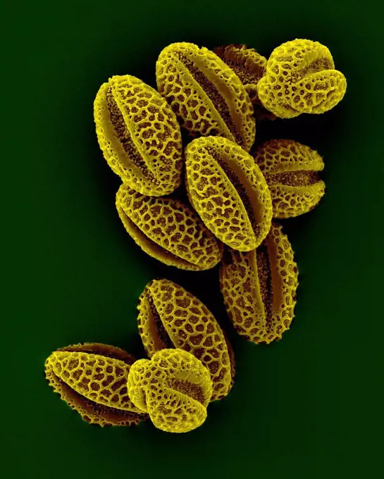
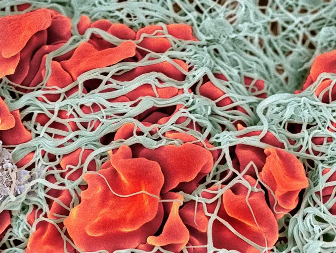
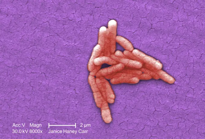
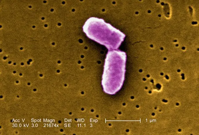
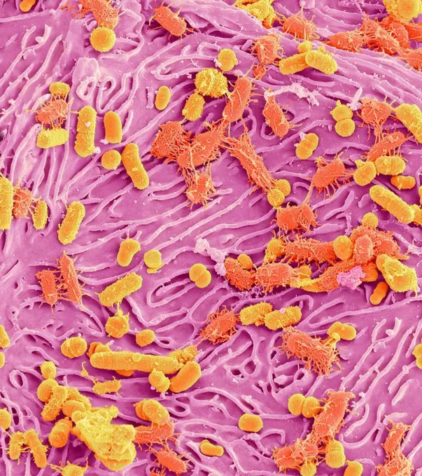
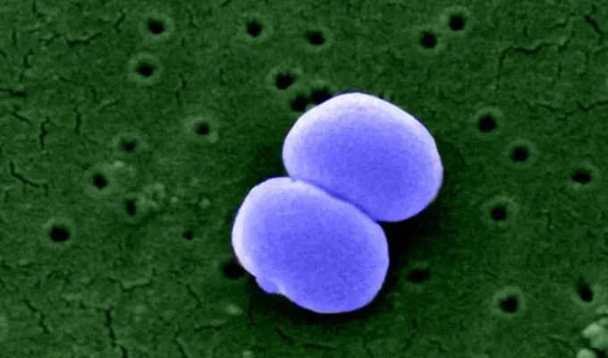
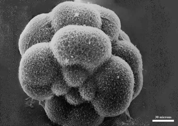
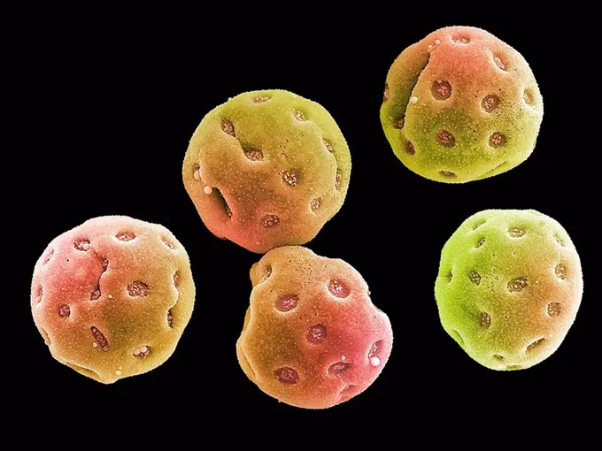
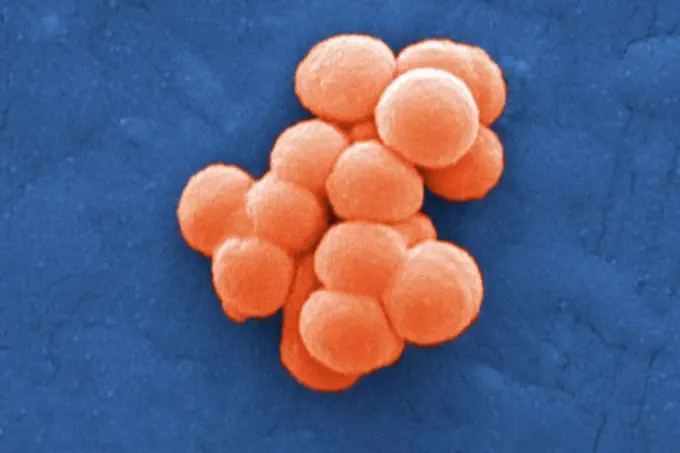
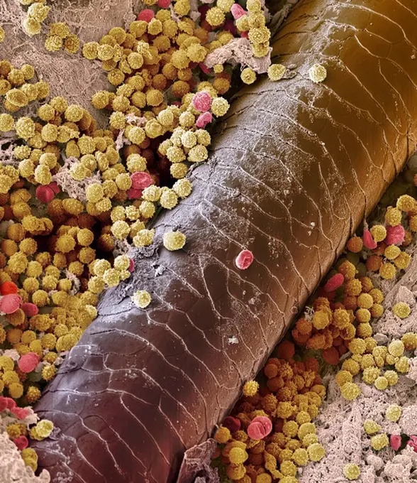
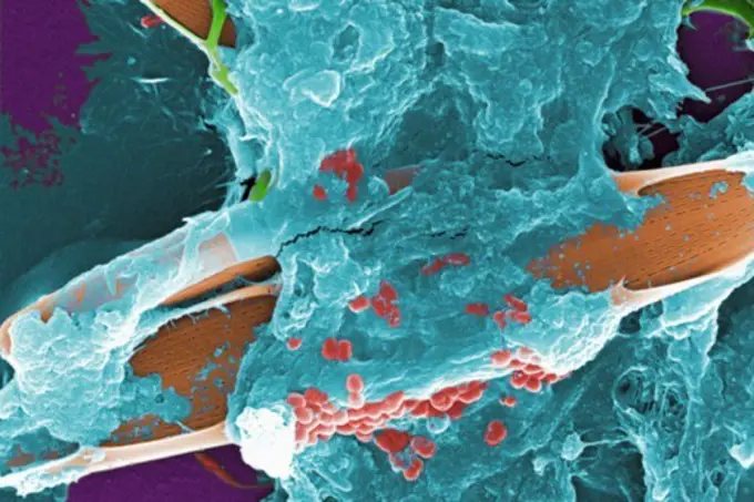
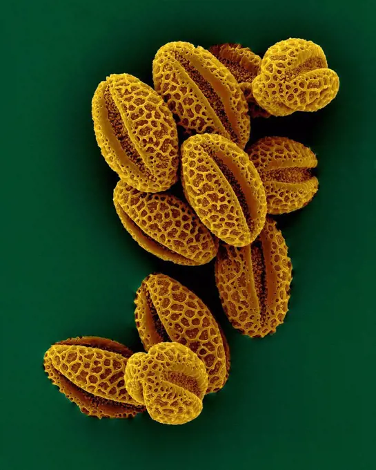
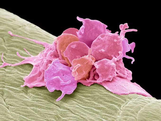
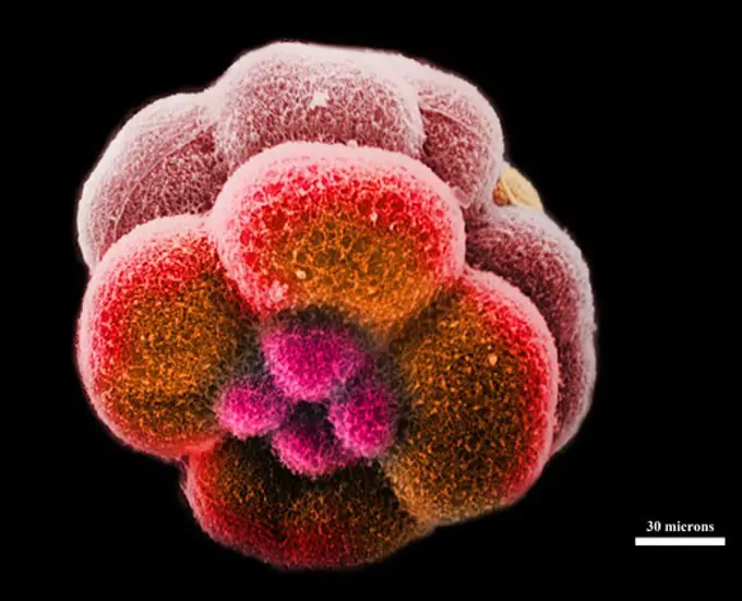
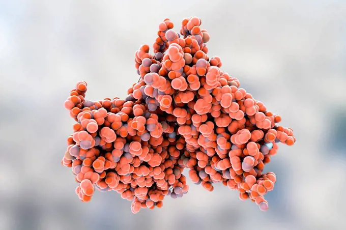
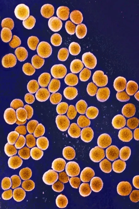
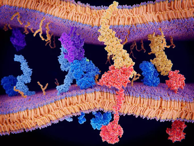
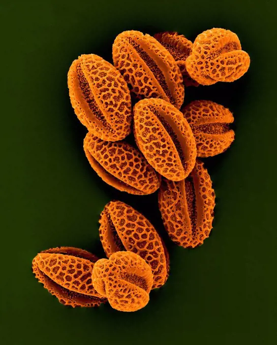
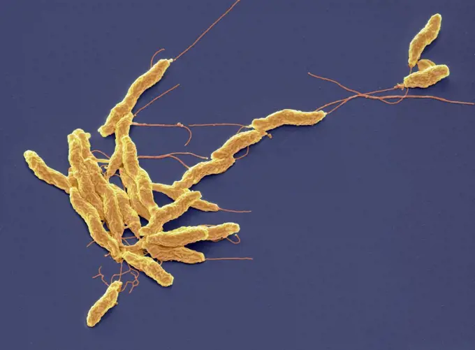
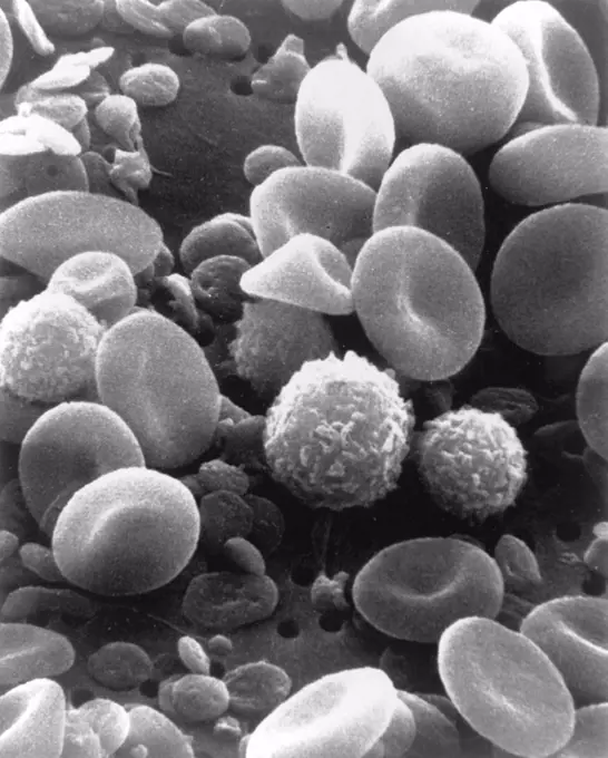
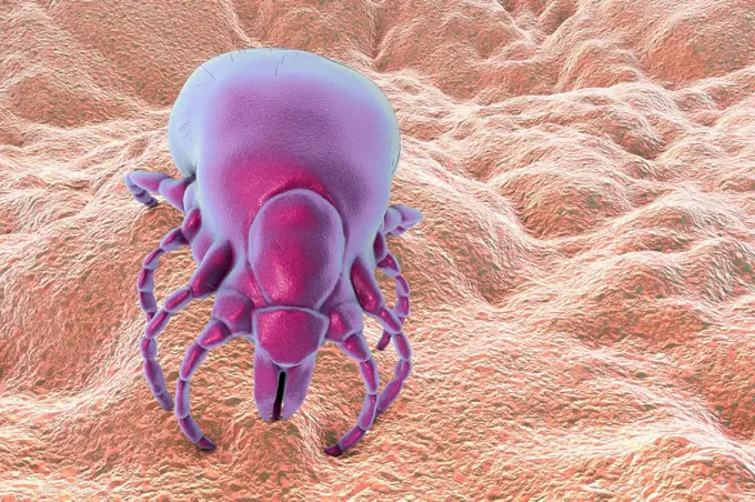
My Boards
View Pictures
  • My Boards

Microscopic Bacteria and Cells

Detailed scanning electron micrographs of bacteria, including Staphylococcus and SARS-CoV-2, showcasing vibrant colors and intricate structures.

Foot bacteria, SEM
Foot bacteria, SEM
173 assets in this story
4128R-8448
Red blood cells, SEM
4269-6808
Scanning electron micrograph (SEM) of stomach wall lined with mucus producing epithelial cells. At center, a drop of secretion from a fundus gland. Maginification 3000x.
4128R-2342
Blood clot, SEM
4128R-11476143
Protein molecules, computer artwork.
4269-6811
Scanning electron micrograph (SEM) of an absorptive epithelial cells lining the surface of the small intestine, showing numerous microvilli. Magnification x20000.
1525-56857797
Deadly Fungal Infection concept as a microbe threat and mucormycosis life threatening fungus as Candida auris fungi spreading as a Mycology health threat as a global outbreak risk as a 3D illustration
1899-53513322
Ebola virus isolated in November 2014 from patient blood samples obtained in Mali. The virus was isolated on Vero cells.
4128R-11476165
Protein molecules, computer artwork.
824-63191221
Scanning electron micrograph of a human neutrophil ingesting MRSA.
4128R-7595
Pancreas tissue, SEM
4384-146
This digitally-colorized scanning electron micrograph (SEM) revealed some of the comparative ultrastructural morphology between normal red blood cells (RBCs), and a sickle cell RBC (left) found in a blood specimen of an 18 year old female patient with sickle cell anemia, (HbSS). People who have this form of sickle cell disease inherit two sickle cell genes (S), one from each parent. This is commonly called sickle cell anemia, and is usually the most severe form of the disease. Sickle cell di
4128R-13620308
Coloured scanning electron micrograph (SEM) of Coral sand (Kailua, Hawaii). Beach sand contains both rock minerals and skeletons that have been derive...
4128R-12699854
Coloured scanning electron micrograph (SEM) of fungal cells. The round structures are sporangia, which house the fungus' reproductive cells (spores). ...
824-65830961
Colorized scanning electron micrograph of mpox virus particles (green) on the surface of infected VERO E6 cells (red).
824-63223158
Scanning electron micrograph of Ebola virus budding from the surface of a Vero cell (African green monkey kidney epithelial cell line). Credit: NIAID.
824-63191209
Scanning electron micrograph of HIV particles infecting a human T cell.
4239-18641821
Conceptual image of virus and bacteria.
4128R-15555
kiwi fruit Actinidia deliciosa pollen grain, coloured scanning electron micrograph SEM. Magnification: x3000 when printed at 10 centimetres wide.
4128R-13048916
Computer illustration of staphylococci bacteria (Staphylococcus aureus).
4128R-11476223
Protein molecules, computer artwork.
824-63223159
Scanning electron micrograph of Ebola virus budding from the surface of a Vero cell (African green monkey kidney epithelial cell line. Credit: NIAID.
4128R-13022174
Campylobacter jejuni bacteria. Coloured scanning electron micrograph (SEM) of a Campylobacter jejuni bacteria. Campylobacter jejuni is a Gram-negative...
824-63194916
This digitally-colorized scanning electron micrograph (SEM) revealed some of the comparative ultrastructural morphology between normal red blood cells (RBCs), and a sickle cell RBC (left) found in a blood specimen of an 18 year old female patient with sickle cell anemia , (HbSS), People who have this form of sickle cell disease inherit two sickle cell genes (S), one from each parent. This is commonly called sickle cell anemia, and is usually the most severe form of the disease.
4201-66268
Leather Fern (Arachniodes adiantiformis) SEM close-up view of a sorus on the underside of a frond is where haploid spores are formed in meiosis
824-63191257
Scanning electron micrograph of Plasmodium gallinaceum invading mosquito midgut.
4128R-15545706
Plaque-forming bacteria, SEM
4128-20039419
Monkeypox virus particles, illustration
4128R-15545699
Fallopian tube, SEM
824-63191233
Scanning electron micrograph of an apoptotic HeLa cell. Zeiss Merlin HR-SEM.
1899-65662308
Colorized scanning electron micrograph of Marburg virus particles (orange) both budding and attached to the surface of an infected VERO E6 cell (purple).
4297-1821
Scanning electron micrograph, SEM, of Leptospira sp. bacteria atop a 0.1. µm polycarbonate filter
4128R-13619962
Activated platelets with channels of the open canalicular system (OCS), coloured scanning electron micrograph (SEM). Platelets are blood cell fragment...
4269-27405
Mycobacterium tuberculosis. Scanning electron micrograph (SEM) of Gram-positive Mycobacterium tuberculosis bacilli, the causative agent for tuberculosis. Magnification 15549x.
4128R-8678
Common daisy anther, SEM
4128R-13620147
Weeping willow pollen (Salix babylonica), coloured scanning electron micrograph (SEM). The willow (and poplar) family, Saliaceae, is characterized by ...
4384-307
Leptospira Bacteria Atop Polycarbonate Filter
4128R-1397
Blood clot, SEM
824-63194413
Under a moderately-high magnification of 8000X, this colorized scanning electron micrograph (SEM) revealed the presence of a small grouping of Gram-negative Salmonella typhimurium bacteria that had been isolated from a pure culture.
4297-1763
Colorized scanning electron micrograph (SEM) showing a highly magnified view of a dividing Escherichia coli bacterium, clearly displaying the point at which the bacteria's cell wall was dividing; Magnification 21674x
4128R-13162328
Oral bacteria. Coloured scanning electron micrograph (SEM) of mixed oral bacteria attached to a buccal cell (pink) from the cheek. The mouth contains ...
1899-61460214
A scanning electron microscope picture of a nerve ending. It has been broken open to reveal vesicles (orange and blue) containing chemicals used to pass messages in the nervous system
824-63128612
Staphylococcus epidermidis seen at the SEM. See image n0642505 on the site.
4128-V58557904
4297-1770
Scanning electron micrograph of Flexispira rappini bacteria, magnified 13,951x
4128-V58557851
4297-1246
Scanning electron microscopic image of Lytechinus pictus (sea urchin) embryo at the 16-cell stage
4128-111584776
Convolvulus pollen grains. Coloured scanning electron micrograph (SEM) of pollen grains from a convolvulus flower.Convolvulus is a genus of about 200 ...
824-63223131
Scanning electron micrograph of filamentous Ebola virus particles replicating from an infected VERO E6 cell (50,000x magnification). Credit: NIAID.
4384-237
Scanning Electron Micrograph of Streptococcus specie Bacteria
4128R-7330
Pyoderma skin disease, coloured scanning electron micrograph SEM. Large numbers of lymphocytes yellow and red blood cells are seen in necrotic tissue ...
4384-253
Scanning Electron Micrograph of Untreated Water Sample
4128R-13620149
Weeping willow pollen (Salix babylonica), coloured scanning electron micrograph (SEM). The willow (and poplar) family, Saliaceae, is characterized by ...
4128R-15465074
Activated platelets, SEM
4297-1247
Scanning electron microscopic image of Lytechinus pictus (sea urchin) embryo at the 16-cell stage
4269-27386
Methicillin-Resistant Staphylococcus Aureus. Scanning electron micrograph (SEM) of methicillin-resistant Staphylococcus aureus bacteria (MRSA); Magnification 9560x. This strain of Gram-positif bacterium has developed wide-ranging antibiotic resistance such as methicillin, and are hospital pathogens.
4269-27464
Clostridium difficile. Scanning Electron Micrograph (SEM) of Clostridium difficile bacteria. C. difficile, an anaerobic gram-positive rod, is the most frequent cause of antibiotic-associated diarrhea (AAC).
4128-20040356
Rhodopsin molecule, illustration
824-63211605
Staphyloccus aureus. Bacteria responsible for food poisoning, localized suppurative infections and some cases of septicemia. Seen under optical microscopy X 1000
824-63227228
The Human Microbiome Project, which was launched by the NIH in 2007, provided the first insight into the microbial life of healthy humans and explores possible relationships between particular human diseases and the microbiome. (Clockwise from top left): Streptococcus (Credit: Tom Schmidt) Mixed species microbial biofilm, from the human body (Credit: A. Earl, Broad Institute/MIT) Bacillus (Credit : Tom Schmidt) Malassezia lopophilis (Credit: JH Carr, CDC).
4128R-15514870
Programmed cell death in immune system, illustration
4128R-13620150
Weeping willow pollen (Salix babylonica), coloured scanning electron micrograph (SEM). The willow (and poplar) family, Saliaceae, is characterized by ...
824-63223148
Colorized scanning electron micrograph of Ebola virus particles (red) in extracellular space between infected African green monkey kidney cells. Image captured and color-enhanced at the NIAID Integrated Research Facility in Ft. Detrick, Maryland. Credit: NIAID.
4128R-13022170
Campylobacter jejuni bacteria. Coloured scanning electron micrograph (SEM) of a Campylobacter jejuni bacteria. Campylobacter jejuni is a Gram-negative...
1525-56160471
Beautiful coral at deep sea 3d illustrated
1899-85573
Blood Cell SEM
6145-29279955
Central nervous system, Zebrafish embryo
4128-19247886
Fusobacterium bacteria, computer illustration. These bacteria are Gram-negative, anaerobic, non-motile rod-shaped prokaryotes (characterized by a long...
4128R-15290693
Lyme disease tick, illustration
4128R-15514885
CAR T cell cancer immunotherapy, illustration
4128R-4612
Chinese hibiscus pollen, SEM
824-63227423
This is a scanning electron microscope image of traumatized muscle tissue taken from a wounded soldier. It shows a red blood cell (false color) entangled in a nanofibrous extracellular matrix. Highly fibrotic regions such as these are thought to precede bone formation during abnormal wound healing, leading to heterotopic ossification, the formation of bone in places outside the skeleton, such as soft tissues.
4128R-15290691
Lyme disease tick, illustration
4384-294
Scanning Electron Micrograph of Borrelia burgdorferi Bacteria
PREVIOUS
of 2
NEXT
2401 S. Ervay, Suite 206
Dallas, Texas 75215
United States
Get Started
Free ResearchMy BoardsMy Cart
For Creators
How To License Your ContentContributor PortalFrame of Mind
Resources
API accessPricing
Contact
+1 866 236 0087help@viewpictures.co.uk Contact form
©2026 View Pictures. All Rights Reserved. -A
This site is protected by reCAPTCHA and Google Privacy Policy and Terms of Service apply.