My Boards
View Pictures
  • My Boards

Microscopic Bacterial Illustrations

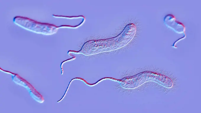
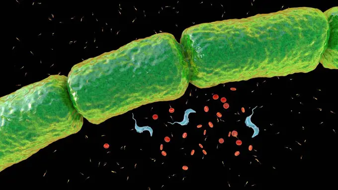
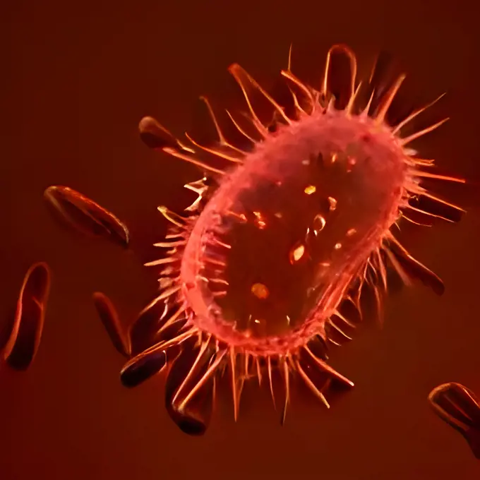
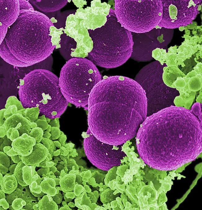
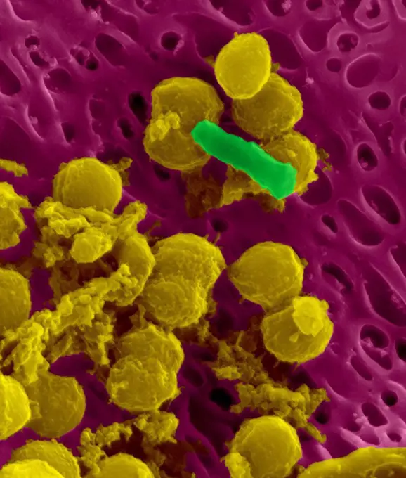
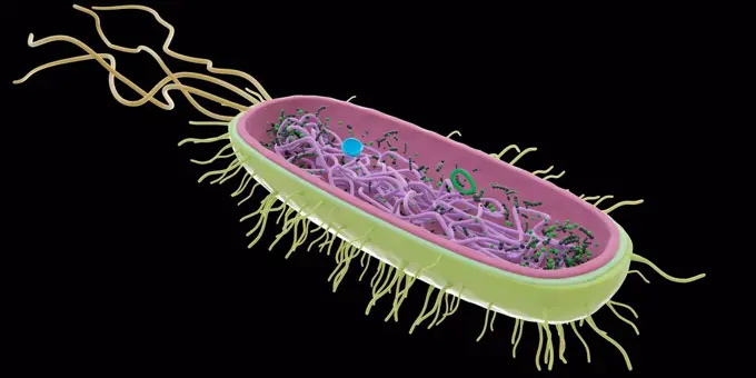
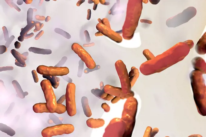
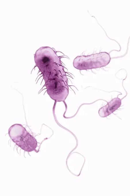
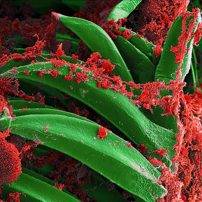
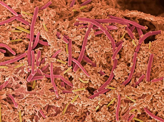
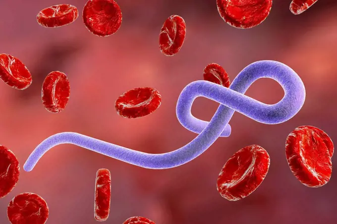
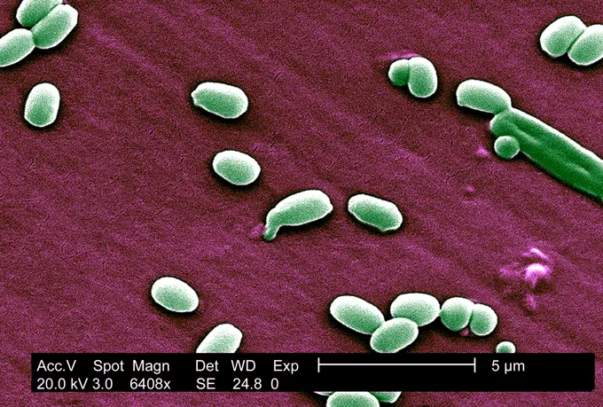
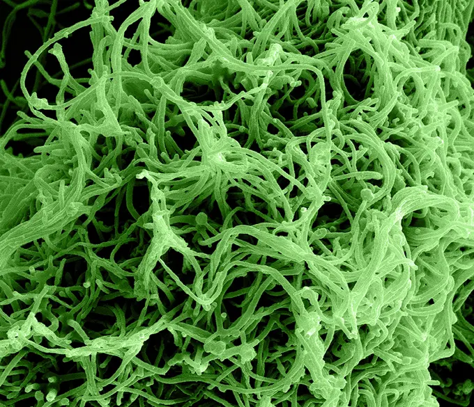
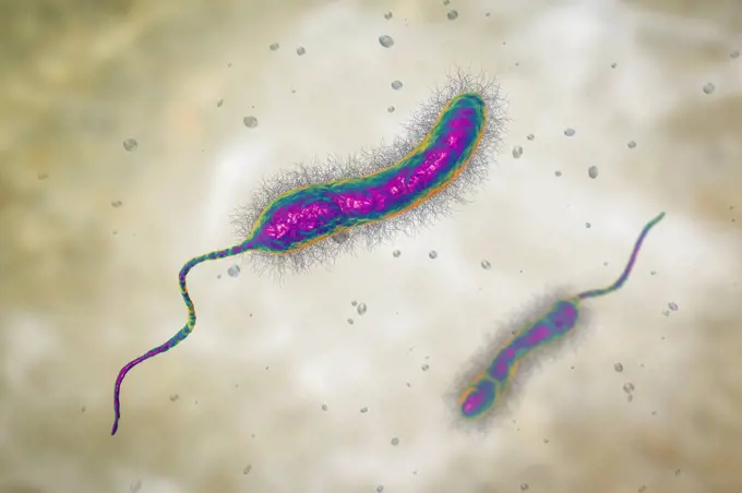
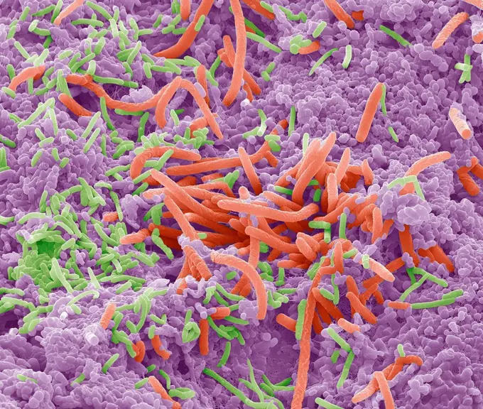
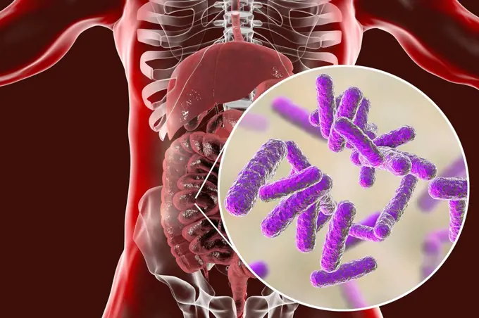
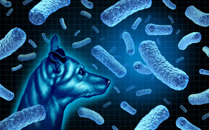
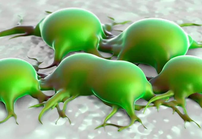
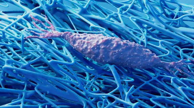
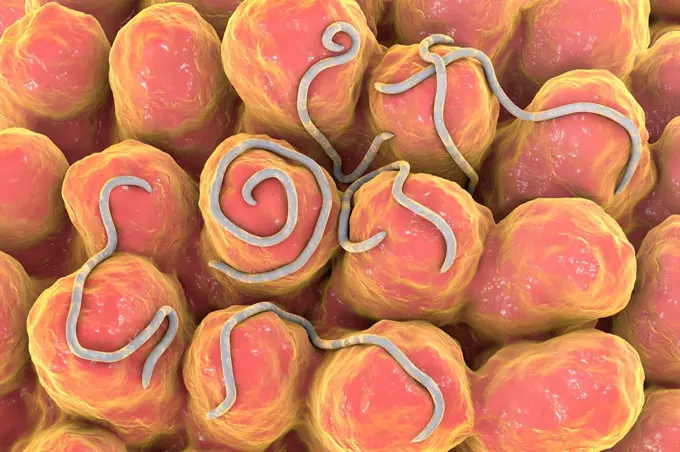
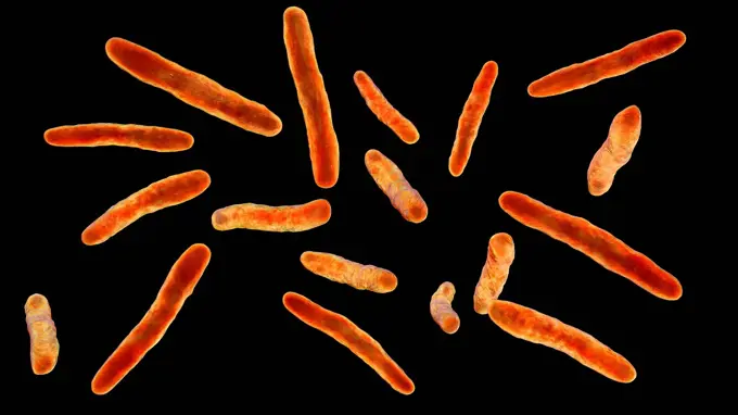
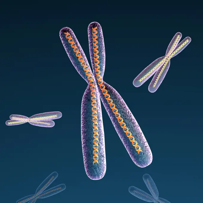
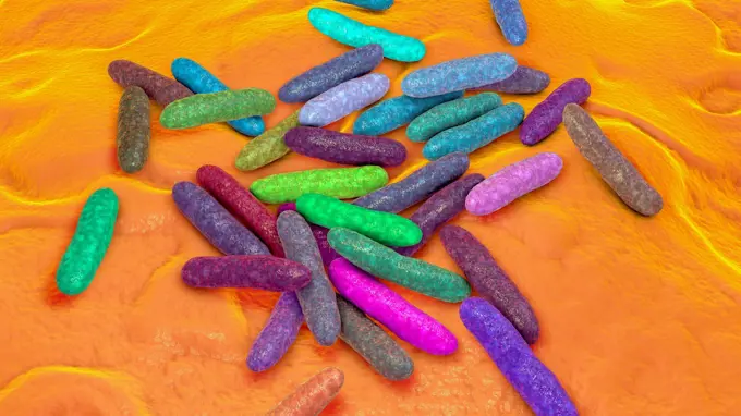
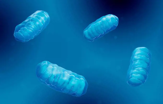
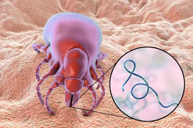
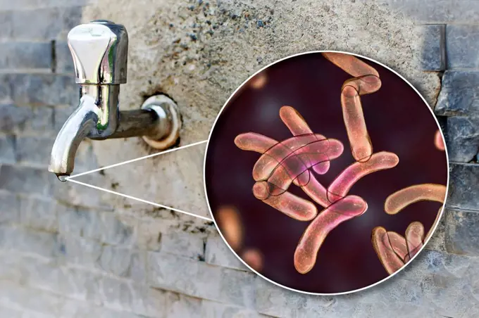
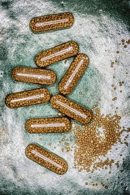
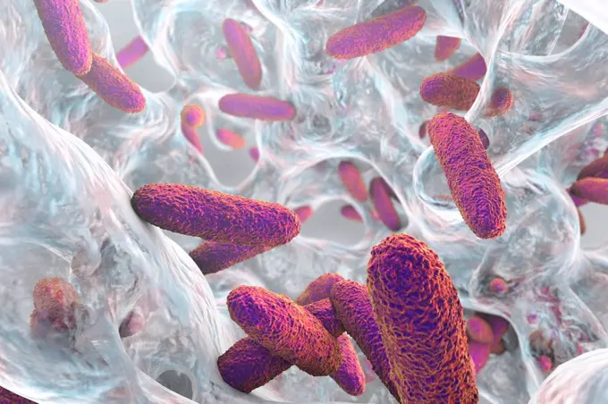
3D rendered images of bacteria in various shades of green and purple, showcasing their structures under microscopic view.

Klebsiella pneumoniae bacteria in biofilm, computer illustration. K. pneumoniae are Gram-negative, encapsulated, non-motile, enteric, rod prokaryote. ...
490 assets in this story
1899-54026851
Meningococcus seen under a scanning electron microscope.
4128R-13682496
Vibrio cholerae and flagella, illustration.
1815-18410349
Three dimensional render of green bacteria on human tissue
4269-27654
Acinetobacter baumannii. Scanning Electron micrograph (SEM) of Acinetobacter baumannii. This aerobic Gram-negative, non-motile bacteria are normal flora on the skin. But because A. baumannii has developed substantial antimicrobial resistance, it is an emerging cause of nosocomial infections in hospitals, responsible for pneumoniae, hemopathic and wound infections. Magnification of 12739x.
4128-28575422
Vibrio mimicus bacteria, illustration
4269-6810
Scanning electron micrograph (SEM) of an absorptive epithelial cells lining the surface of the small intestine, showing numerous microvilli. Magnification x20000.
4128-V58570351
4128-20042776
Thiomargarita magnifica, illustration
824-63123124
A view at SEM x 6000.
4128R-3136
Plaque_forming bacteria, SEM
824-63225896
Colorized scanning electron micrograph of a cell (green) infected with SARS-CoV-2 virus particles (orange), isolated from a patient sample. Image captured at the NIAID Integrated Research Facility (IRF) in Fort Detrick, Maryland.
6019-28586250
microscopic bacteria. Legionella pneumophila illustration
4128R-3547
Plaque_forming bacteria, SEM
824-63211079
Legionella pneumophila is the bacteria responsible for legionellosis (pneumonia known as Legionnaires' disease). Optical microscopy X 1000.
4128R-4999
Climbing lily pollen, SEM
4128R-8115
Blood clot, SEM
4413-174723
Coralline red algae by light microscopy
824-63194633
Scanning electron micrograph of S. aureus bacteria escaping destruction by human white blood cells.
824-63211078
Legionella pneumophila is the bacteria responsible for legionellosis (pneumonia known as Legionnaires' disease). Optical microscopy X 1000.
4128R-13619988
Bacteria on an epithelial cell from the human tongue filiform papilla, coloured scanning electron micrograph (SEM). Coccoid bacteria (likely Streptoco...
824-63123764
This scanning electron micrograph depicts a grouping of Gram-negative Flexispira rappini bacteria, magnified 13,951x. Its name F. rappini is considered provisional, for it was never formally proposed or accepted. Subsequently determined to be closely related to Helicobacter spp. it is referred to as Helicobacter sp. flexispira in the literature.
1899-54026846
Clostridium difficile (bacteria responsible for hospital acquired diarrhoea) seen under optical microscopy.
4269-27406
Mycobacterium tuberculosis. Scanning electron micrograph (SEM) of Gram-positive Mycobacterium tuberculosis bacilli, the causative agent for tuberculosis. Magnification 15549x.
4128R-13587515
Various microbes of different shapes, computer illustration. Mixture of microorganisms including spherical (cocci) and rod-shaped bacteria together wi...
824-63211037
Cholera bacilli or cholera vibrio (Vibrio cholerae). Cholera is an infectious and contagious disease characterized by sudden and very abundant diarrhoea. In the absence of treatment, the classic major form is fatal in more than half of the cases. Optical microscopy X 1000.
4128R-13620037
Coloured scanning electron micrograph (SEM) of Photocomposite, E. coli on the surface of human skin. Escherichia coli is a Gram-negative, facultativel...
4128R-13047360
Tongue bacteria. Coloured scanning electron micrograph (SEM) of bacteria on the surface of a human tongue. Large numbers of bacteria can form a visibl...
4128-111578639
Bacterium, computer illustration.
4128R-13587566
Plague bacteria (Yersinia pestis), computer illustration. Y. pestis are gram-negative bacteria from Enterobacteriaceae family, they have ovoid shape w...
1525-26235515
An abscess is a collection of pus that has built up within the tissue of the body. 3D illustration. Animatied of infected wound ,bacteria , pus, abscess
1525-27535058
Probiotics. Microbacterium and therapeutic microscopic bacteria organism, lactobacillus for flora stomach, microbiology scientific medicine blue organic 3d macro spheres in liquid vector background. Probiotics. Microbacterium and therapeutic microscopic bacteria organism, lactobacillus for flora stomach, microbiology blue organic 3d macro spheres in liquid vector background
4128-111576786
Yeast cells. Coloured scanning electron micrograph (SEM) of cells of brewer's, or baker's, yeast (Saccharomyces cerevisiae). This fungus consists of s...
1899-61461207
Cholera bacilli or cholera vibrio (Vibrio cholerae). Cholera is a contagious epidemic enteric poisoning infection. If left untreated, the classic major form is fatal in more than half of cases. Image taken from an X 1000 optical microscopy
824-63227067
This scanning electron microscope image shows SARS-CoV-2 (round gold particles) emerging from the surface of a cell cultured in the laboratory. SARS-CoV-2, also known as 2019-nCoV, is the virus that causes COVID-19. Image captured and colorized at Rocky Mountain Laboratories in Hamilton, Montana. Credit: NIAID
1525-23490829
Orange 3d bacteria cell culture with flagellums under microscope. Vector EPS10. Biology, Microbiology, Science.
4269-27411
Marburg virus. Transmission electron micrograph (TEM) of filamentous Marburg virus, magnified 100,000x. This RNA virus of the Filovirus (like Ebola) is known to cause Marburg hemorrhagic fever, which can be fatal.
4128R-13476
E. coli bacteria. Coloured scanning electron micrograph SEM of Escherichia coli bacteria orange on a gecko´s tongue. Magnification: x8000 when printed...
4269-27370
Vibrio cholerae. Scanning electron micrograph (SEM) of Vibrio cholerae bacteria. This Gram-negative bacterium causes cholera. Magnified 22399x.
4128-19055997
Citrobacter bacteria, illustration
4128R-13763257
Pseudomonas aeruginosa bacteria inside biofilm, computer illustration. This is a Gram-negative, aerobic, enteric, rod prokaryote. P. aeruginosa causes...
4201-66346
Common Camellia (Camellia japonica) SEM close-up view of pollen at 725x magnifcation
4239R-7975
Conceptual image of chromosome.
4378-969
Scanning electromicrograph styled rod shaped bacteria.
824-63128434
Pneumococcus or streptococcus pneumoniae is a common and harmless host of the oropharyngeal mucosa. Under certain conditions, it is responsible for pneumonia, meningitis, septicemia and otitis. Pneumococci are small rounded bacteria that take on the Gram stain. They are often associated by 2 (diplococci) forming an 8. They are surrounded by a large capsule which prevents phagocytosis. SEM x 7,800. See image n0219805 on the site.
1525R-243339
3d rendered scientific illustration of some bacteria
4128-28575345
Eikenella bacteria, illustration
4269-27462
Clostridium difficile. Scanning Electron Micrograph (SEM) of Clostridium difficile bacteria. C. difficile, an anaerobic gram-positive rod, is the most frequent cause of antibiotic-associated diarrhea (AAC).
4128-18679877
Threadworm, illustration
824-63194448
Scanning electron micrograph of Yersinia pestis, which causes bubonic plague, on proventricular spines of a Xenopsylla cheopis flea.
4128R-6020
Dental plaque, SEM
4128R-13048942
Ebola virus, illustration.
824-63123771
An electron micrograph of spores from the Aimes strain of Bacillus anthracis bacteria. A key characteristic of the Aimes strain of Bacillus anthracis is the smooth surface of the protein coat of these bacterial spores. These spores can live for many years
1570R-137632
Microscopic view of bacilli bacterium swimming through body
824-63194384
Colorized scanning electron micrograph of filamentous Ebola virus particles budding from a chronically infected VERO E6 cell (35,000x magnification).
4128-20045004
Cholera bacteria, illustration
4128R-13047351
Tongue bacteria. Coloured scanning electron micrograph (SEM) of bacteria on the surface of a human tongue. Large numbers of bacteria can form a visibl...
4128R-11477265
Computer artwork of a number of human chromosomes.
1525-24904581
Capsule
4239R-20483424
Conceptual image of salmonella typhi causing typhoid.
4128R-14939821
Intestinal microbes, illustration
4239R-20483558
Conceptual image of bacteria.
1525-27940782
Canine brucellosis contagious bacterial infection from dogs with the danger of the dog bacteria disease infecting humans as a public health risk with 3D illustration.
5507-47377036
Vector black sketch bacteria isolated on white backgtound. Microbe in medical therapy. Germ illness element. Hand painted bacterium for medicine concept.
4128R-12577787
Fat cells, computer illustration.
4128R-13023005
Illustration of rod-shaped bacteria. Rod-shaped bacteria include Escherichia coli, Salmonella, Shigella, Klebsiella, Citrobacter, Enterobacter, Yersin...
4128R-13928445
Dust mite (Dermatophagoides pteronyssinus), computer illustration. This mite is eight legged. Its rounded, unsegmented body has a few touch-sensitive ...
4128-111493743
Computer generated illustration of a fibroblast embedded within the extracellular matrix (ECM). The ECM consists of a network of macromolecules such a...
4128R-14324193
Threadworms in the gut, illustration
824-65830909
Three influenza A (H5N1/bird flu) virus particles (rod-shaped orange). Note: Layout incorporates two CDC transmission electron micrographs that have been repositioned and colorized by NIAID.
4269-27424
Klebsiella pneumoniae. Scanning electron micrograph (SEM) of Klebsiella pneumoniae. K. pneumoniae is a gram-negative, rod-shaped bacterium commonly found in the human gastrointestinal tract. It is responsible for various infections of the urinary tract and lungs (pneumonia), and are often involved in nosocomial infections.
4128R-13928420
A bacterium with flagella and pili, computer illustration. Such structures are characteristic of many bacteria, including Escherichia coli, Salmonella...
4128-28575409
Mycobacterium bovis bacteria, illustration
4292-76690
Chromosome and dna
1525-56722659
A blood clot in a vein, or venous thrombus, is the clumping of blood in vessels that bring blood back to the heart. 3D rendering. A blood clot in a vein, or venous thrombus, is the clumping of blood in vessels that bring blood back to the heart.
4128-28767100
Cardiobacterium hominis bacteria, illustration.
4128-20041793
Ecoli bacteria, SEM
1899-61461206
Cholera bacilli or cholera vibrio (Vibrio cholerae). Cholera is a contagious epidemic enteric poisoning infection. If left untreated, the classic major form is fatal in more than half of cases. Image taken from an X 1000 optical microscopy
4239R-8321
Microscopic view of a group of macrophages. Macrophages contribute to tumor growth and progression. Attracted to oxygen-starved (hypoxic) and necrotic tumor cells they promote chronic inflammation.
4128R-15545721
C elegans, SEM
4378-4559
Salmonella Bacteria
4128-18680788
Mitochondria, illustration
4128R-15290692
Lyme disease tick, illustration
1525-75679726
Bacteria floating virus infection background
4128R-12572050
Bacteria, computer illustration.
4239-V53647322
4128R-11286265
Haemophilus bacteria, computer artwork.
4128-V58569664
4128R-15465577
Safety of drinking water, conceptual illustration
4269-21220032
Brewer's yeast capsules.
4128R-15356
Salmonella sp. bacteria, computer artwork.
PREVIOUS
of 5
NEXT
2401 S. Ervay, Suite 206
Dallas, Texas 75215
United States
Get Started
Free ResearchMy BoardsMy Cart
For Creators
How To License Your ContentContributor PortalFrame of Mind
Resources
API accessPricing
Contact
+1 866 236 0087help@viewpictures.co.uk Contact form
©2026 View Pictures. All Rights Reserved. -A
This site is protected by reCAPTCHA and Google Privacy Policy and Terms of Service apply.