My Boards
View Pictures
  • My Boards

Microscopic Biological Samples

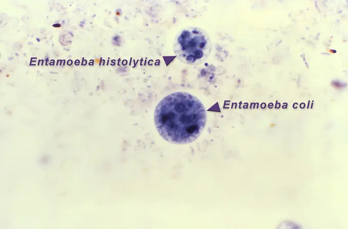
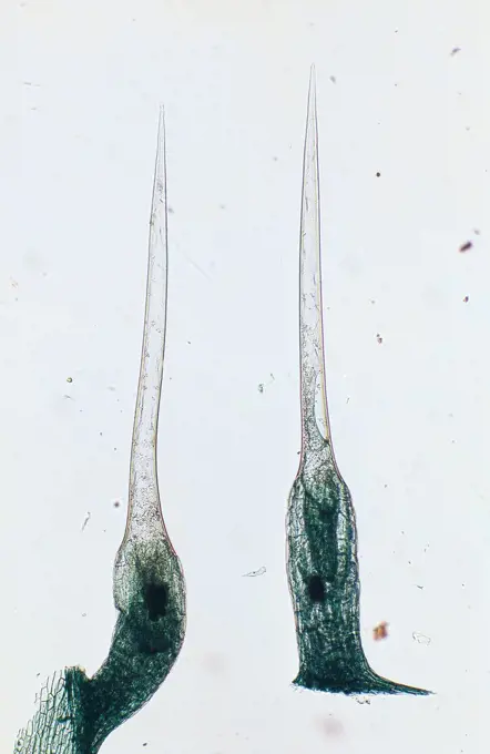
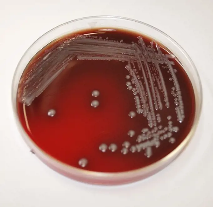
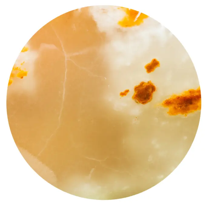
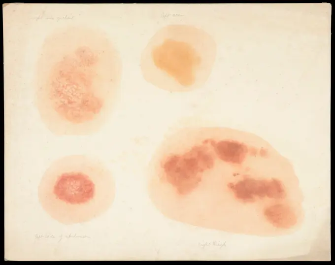
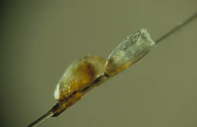
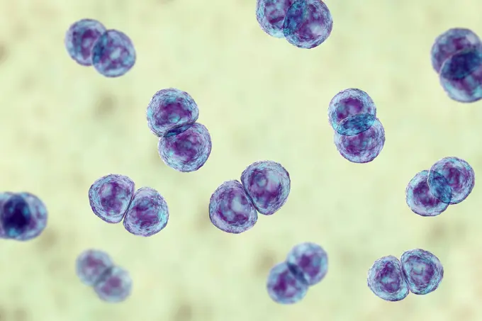
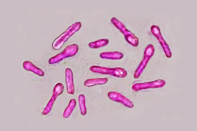
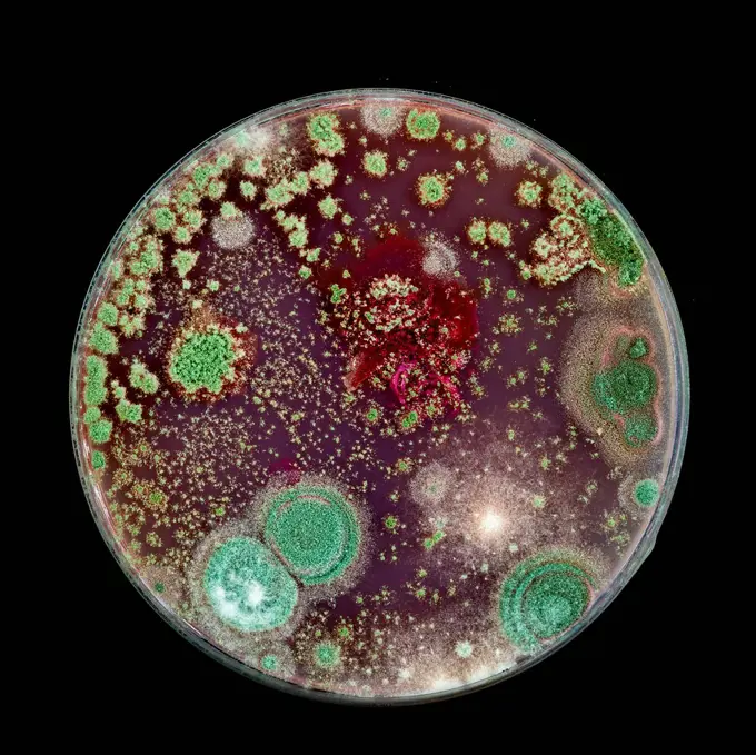
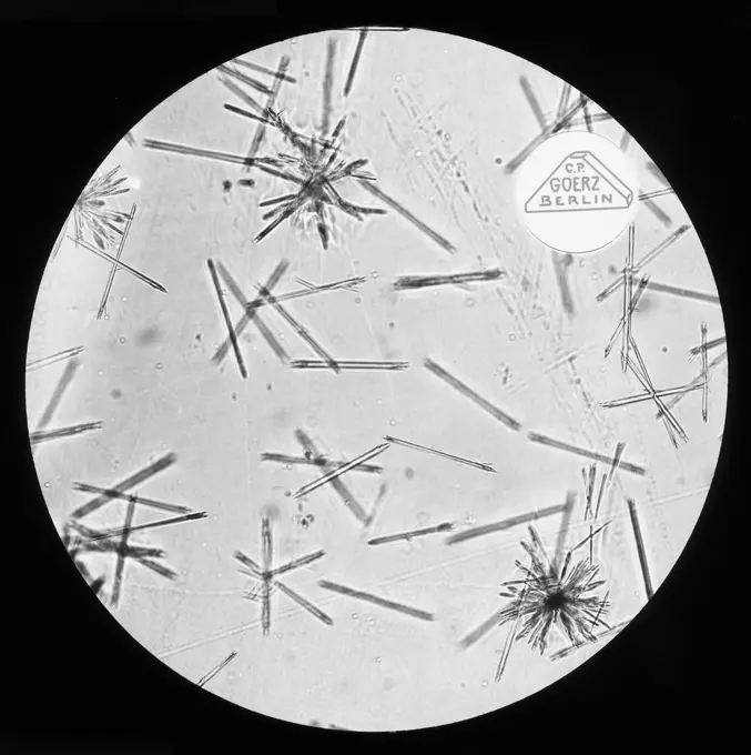
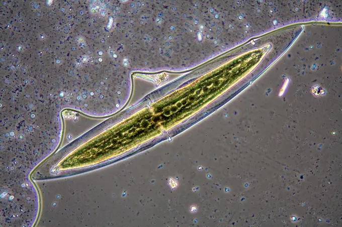
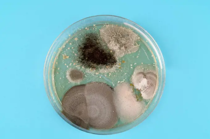
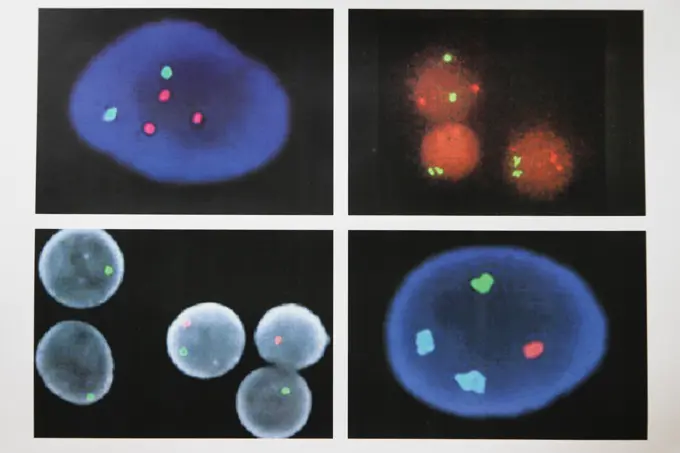
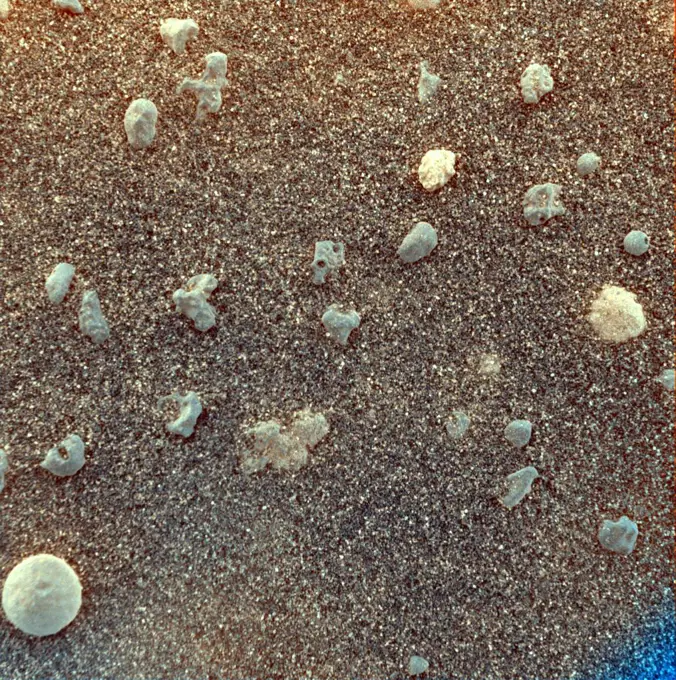
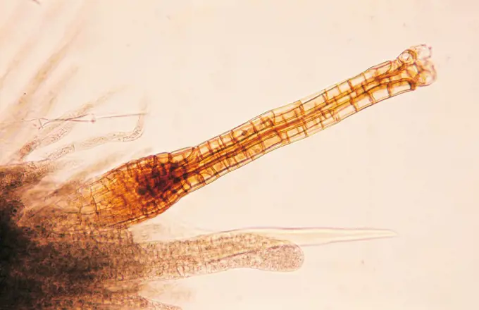
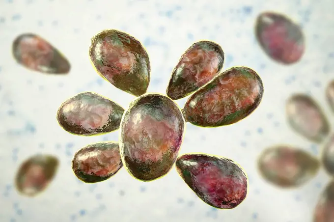
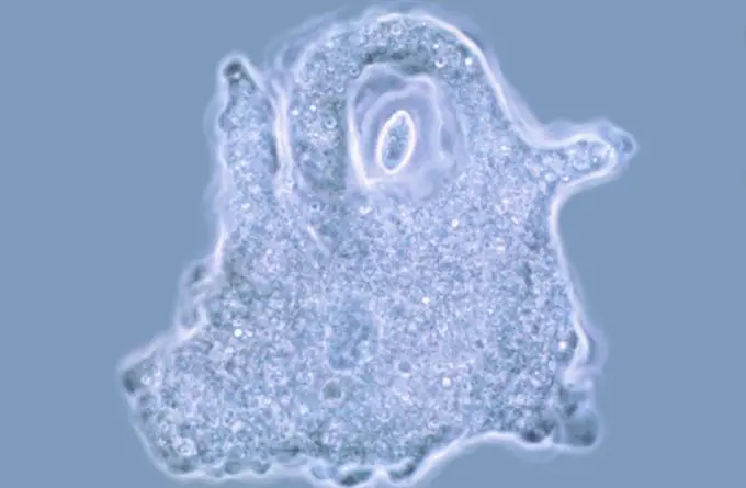
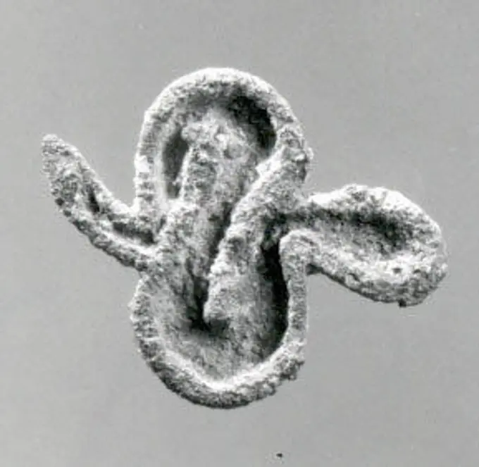
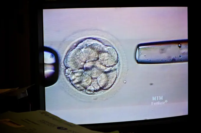
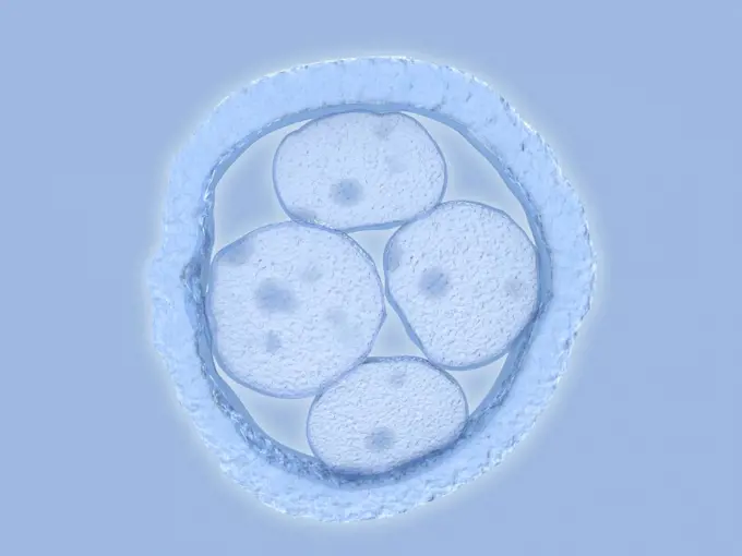
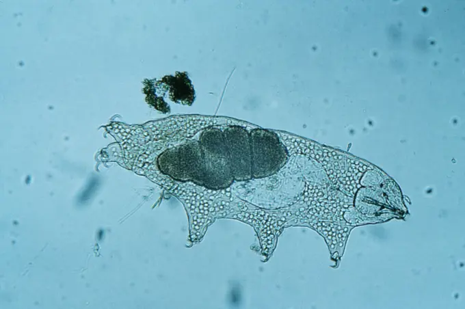
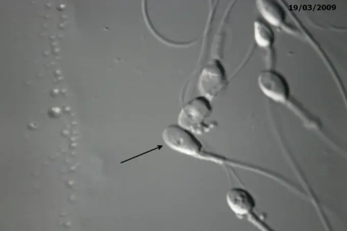
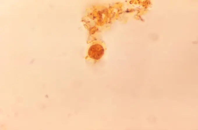
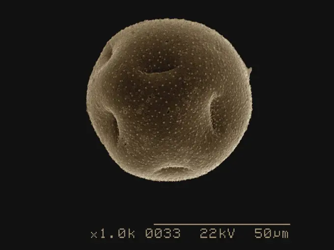
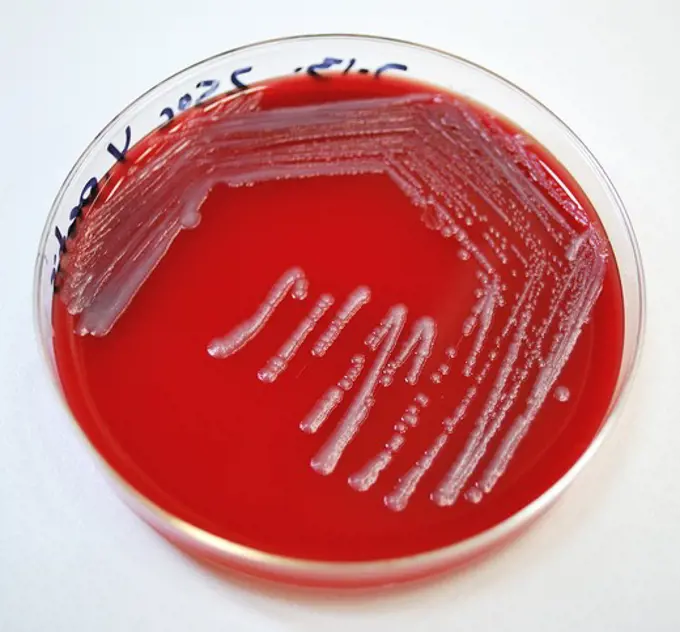
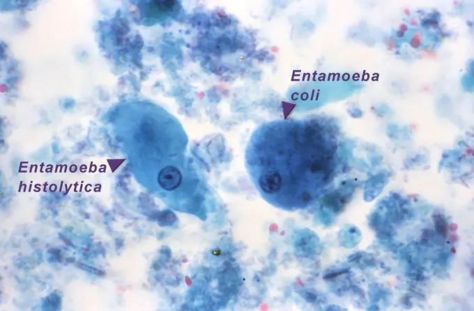
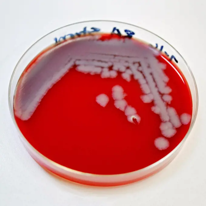
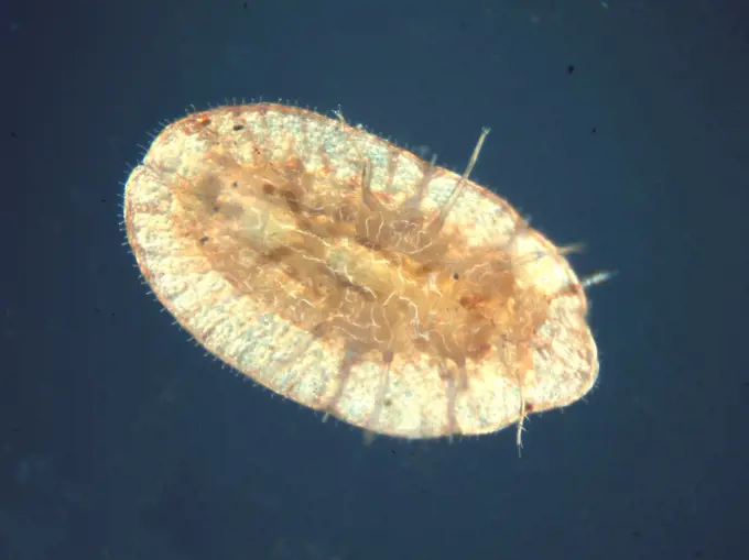
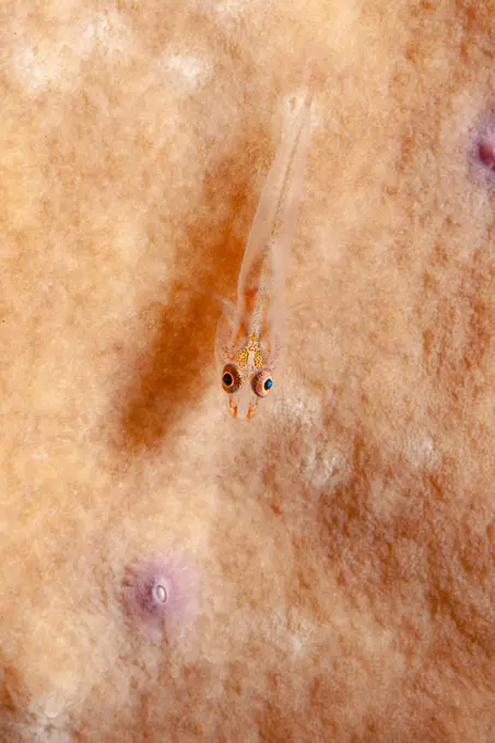
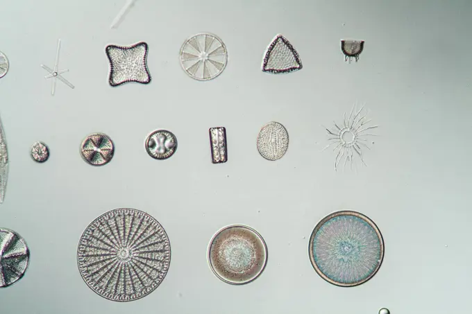
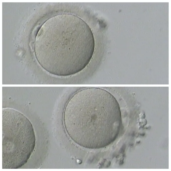
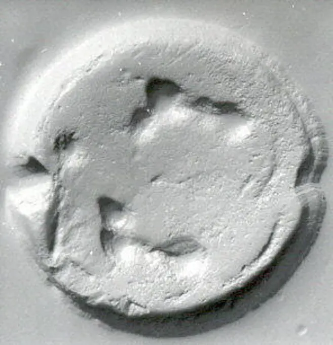
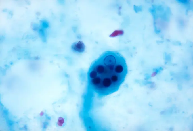
Close-up images of various microorganisms and cellular structures, revealing intricate details of parasites and mold growth in a scientific context.

This photomicrograph depicts an Entamoeba histolytica parasitic trophozoite, in which you can clearly see a single nucleus with its centrally placed karyosome, along with six ingested erythrocytes, or red blood cells (RBCs). CDC 1970
276 assets in this story
6188-55645573
Zieralge unter dem Mikroskop 100x Zieralge unter dem Mikroskop 100x Copyright: xZoonar.com/Dr.xNorbertxLangex 13915974
824-57659373
This photomicrograph of an iron-hematoxylin-stained specimen, revealed the presence of two amoebic organisms in their cystic stages of their development. In this focal plane, the smaller Entamoeba histolytica cyst (top), contained four nuclei, and two bluntly-tipped chromatoid bodies, while six nuclei could be detected in the larger Entamoeba coli cyst (bottom), which also contained two chromatoid bodies as well. CDC/ Dr. Mae Melvin 1977
6188-55645054
Radiolarien Radiolarien Schalen unter dem Mikroskop 100x Copyright: xZoonar.com/Dr.xNorbertxLangex 13785124
824-63123767
This scanning electron micrograph (SEM) depicts a Vibrio parahaemolyticus bacterium, Mag. 21851x. Vibrio parahaemolyticus is a bacterium in the same family as those that cause cholera. It lives in brackish saltwater, and causes gastrointestinal illness in humans. V. parahaemolyticus is a halophilic, or salt-requiring organism, naturally inhabiting coastal waters in the United States and Canada, and is present in higher concentrations during summer. When ingested, V. parahaemolyticus causes watery diarrhea often with abdominal cramping, nausea, vomiting fever and chills. Usually these symptoms occur within 24 hours of ingestion. Illness is usually self-limited and lasts 3 days. Severe disease is rare and occurs more commonly in persons with weakened immune systems.
6177-V53477226
1788-111173104
Stinging hairs of a nettle (Urtica kind), Urticaceae, seen under a microscope.
6176-59553780
amobas, amoebas (Amoebozoa), amoeba
4297-1843
Petri dish containing a sheep's blood agar medium which had been inoculated with Gram-negative Yersinia pestis bacteria the pathogen responsible for causing human plague
6188-55497230
cabochon from selenite natural mineral gem stone cabochon from selenite natural mineral gem stone isolated on white background Copyright: xZoonar.com/ValeryxVoennyyx 8635837
4413-109844
Microscopic view of Sporangia Bryum Moss
6145-29274540
Rashes on different parts of a body: four figures. Watercolour, ca. 189-.
4141-28656
head louse eggs pediculus humanus capitis a human parasite
4128-30422605
Streptococcus pneumoniae bacteria, illustration
824-57660004
Clostridium botulinum bacteria (botulism bacillus). This bacteria secretes a very powerful toxin (botox), which inhibits the motor neurons of muscle contraction. Image taken from an X 1000 optical microscopy.
1788-20761806
Linen textile fibers, viewed under a microscope with x50 magnification.
4128-28515229
Bacteria and fungi cultured on Petri dish
4413-9677
Diatoms seen under the optical microscope
6188-55645476
Kiesel algae from the sea under the microscope 100x pebble algae from the sea under the microscope 100x copyright: xzoonar.com/dr.xnorberbertxlangex 13865949
6188-66549291
Gesichtsserumtropfen in einer Petrischale auf weißem Hintergrund. Gesichtsöl Pipette Tropfer. Schönheit Gesicht kosmetische Produkt Textur Draufsicht. Körperpflege Lotion. Face serum drops in a petri dish on white background. Face oil pipette dropper. Beauty facial cosmetic product texture top view. Body care lotion Copyright: xZoonar.com/IrinaxKvyatkovskayax 22748521
824-63190240
Meningococcus or Neisseria meningitidis, is a bacterium that can cause meningitis. The bacteria are germs that can be found in the rhinopharynx, where it can cause benign rhinopharyngitis or an asymptomatic state. Only one in 400 carriers become victim to a serious meningococcal disease, mostly commonly acute purulent meningitis. Seen through an optical microscope X 1000.
6219-67422593
Serum or cosmetic oil flows into a transparent bowl. High quality photo
1439-V18029450
6145-46756591
Skiopticon image from the Department of Photography at the Royal Institute of Technology. Use by Professor Helmer Bäckström as teaching material. Bäckström was Sweden's first professor in photography at the Royal Institute of Technology 1948-1958. Optische Anstal C. P. Goerz, Berlin.
1848-53877849
Mould, known as isolate, cultured in the laboratory for research purposes, Germany, Europe
4413-88860
Green microalgae freshwater Closterium
6145-46860321
Basin withPoems
4128R-15221318
Microbes growing on agar plate
824-28705588
Cytogenetics laboratory, prenatal diagnosis by medical imaging FISH (Fluorescent In Situ Hybridization / Hybridization par Fluorescence In Situ). Imaging results with evidence of trisomy 21 in the upper left.
6145-44608076
Mars Under the Microscope (stretched)
4128-111495268
Colonies of fungi and bacteria growing on agar in a petri dish.
4048-5870
Microscopic view of a malaria parasite painted from microscopic view from a manual to assist in malaria diagnosis among U.S. troops serving in tropics during World War II. 1943.
6188-63949561
Spices, saffron thread in water on lavender background Copyright: xHarixMahidhar/DinodiaxPhotox
4128-V58562672
4128R-14940326
Cultures growing on Petri dish
4421-35845
Echinus esculentus, the European edible sea urchin or common sea urchin, is a species of marine invertebrate in the Echinidae family - 2nd cleavage x30
1788-111173056
Archegonium of moss, Bryophytes, seen under a microscope.
6188-55645494
Kieselalgen aus dem Meer unter dem Mikroskop 100x Kieselalgen aus dem Meer unter dem Mikroskop 100x Copyright: xZoonar.com/Dr.xNorbertxLangex 13866029
4128-19056131
Fungi Paracoccidioides braziliensis, illustration
6145-45266155
This series of images shows carbon dioxide ice sublimating (going directly from a solid to a gas) inside a pit at Mars' south pole. As ice is lost from the steep walls of pits like this, it reforms on nearby flat surfaces. Each frame of the animation was taken by the High Resolution Imaging Science Experiment camera (HiRISE) on NASA's Mars Reconnaissance Orbiter. The images used in this animation were taken between 2007 and 2013. The walls of the pit are about 656 feet (200 meters) across. Animation available at
6188-55643745
Cat small brain cat clever brain tissue under the microscope 100x copyright: xzoonar.com/DR.XNORBERBERTXLANGEX 13644950
6145-44282822
Stud. Unknown
4141-32799
amoeba feeding
4128-15978897
Bacteria on petri dish.
6145-29708754
Stamp seal ca. late 3rd-early 2nd millennium B.C. Bactria-Margiana Archaeological Complex. Stamp seal. Bactria-Margiana Archaeological Complex. ca. late 3rd-early 2nd millennium B.C.. Copper alloy. Bronze Age. Bactria-Margiana
6188-55645661
Orteralge Micrasterias under the microscope ornamental micrastias under the microscope copyright: xzoonar.com/DR.XNORBERBERTXLANGEX 14094896
6145-45133954
The bright landing platform left behind by NASA's Mars Exploration Rover Opportunity in 2004 is visible inside Eagle Crater, at upper right in this April 8, 2015, observation by NASA's Mars Reconnaissance Orbiter.
824-63169425
Report from the Medical Assistance to Procreation (AMP) service at Antoine-Béclère hospital (92). In Vitro Fertilization (IVF). Embryo biopsy in the biology laboratory.
6145-44017818
Europa. Quecke (Elymus)
4286-21398
Growth of Bacillus Cereus day 4 of 5 on nutrient agar
5514-53894989
HIV vaccine experiment, conducted by Swedish professor Eric Sanstram, in Dar es Salaam, Tanzania.
4128R-11291627
Artwork of a human embryo with four cells.
4401R-2586
Eggs in water
4220-20164027
Freshwater TARDIGRADE - Water Bear / Tardigrada (Macrobiotus macronyx). Freshwater tardigrade x 25.
824-63168541
Report at the Rouen University Hospital, PMA (Medically Assisted Procreation) service. Biology laboratory. IVF by ICSI.
824-57659402
Under 1187.5X magnification, this photomicrograph of a trichrome-stained specimen revealed the presence of an amoebic trophozoite Entamoeba hartmanni. CDC/Dr LLC Moore, Jr. 1969
4128-19357712
Illustration of a syphilis infection. Syphilis is caused by the spiral-shaped bacterium Treponema pallidum (green).
6177-V53653508
6188-55644161
Sarcocysteis beim Affen Sarcocysteis beim Affen Sporentiere im Muskel, 200x Copyright: xZoonar.com/Dr.xNorbertxLangex 13698985
4128R-13591233
Petri dish with fungi.
4413-9681
Dinoflagellata Ceratium under the optical microscope
1439-57949045
Closeup of a microscopic spherical object with a textured surface against a black background.
6145-45242352
STS-47 Spacelab-J - post flight tadpoles
4297-1841
Petri dish containing a sheep's blood agar medium which had been inoculated with Gram-negative Yersinia pestis bacteria the pathogen responsible for causing human plague
4128R-13818698
Petri dish with bacteria.
1899-61460906
This photomicrograph of a trichrome-stained specimen, displayed two amoebic trophozoites. On the left, was an Entamoeba histolytica trophozoite, and on the right, was an Entamoeba coli trophozoite. CDC/ Dr. Mae Melvin 1977
4384-166
Numbers of Bacillus anthracis bacterial colonies, which had been allowed to grow on sheeps blood agar (SBA) for a 24 hour period. Note the classical appearance exhibited in the colonial morphology including a ground-glass, non-pigmented texture with accompanying comma projections from some of the individual rough-edged colonies. There is evidence that a tenacity test had been performed using an inoculating loop, which proved positive for  B. anthracis, causing the colony to stand up like
1525-20334783
Human IVF
4413-43386
Female Brown Soft Scale
1788-20761846
Sporangia of Shield Fern or Male Fern (Dryopteris filix-mas), Dryopteridaceae.
6188-64721290
Two eyes on the coral tiny basic on a coral block copyright: xzoonar.com/haraldxschottnerx 17839803
4128R-15466551
Microbiology, conceptual image
6188-55644661
Kiesel algae Different types of pebbles, 200x copyright: xzoonar.com/DR.XNORBERBERTXLANGEX 13747315
6145-52771007
Photographs of light spectra produced by interference, captured by Eduard Valenta to analyze color patterns and optical phenomena.
1525-20334879
Human IVF
6177-V53472005
6145-29822711
Drilled hemispheroid seal ca. 3500-2900 B.C.. Drilled hemispheroid seal 326989
PREVIOUS
of 3
NEXT
2401 S. Ervay, Suite 206
Dallas, Texas 75215
United States
Get Started
Free ResearchMy BoardsMy Cart
For Creators
How To License Your ContentContributor PortalFrame of Mind
Resources
API accessPricing
Contact
+1 866 236 0087help@viewpictures.co.uk Contact form
©2026 View Pictures. All Rights Reserved. -A
This site is protected by reCAPTCHA and Google Privacy Policy and Terms of Service apply.