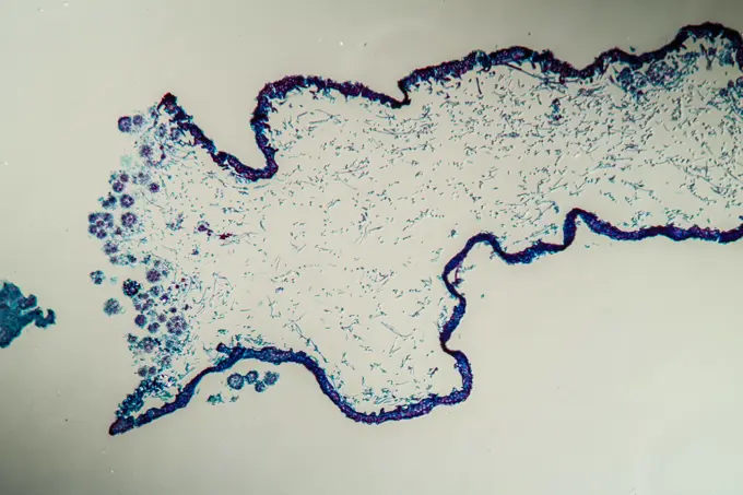
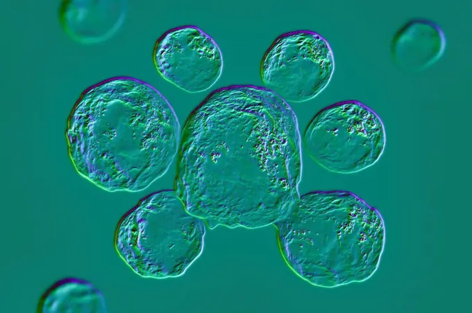
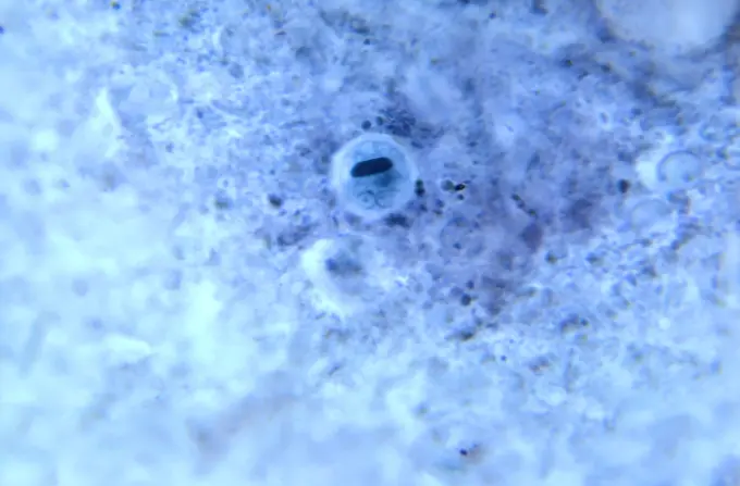
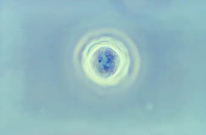
My Boards
View Pictures
  • My Boards

Microscopic Biological Specimens

Various lab-cultured biological specimens viewed under a microscope, showing microbes, cells, and fungi in differing states and preparations.

Indonesia, Papua (formerly Irian), Misool, Pleurobranch detail
259 assets in this story
4413-135001
Microscopic view of Closterium acerosum Algae
1788-111173080
Cross section of the stem of a horsetail (genus Equisetum) seen under a microscope.
4239-18641431
Microscopic view of Paramecium bursaria, a species of ciliate protozoan that has a mutualistic symbiotic relationship with green algae called Zoochlorella.
6188-55645616
Bärtchen eggs in old skin bearing eggs in old skin copyright: xzoonar.com/dr.xnorbertxlangex 13928234
6177-V65999626
1899-61460943
This photomicrograph shows two Entamoeba histolytica cysts. The cyst on the left contains a single nucleus and is therefore called a mononuclear cyst, while the cyst shown on the right contains two separate nuclei and is called a binuclear cyst. CDC/Dr Mae Melvin 1977
6188-55645611
Radder animal under the microscope 100x wheel animal under the microscope 100x copyright: xzoonar.com/DR.XNORBERBERTXLANGEX 13928209
6188-55644768
Lichen cross -section through the symbiodic body 100x copyright: xzoonar.com/dr.xnorbertxlangeex 13762676
1788-111174443
Microphotograph of a smear of human blood in which erythrocytes, platelets, and two neutrophil leukocytes are visible, at x800 magnification, counterstained.
6145-45252294
(Rabbit) Converts the major storage form of high energy phosphate (creatine phosphate) to usable energy form (ATP). A major muscle enzyme and implicated in some muscle diseases.
4128-19056186
Fungi Paracoccidioides lutzii, illustration
824-57659469
Under 900X magnification, this photomicrograph revealed the presence of an Entamoeba hartmanni microorganism at the cyst stage, in a specimen prepared using the Kohn staining method. In this focal plane you can see two distinct nuclei, each containing a single centrally located karyosome and a cigar-shaped, dark-colored chromatoid body. CDC/Dr Healy 1964
6145-44594502
KENNEDY SPACE CENTER, FLA. - C. elegans nemotodes (round worms) undergo examination by project scientists. These specimens were found in a Biological Research in Canisters (BRIC) container, a middeck experiment that was among the Columbia debris recovered in East Texas. The worms are descendants of those that were part of an experiment that flew on Columbia's last mission, STS-107. The experiment was designed to verify a new synthetic nutrient solution for an International Space Station (ISS) "model" specimen planned to be used extensively for ISS gene expression studies and was sponsored by the NASA Ames Research Center. Scientists are now looking over the experiment at KSC to determine if it will yield any scientific results. The investigation into the cause of the Columbia accident is ongoing. (
6145-44594501
KENNEDY SPACE CENTER, FLA. - C. elegans nemotodes (round worms) undergo examination by project scientists. These specimens were found in a Biological Research in Canisters (BRIC) container, a middeck experiment that was among the Columbia debris recovered in East Texas. The worms are descendants of those that were part of an experiment that flew on Columbia's last mission, STS-107. The experiment was designed to verify a new synthetic nutrient solution for an International Space Station (ISS) "model" specimen planned to be used extensively for ISS gene expression studies and was sponsored by the NASA Ames Research Center. Scientists are now looking over the experiment at KSC to determine if it will yield any scientific results. The investigation into the cause of the Columbia accident is ongoing.  (
824-57659334
Using phase contrast imaging and magnified 1125X, this photomicrograph depicts a cyst of the protozoan Acanthamoeba polyphaga. CDC/Dr George Healy 1973
1788-20761622
Bladderwrack (Fucus vesiculosus) viewed under a microscope, Fucaceae.
1788-111176126
Microphotograph of the segmentation process of the fertilized egg of a frog, beginning of the first division of the egg.
4201-66199
Copepod nauplius larval stage of development at 120x magnification, Spain
824-57659343
This photomicrograph of a trichrome-stained specimen revealed the presence of a mononuclear cyst of Entamoeba hartmanni (arrow). The cysts of E. hartmanni are similar to those of E. histolytica, but smaller, measuring 5 to 10 m. Mature cysts contain four nuclei which have a small centrally located discrete karyosome and evenly distributed peripheral chromatin. Cysts may not be visible on unstained specimens. The cytoplasm of mature cysts may contain diffuse glycogen and rounded or elongated chromatoid bodies with rounded ends. CDC/Dr Mae Melvin 1977
6188-66549589
Abstrakte Öl Kreis Blasen in einer Petrischale Ansicht von oben. Labor Kosmetik oder Medizin Hintergrund. Abstract oil circle bubbles in a petri dish top view. Laboratory cosmetic or medicine background Copyright: xZoonar.com/IrinaxKvyatkovskayax 22748703
4220-21334603
Mouse Ovum being fertilized.     Date:
824-63227386
Scanning electron micrograph of just divided HeLa cells.
6177-V54021736
824-57657653
Variola virus (poxviruses are large viruses of 300/200/100 nm. They are pathogenic for humans such as smallpox and vaccinia viruses as well as animal viruses that infect humans such as viruses monkeypox, cowpox, milker's nodule, pustular dermatitis in sheep Transmission electron microscopy.
4413-174719
Epidermal cells of leek bulb
6145-44666547
This anaglyph from NASA's Mars Reconnaissance Orbiter spacecraft, is of East Mareotis Tholus, a small volcano in Tempe Terra, Mars. 3D glasses are necessary to view this image.
4269-5525
Photomicrograph of Borrelia burgdorferi, gram negative spirochete bacterium agent of the Lyme disease, passed on to human by the ticks.
4128-20045020
West Nile virus particles, illustration
1899-61460816
Under 100X magnification, this photomicrograph of a direct mount unstained specimen revealed the presence of three Entamoeba coli protozoan parasitic cysts, as well as what may be an E. coli trophozoite. CDC/Dr Moore 1972
4201-14279
Arctic Copepod group swimming, Arctic
1525-28067492
Six stages of division of a gill lamella cell of the spotted salamander larva, vintage engraved illustration. From the Universe and Humanity, 1910.
4239-69154290
Conceptual biomedical illustration of Trichophyton.
1899-61460978
Under 900X magnification, this photomicrograph revealed the presence of a parasitic microorganism Entamoeba coli, at the trophozoite stage, in a specimen prepared using the Kohn staining method. CDC/Dr Healy 1964
4413-43440
Eucalyptus gall wasp larva
4128-V58573345
6145-45536825
Bliżej Kultury
4128R-13818526
Petri dish with bacteria.
824-57659404
Under 500X magnification, this photomicrograph revealed the presence of two parasitic cysts, Entamoeba histolytica. CDC/Dr LLC Moore, Jr. 1969
4413-43438
Eucalyptus gall wasp larva
6145-44594503
KENNEDY SPACE CENTER, FLA. - C. elegans nemotodes (round worms) undergo examination by project scientists. These specimens were found in a Biological Research in Canisters (BRIC) container, a middeck experiment that was among the Columbia debris recovered in East Texas. The worms are descendants of those that were part of an experiment that flew on Columbia's last mission, STS-107. The experiment was designed to verify a new synthetic nutrient solution for an International Space Station (ISS) "model" specimen planned to be used extensively for ISS gene expression studies and was sponsored by the NASA Ames Research Center. Scientists are now looking over the experiment at KSC to determine if it will yield any scientific results. The investigation into the cause of the Columbia accident is ongoing. (
824-63222232
Coronavirus (CoV). viruses of the Coronaviridae family and the Orthocoronavirinae subfamily. It is a pathogen of respiratory syndromes. Viewed from a transmission electron microscopy (TEM) image. Viral diameter 80nm to 100nm.
1746-19671662
Voyager 1 took this photo of Jupiter and two of its satellites (Io, left, and Europa) on Feb. 13, 1979.
1899-61460927
Under 1187.5X magnification, this photomicrograph of a trichrome-stained specimen revealed the presence of an amoebic trophozoite Entamoeba hartmanni. CDC/Dr LLC Moore, Jr. 1969
4413-20028995
Aquaculture, Grouper egg eclosion (Epinephelus sp), Aru Islands, Arafura Sea, Moluccas, Indonesia
4220-20096034
Gall-wasp - Silk Button Spangle Galls on Oak (Neuroterus numismalis)
4220-21334597
Human Sperm.     Date:
6145-44289712
Stud. Unknown
1532-V111198248
6177-V53467098
6145-29297751
Allergy research, autoradiograph, DNA
4269-27426
Klebsiella pneumoniae. Colonies of Klebsiella pneumoniae bacteria. K. pneumoniae is a gram-negative, rod-shaped bacterium commonly found in the human gastrointestinal tract. It is responsible for various infections of the urinary tract and lungs (pneumonia), and are often involved in nosocomial infections.
4128-V58573551
824-57659377
This photomicrograph of a trichrome-stained specimen, displayed two amoebic trophozoites. On the left, was an Entamoeba histolytica trophozoite, and on the right, was an Entamoeba coli trophozoite. CDC/ Dr. Mae Melvin 1977
824-63184494
Reportage in the Medically Assisted Procreation (MAP) service in Antoine-Béclère hospital in France. IVF (In Vitro Fertilisation). Analysing sperm.
4128-V58573267
4128-V58573548
4421-20037172
Larval scales of cabbage whitefly, Aleyrodes proletella, parasitised by a parasitoiid wasp, Encarsia tricolor, for commercial biological control
824-65830717
Colorized transmission electron micrograph of negative-stained Zika virus particles (yellow).
1899-54027722
Human immunodeficiency virus (HIV), the retrovirus responsible for AIDS. Image produced with transmission electron microscope (viral diameter ranges from around 110 to 125 nm).
PREVIOUS
of 3
NEXT
2401 S. Ervay, Suite 206
Dallas, Texas 75215
United States
Get Started
Free ResearchMy BoardsMy Cart
For Creators
How To License Your ContentContributor PortalFrame of Mind
Resources
API accessPricing
Contact
+1 866 236 0087help@viewpictures.co.uk Contact form
©2026 View Pictures. All Rights Reserved. -A
This site is protected by reCAPTCHA and Google Privacy Policy and Terms of Service apply.