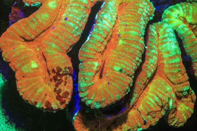
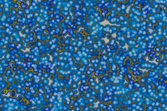
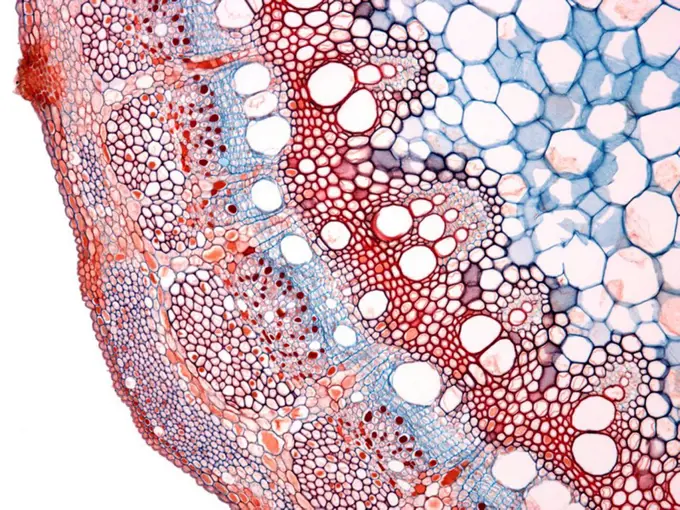
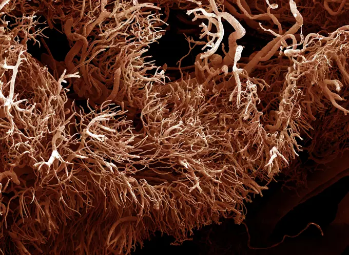
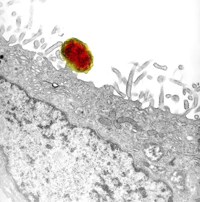
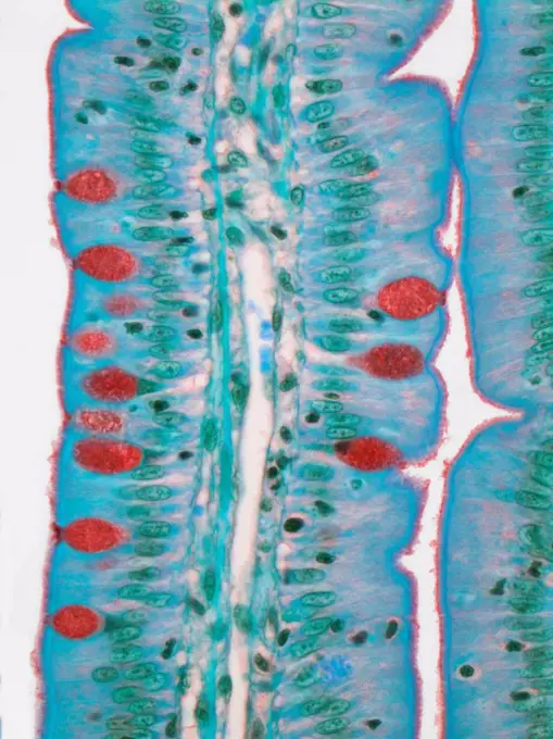
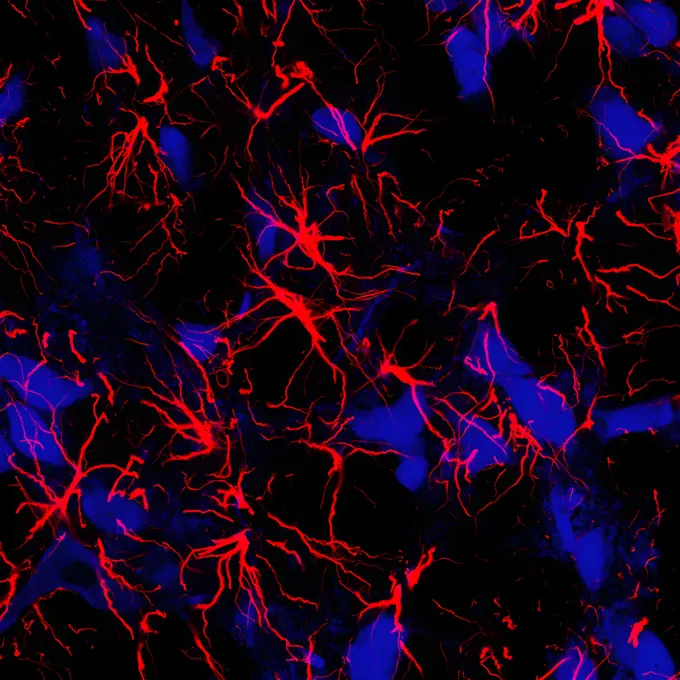
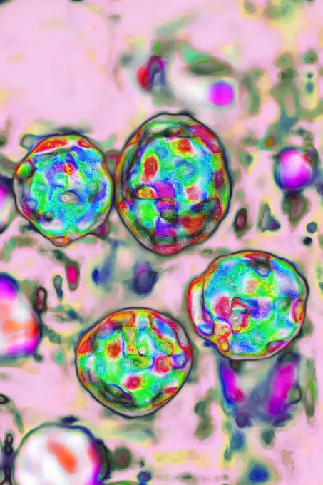
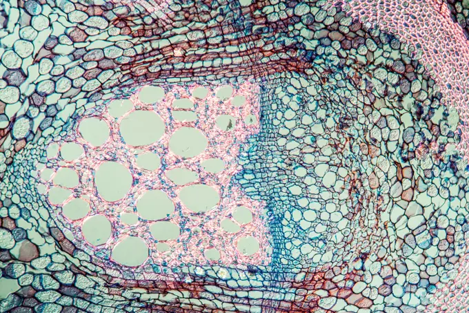
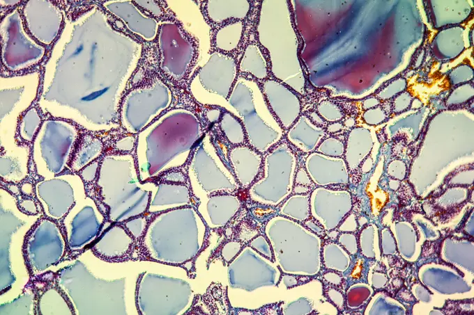
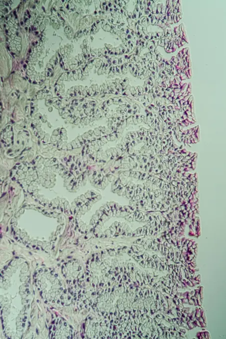
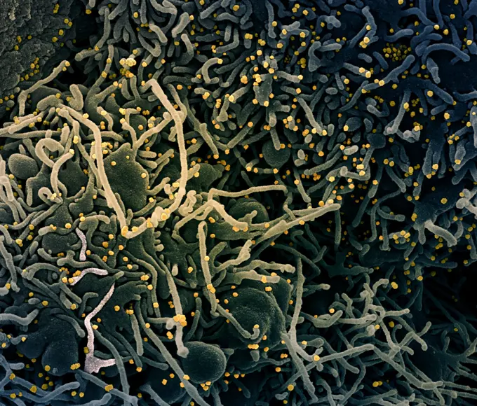
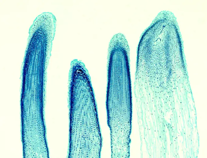
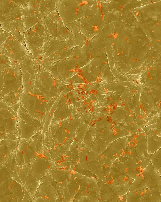
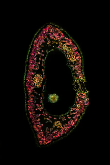
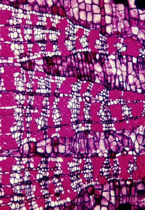
My Boards
View Pictures
  • My Boards

Microscopic Biological Textures

A collection of colorful microscopic images showcasing lung tissue, butterfly wings, and plant structures, highlighting intricate patterns and textures in vibrant hues.

X-ray image of human lungs
317 assets in this story
4341-130
Genetically Engineered Cells Producing Too Much Protein, Microscopic View
4269-25451
Histology. Infection of stomach lining by Helicobacter pylori bacteria causing interstitial gastritis. Light microscopy
4201-81423
Brazilian Free-tailed Bat (Tadarida brasiliensis) group flying from Carlsbad Caverns at dusk New Mexico
1899-61460377
Colorized transmission electron micrograph of monkeypox particles (green) found within an infected cell (pink and purple), cultured in the laboratory
1848-20178692
Conspicuous patterns at the bottom of the shallow lagoon Salinas de Cerrillos, aerial view, drone shot, Almería province, Andalusia, Spain, Europe
4491-V111125337
4341-121
Cells Undergoing Heat Shock, Microscopic View
1848-49871403
Detail of the Rio Tinto (Red river) with its deep reddish hue due to oxidised iron minerals in its water, aerial view, drone shot, Huelva province, Andalusia, Spain, Europe
1848-61039019
Fake microorganisms. microbiology. Bacteria, cells, viruses, germs, microorganisms
824-63218206
Flavivirus (family Flaviviridae), these viruses are responsible for yellow fever, dengue fever, Japanese encephalitis, Zika virus, West Nile encephalitis. They are transmitted by mosquitoes or ticks. image produced from transmission electron microscopy (viral diameter approximately 40 to 60 nm).
824-63227296
This image shows mouse neurons (purple) with their nuclei (blue) and primary cilia (green).
1899-65662320
Colorized transmission electron micrograph of mpox virus particles (pink/purple) found within an infected cell (green), cultured in the laboratory.
7203-70647275
Sinuous Coral, Day Fluorescing, Palau, Rock Islands-World Heritage Site, Micronesia
6188-55644808
Margarite Margarite Blüte im Querschnitt, 100x Copyright: xZoonar.com/Dr.xNorbertxLangex 13762731
4128-111493675
Biofilm, conceptual illustration.
4197-V71961848
1525-22156844
1525-25708197
Nerve cell under the microscope - Abstract blue dots on white background.
1525-28109986
Shape of bacterial cell: cocci, bacilli, spirilla bacteria background
4128R-2793
Hair follicle
6058-V28775096
4128R-10343
Epididymis, light micrograph
824-63178992
Bacteria Escherichia coli, also called colibacillus, from the Enterobacteriaceae family. Certain Escherichia coli strains are responsible for various pathologies, diarrhoea, gastroenteritis, urinary tract infections, meningitis, septicemia. Visualization by Optical Microscopy
824-63223135
Ebola virus isolated in November 2014 from patient blood samples obtained in Mali. The virus was isolated on Vero cells. Credit: NIAID.
1788-111173020
Section of grape leaf with peronospora (Peronospora parasitica), Phycomycetes, producing sporangiophores seen under a microscope.
1848-49164570
Painted turquoise water with small star spangles
4298-1021
Hydrodictyon algae, magnification 200x, This algae has become a pest on some parts of planet, forming large nets over areas of water, Magnification 200x
4269-24802
Biomaterial. Osseous growth in the macropores of the ceramic
7203-70647232
Fluorescence emitted in corals, captured using special barrier filter. Milne Bay, Papua New Guinea
4413-88858
Tangential section of wood Kapoktree
6188-55644564
Lungs of an aid patient lung of an aid patient pneumocyctis 100x copyright: xzoonar.com/dr.xnorbertxlangex 13735504
824-63212856
Osseous edema of the knee. Subcortical lesion of the condyle, radial cross-section MRI scan of the knee.
6188-55644054
Wasserpest Wasserpest Wurzel quer 100x Copyright: xZoonar.com/Dr.xNorbertxLangex 13683674
4269-27757
Human herpes virus-6. Transmission electron micrograph (TEM) of human herpes virus-6 (HHV-6) previously called HBLV (human b-lymphotropic virus). It infects lymphocyte T cells as well. It is the cause of the childhood rash 'roseola' and some cases of mononucleosis.
6188-55643566
Narzisse Narzisse Samenkapsel quer 100x Copyright: xZoonar.com/Dr.xNorbertxLangex 13622869
1788-111175368
Section of embryo of peanut (Arachis hypognea) high in fat, seen under a microscope, coloured with Sudan III.
4128R-10349
Grape stem, light micrograph
1848-61039785
Fake microorganisms. microbiology. Bacteria, cells, viruses, germs, microorganisms
6145-45246204
Isolation of human mammary epithelial cells (HMEC) from breast cancer susceptible tissue. Same long-term growth human mammary epithelial cells (HMEC), but after 3 weeks in concinuous culture. Note attempts to reform duct elements, but this time in two dimensions in a dish rather that in three demensions in tissue. NASA's Marshall Space Flight Center (MSFC) is sponsoring research with Bioreactors, rotating wall vessels designed to grow tissue samples in space, to understand how breast cancer works. This ground-based work studies the growth and assembly of human mammary epithelial cell (HMEC) from breast cancer susceptible tissue. Radiation can make the cells cancerous, thus allowing better comparisons of healthy vs. tunorous tissue.
4220-21334676
Scanning Electron Micrograph (SEM): a corrosion cast of a gut tumour - Liquid plastic is injected into the blood vessels; it solidifies and the tissues are dissolved away leaving a cast of the blood supply.     Date:
4297-1447
Transmission electron micrograph of a murine peritoneal mesothelial cell that was infected with multiple free cytoplasmic Gram-negative Orientia tsutsugamushi rickettsial microorganisms
4128R-13447187
Small intestine. Light micrograph (LM) of a section through the finger-like projections (villi) of the duodenum, the uppermost part of the small intes...
1899-65662441
Colorized transmission electron micrograph of HIV-1 virus particles (pink) budding and replicating from an H9 T cell (teal). The virus particles are in various stages of maturity, which accounts for differences in shape.
824-63223178
Transmission electron micrograph of Middle East Respiratory Syndrome virusCoV particles found in the lumen of the endoplasmic reticulum in an infected MRC-5 cell. Credit: NIAID.
824-63222954
Prevotella bacteria of the Prevotellaceae family. Bacteria responsible for respiratory infections (pneumonia, lung abscess, pulmonary empyema, chronic otitis media, sinusitis, periodontal disease and periodontal abscess. Image taken from an optical microscope.
4141-16157
marine diatom triceteratum puturatum two displayed under microscope
6188-55644037
Eibe Eibe Wurzel quer 100x Copyright: xZoonar.com/Dr.xNorbertxLangex 13683649
824-63227102
An NIH study in rats shows that star-shaped brain cells called astrocytes (red) can play an active role in respiration.
1899-54027427
The measles virus, paramyxoviridae from the Morbillivirus family, transmission microscopy view. (Photo by: BSIP/UIG via Getty Images)
6188-55644084
Pipe shrub pipe bush Stamm cross 100x copyright: xzoonar.com/dr.xnorberbertxlangeex 13688309
824-63227155
Red: TUJ1 neuronal marker (beta III tubulin).
6188-55643960
Kropf Kropf Schilddrüsenerkrankung 100x Copyright: xZoonar.com/Dr.xNorbertxLangex 13667466
1899-61460836
This scanning electron microscopic (SEM) image of an untreated water specimen extracted from a wild stream mainly used to control flooding during inclement weather, revealed the presence of unidentified organisms, which included bacteria, protozoa, and algae. In this particular view, a mass of gelatinous biofilm had enmeshed numbers of microorganisms, including amoebae and bacteria. See PHIL 11706 for a colorized version of this image. CDC/ Janice Haney Carr 2009
824-63227208
NIAMS-funded researchers have found that two opposing signaling pathways determine the formation of body hair and sweat glands. In developing (embryonic) human skin, hair follicles (pink) form first and sweat glands (arrow) emerge later. The study may help researchers improve skin grafts for burn victims and people with certain genetic disorders.
6145-29245366
Stress fibres & focal adhesions
1899-54027138
Cancer of the liver, seen in a radial cross-section abdominal scan.
824-63208105
Human herpes virus (HHV), image made from transmission electron microscopy. VHH 1: causes cold sores. VHH 2: causes genital herpes. VHH 3: causes chickenpox and shingles. VHH 4 or EBV: causes infectious mononucleosis and Burkitt's lymphoma. VHH 5 or cytomegalovirus (CMV): causes mononucleosis syndrome. VHH 6: causes roseola. VHH 7: also causes roseola. VHH 8: causes Kaposi's sarcoma and lymph node cancer.
4341-116
Oxygen-Starved Cancer Cells, Microscopic View
4128-V58564300
824-63123152
Lymphocyte with a centriole in a fold of the nucleus viewed under TEM. Magnification: 15000x.
4269-27738
Leptospira. Scanning electron micrograph (SEM) of the Gram-negative bacteria Leptospira sp. Leptospira is a type of spirochete. Many species are pathogenic, causing leptospirosis (Weil's disease) or other diseases with similar symptoms, attacking the liver (jaundice) or the meninges of the brain (meningitis).
4341-115
Mysterious Cell Vaults, Microscopic View
4269-25221
Breast tumour cell. Atypical cell division (multipolar mitosis) of 3 mitotic spindles (green) and DNA (red). Confocal microscopy.
4128R-3058
Coloured SEM of a nerve cell in brain tissue
4384-408
Microscopic Detail of Mosquito's Exoskeletal Surface
824-63227244
A parvalbumin (green) containing an interneuron is stained for the glutamate receptor subunit GluA4 (red).
6188-55643706
Hypophyse Hypophyse Gewebe unter dem Mikroskop 100x Copyright: xZoonar.com/Dr.xNorbertxLangex 13644852
1899-53511333
Atheroma reducing the internal diameter of an artery. View of a histological section of an artery suffering from atherosclerosis.
824-63224534
Atheroma reducing the internal diameter of an artery. View of a histological section of an artery suffering from atherosclerosis.
4341-124
Bloated Red Cells with Mutant Protein, Microscopic View
824-63227237
An NIH-funded mouse study suggests that a high-salt diet may impair the brain. A high-salt diet in mice can increase the number of immune cells releasing IL17 (green), which can negatively affect blood flow in the brain.
6145-43965759
Hardystonite. minerals. North America; USA; New Jersey; Sussex County; Franklin District
6188-55644574
Lungs of an aid patient lung of an aid patient lung tissue 200x copyright: xzoonar.com/dr.xnorbertxlangex 13735521
6188-55644532
Lepra Lepra Haut unter dem Mikroskop 200x Copyright: xZoonar.com/Dr.xNorbertxLangex 13732777
7203-70648795
Underwater Fluorescence, Blue Heron Bridge, Intercoastal Waterway, West Palm Beach, Florida, USA
6188-55644962
Leaf cross -section leaf cross -section under the microscope in the dark field 100x copyright: xzoonar.com/DR.XNORBERBERTXLANGEX 13776007
1525-24224238
Electric flash effect. Thunderbolt light and storm sparks, realistic Energy lighting voltage power charge and electricity strike
6188-55644374
Mardon tongue marten tongue with taste buds cross 100x copyright: xzoonar.com/dr.xnorbertxlangex 13718068
6188-55644234
Affenzunge Affe Zunge quer, Papillen 100x Copyright: xZoonar.com/Dr.xNorbertxLangex 13702127
824-63208113
Osteoporosis with compression of L1-L2 vertebrae, seen on a sagittal plane MRI scan.
824-63227364
Researchers at the University of North Carolina at Chapel Hill, Carolina Center of Cancer Nanotechnology Excellence (CCNE) are synthesizing particles that mimic red blood cells (RBCs) using a technology called PRINT (Particle Replication In Non- wetting models). These imitation red blood cells (RBCM) are the same size, shape and softness as real red blood cells. Spongy microparticles can easily deform from their original disc-like shapes into irregular shapes when densely packed. The soft, spongy nature of the particles allows them to serve as carriers of hemoglobin, providing new opportunities for delivering oxygen and creating blood substitutes. In addition to RBCMs, researchers are using PRINT to create nanoparticles for new cancer treatments.
6058-V28775210
6188-55644986
Rootstock of the lily of the valley root of the lily of the lily
1848-65798671
Beautiful abstract art of Ebru marbling painting techniques on water with acrylic paints
6188-55644208
Tongue toad toads cross -section through the tongue 200x copyright: xzoonar.com/DR.XNORBERBERTXLANGEX 13702079
4201-47144
Bioluminescent fungi in decaying wood, Atewa Range, Ghana
4128R-13620226
Coloured scanning electron micrograph (SEM) of Plastic wrap, Glad Press'n Seal. Plastic wrap is a thin plastic film (less than 0.01mm in thickness) th...
4413-88863
Structure of mesophyll leaf Boxwood
1848-61033454
Ebru marbling handmade art, traditional Ebru technique. abstract background of marble liquid ink art
824-63223239
Colorized scanning electron micrograph of a VERO E6 cell (blue-green) exhibiting elongated cell projections and signs of apoptosis, after infection with SARS-COV-2 virus particles (yellow), which were isolated from a patient sample. Image captured at the NIAID Integrated Research Facility (IRF) in Fort Detrick, Maryland. Credit: NIAID.
4413-109768
Microscopic view of veins of a leaf Boxwood
4141-111428911
Light micrograph longitudinal section through the tips of an onion root showing mitosis, X30, UK.
4128R-13620034
Coloured scanning electron micrograph (SEM) of Photocomposite, E. coli on the surface of human skin. Escherichia coli is a Gram-negative, facultativel...
4413-20032303
Eggs of Colomerus vitis (Pagenstecher, 1857) on the reverse side of a vine leaf. Species of the family Eriophyidae. These mites are microscopic in size, measuring barely one tenth of a millimetre. The Colomier mites live on the underside of the vine leaves where their stings cause a reaction in the form of blisters due to the formation of numerous filaments. The mites live in the middle of these filaments and are invisible. This disease is called grapevine erinosis.
4128-18929792
Chick development, LM
4201-98261
Coral from Guadalupe, Senckenberg Museum, Frankfurt, Germany
4341-125
Modified Cells, Microscopic View
6188-55643012
Crowberry leaves cross section 100x crowberry leaves cross section 100x copyright: xzoonar.com/dr.xlangeex 13391208
4201-21258824
Adonis Blue (Lysandra bellargus) butterfly wing, Hessen, Germany
1788-111173308
Microphotograph of dilatation cones of the secondary growth from the trunk of a linden trees (genus Tilia), Tiliaceae.
PREVIOUS
of 4
NEXT
2401 S. Ervay, Suite 206
Dallas, Texas 75215
United States
Get Started
Free ResearchMy BoardsMy Cart
For Creators
How To License Your ContentContributor PortalFrame of Mind
Resources
API accessPricing
Contact
+1 866 236 0087help@viewpictures.co.uk Contact form
©2026 View Pictures. All Rights Reserved. -A
This site is protected by reCAPTCHA and Google Privacy Policy and Terms of Service apply.