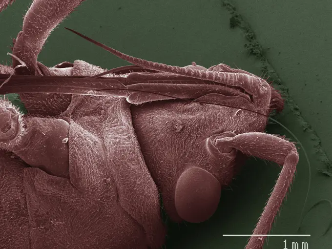
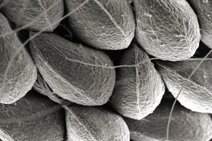
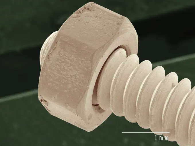
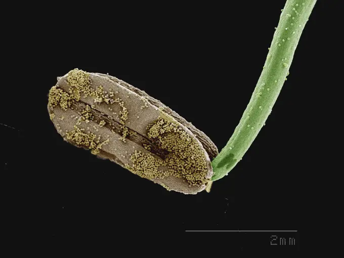
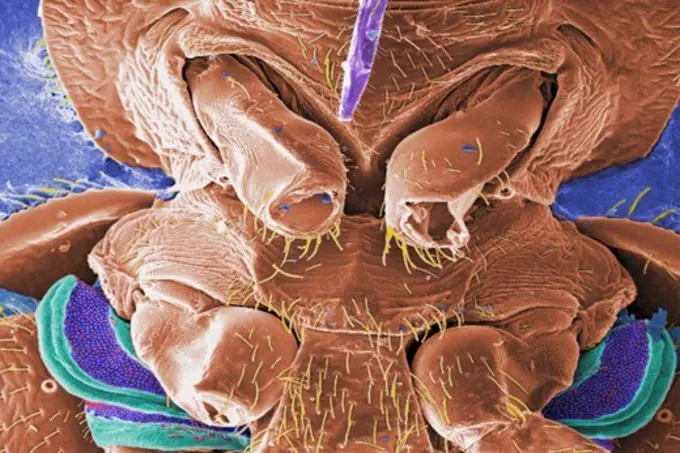
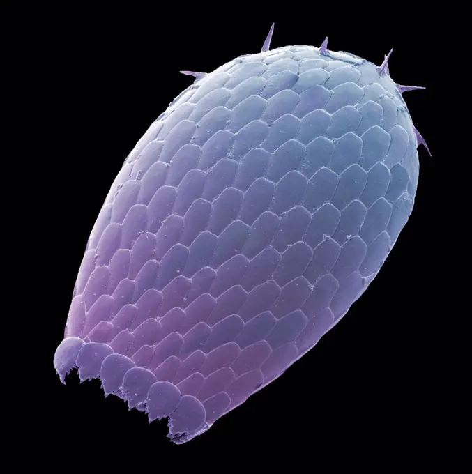
My Boards
View Pictures
  • My Boards

Microscopic Closeups of Structures

Detailed electron microscope images showing intricate structures of insects, plants, and various materials, highlighting their textures and details.

Extreme closeup of an ant captured through an electron microscope.
138 assets in this story
4384-429
Microscopic Detail of Wild Strawberry
1439-57940055
Closeup of a micro screw under high magnification
4384-372
Microscopic Detail of a Roach
1439-57949034
Closeup of a flower's stamen covered in pollen against a black background.
824-63227232
Spinal nerves are part of the peripheral nervous system. They run through the spine to carry nerve signals to and from all parts of the body. Spinal nerves allow all the movements we make, from turning our heads to wiggling our toes, control the movements of our internal organs, such as the colon and bladder, as well as allow us to feel the touch and location of our members.
4128R-5639
Foetal sense organ, SEM
4128R-9191
Water bear, SEM
4128R-13619958
Human hair with dandruff, coloured scanning electron micrograph (SEM). The outer layer of hair (the cuticle) has overlapping scales of keratin. These ...
4378-872
The earth as if seen under scanning electron microscope (conceptualization)
6145-45258890
This image shows the central part of the dune field on the floor of Russell Crater. Russell Crater is located in Noachis Terra. A spectacular dune ridge and other dune forms on the crater floor have caused extensive imaging. The Odyssey spacecraft has spent over 15 years in orbit around Mars, circling the planet more than 69000 times. It holds the record for longest working spacecraft at Mars. THEMIS, the IR/VIS camera system, has collected data for the entire mission and provides images covering all seasons and lighting conditions. Over the years many features of interest have received repeated imaging, building up a suite of images covering the entire feature. From the deepest chasma to the tallest volcano, individual dunes inside craters and dune fields that encircle the north pole, channels carved by water and lava, and a variety of other feature, THEMIS has imaged them all. For the next several months the image of the day will focus on the Tharsis volcanoes, the various chasmata o
4384-260
Scanning Electron Micrograph of Bed Bug's Mouthparts
4128R-8942
Euglypha amoeba shell, SEM
1439-57949002
Closeup of a microscopic texture in green hues.
4413-20032306
Colomerus vitis (Pagenstecher, 1857) A species of the family Eriophyidae. These mites are microscopic in size. The one in this photograph measures only 43 µm (1/2 millimetre) and is therefore completely invisible without optical equipment such as a microscope. The Colomier mites live on the underside of the vine leaves where their bites cause a reaction of the plant in the form of blisters due to the formation of numerous filaments. The mites live in the middle of these filaments and are almost invisible. This disease is called grapevine erinosis.
4128R-32072
Phospholipid bilayer. Computer artwork of the phospholipid bilayer that forms the membrane around all living cells. The cell membrane is made of phosp...
4128R-12571802
Radiolarian. Coloured scanning electron micrograph (SEM) of a radiolarian. Radiolarians are amoeboid protozoa that produce intricate mineral skeletons...
1439-57952414
A closeup view of a lily anther coated with pollen grains
255-32335
Vintage Photograph. Close-up view of Venus Fly-trap plant.
4128R-11288968
Dust mite, computer artwork.
4297-1892
Scanning electron microscopic image of a Klebsiella pneumoniae bacterium
4141-32824
freshwater bryozoan plumatella repens portion of a colony
1439-57940101
Close-up view of a microstructure under an electron microscope
4128R-15324
Dust mite. Computer artwork of a house dust mite Dermatophagoides pteronyssinus.
4128R-13620138
Coloured scanning electron micrograph (SEM) of Woodland skipper butterfly compound eye and proboscis (Ochlodes sylvanoides). (Ochlodes sylvanoides). S...
1848-53914854
Bacteria, illustration
4384-351
Honey Bee's Exoskeletal Surface
4141-32837
spirogyra cell
1439-57940005
Closeup of textured microscopic structures in sepia tones
4128R-11291603
Artwork of dust mites.
4128R-13620537
Coloured scanning electron micrograph (SEM) of Dust mite (Dermatophagoides sp.). Millions of dust mites inhabit the home, feeding on dead human skin t...
4201-22092
Rotifer (Rotaria rotatoria) a microscopically small animal with a length of 004 to 240 mm, showing cilia on its trochal discs, waterborne and can inhabit highly acidic environments
1439-57939996
Closeup view of microscopic structures with a scale indicator
1773-95772
Science: SEM of chemically deposited Antimony on metallic substrate
4201-66381
Stone Centipede (Lithobius sp) SEM close-up view at 21x magnification
1439-57952403
A closeup view of microchip circuitry with vivid green highlights
1848-49235415
Hollow stem at insect hotel for solitary bees occupied by spider
4128R-13620137
Coloured scanning electron micrograph (SEM) of Woodland skipper butterfly compound eye and proboscis (Ochlodes sylvanoides). (Ochlodes sylvanoides). S...
1439-57943353
Closeup of a crosssection of a yellow fibrous material against a black background.
PREVIOUS
of 2
NEXT
2401 S. Ervay, Suite 206
Dallas, Texas 75215
United States
Get Started
Free ResearchMy BoardsMy Cart
For Creators
How To License Your ContentContributor PortalFrame of Mind
Resources
API accessPricing
Contact
+1 866 236 0087help@viewpictures.co.uk Contact form
©2026 View Pictures. All Rights Reserved. -A
This site is protected by reCAPTCHA and Google Privacy Policy and Terms of Service apply.