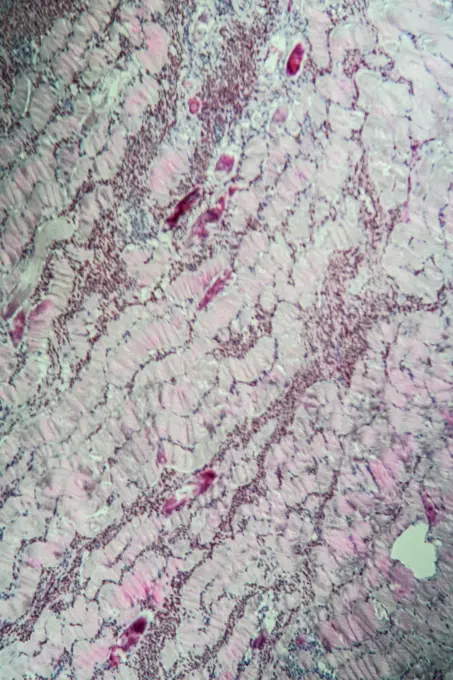
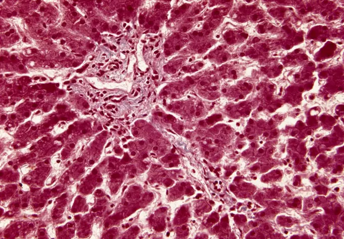
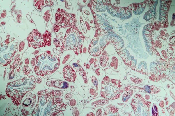
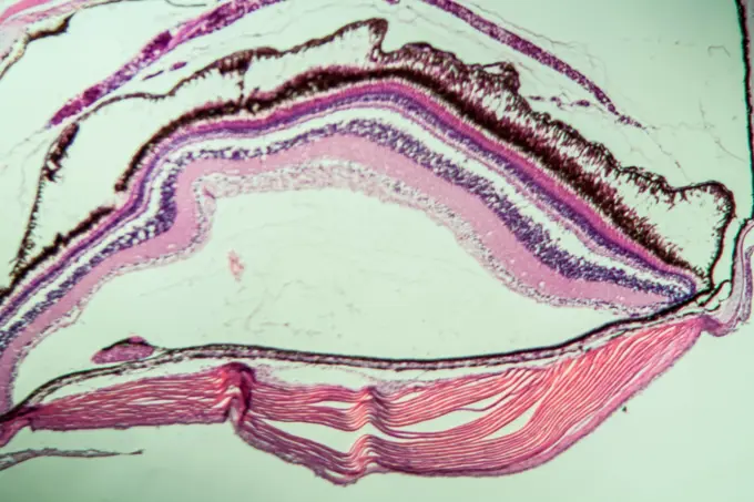
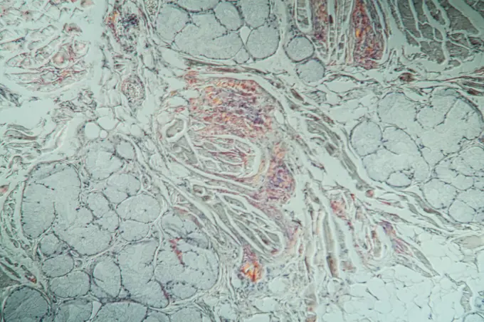
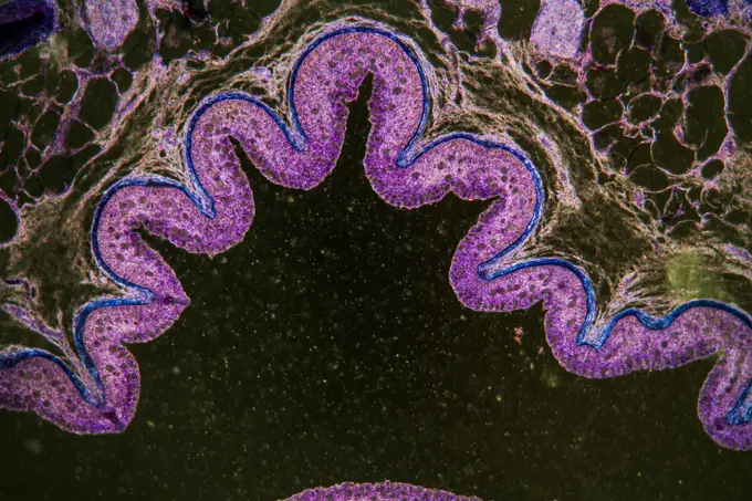
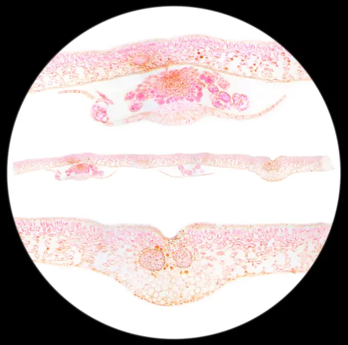
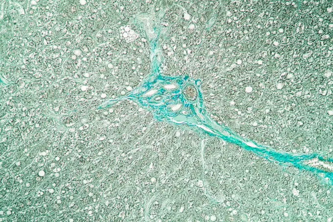
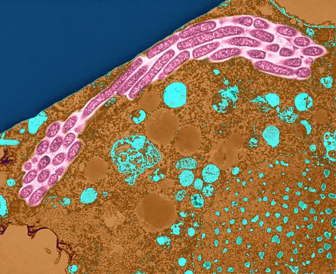
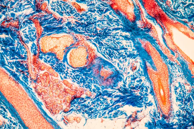
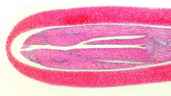
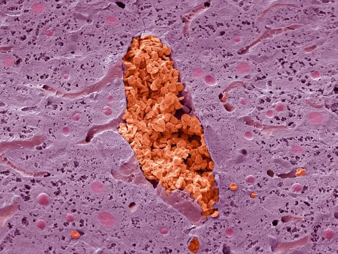
My Boards
View Pictures
  • My Boards

Microscopic Medical Conditions

Detailed microscopic views of various diseases, including leprosy and cancer, highlighting medical research.

Prostate
Prostate
340 assets in this story
4413-135012
Microscopic view of Euglena
824-57657906
A section of the cerebral portion of the brain of a patient with primary amoebic meningoencephalitis, stained with hematoxylin and eosin, showing large clumps of Naegleria fowleri trophozoites and destruction of normal tissue architecture cerebral. The cysts are not visible. Magnification: 100x. Inset: Higher magnification (1000x) of Naegleria fowleri trophozoites (arrows) with characteristic nuclear morphology.
4128R-13573027
Taste buds. Coloured light micrograph of a section through the tongue, showing taste buds (round, yellow). The taste buds are within papillae (project...
6188-55644167
Sarcocysteis at beef sarcocysteis at beef spars in the muscle, 100x copyright: xzoonar.com/DR.XNORBERBERTXLANGEX 13698993
1788-111177728
Section of liver tissue, liver, seen under a microscope.
6188-55643904
Water snail with liver sail water snail infection with liver sail 100x copyright: xzoonar.com/DR.XNORBERBERTXLANGEX 13662845
6188-55644386
Black Amazon Black Amazon Fishing Avicity with retina 100x copyright: xzoonar.com/DR.XNORBERBERTXLANGEX 13718087
6188-55644636
Amyloid deposits in the tongue amyloid deposits in the tongue sick tissue under the microscope 200x
1848-18101100
Winter truffle, Muscat truffle (Tuber brumale) Spores
6188-55644979
Darm with cup cells with cup cells with cup cells under the microscope 100x copyright: xzoonar.com/dr.xnorbertxlangex 13778340
4128-V58573268
1525-19842957
Slice fern leaf under a microscope, (Fern Leaf Sec.) 100x
6188-55644890
Rat liver rat liver tissue cut under the microscope 100x copyright: xzoonar.com/DR.XNORBERBERTXLANGEX 13772899
6188-55644330
Tongue of the ratte rat tongue with taste buds cross 200x copyright: xzoonar.com/dr.xnorberbertxlangex 13712913
1525-19882738
Smooth muscle teased preparation under a microscope, Smooth Muscle W.M., 400x

4269-24641
Fibroblast. Fibroblasts on the external surface of the intestine SEM (Scanning Electron Microscope)
824-65830833
Colorized transmission electron micrograph of numerous HIV-1 virus particles (purple) replicating from a segment of a chronically infected H9 T cell (pink).
4269-25178
Histology. Light microscopy of cervical smear showing normal epithelial cells.
4128-V58573211
6188-55644248
Spinaliom spinalioma carcinoma in guinea pigs, fabric 200x copyright: xzoonar.com/dr.xnorberbertxlangex 13702184
1788-20761741
Tonsillar crypt with gram-positive bacteria, at x300 magnification.
4128R-10340
Bone marrow, light micrograph
6188-55643701
Gallertkrebs des Mastdarms Gallertkrebs des Mastdarms Erkranktes Gewebe 100x Copyright: xZoonar.com/Dr.xNorbertxLangex 13644844
1848-49284470
Painted pink water with star spangles
6188-55644405
Reworm earthworm cross section 10. Segment 100x
6188-55648416
Stäbchenförmige Bakterien unter dem Mikroskop 400x Stäbchenförmige Bakterien unter dem Mikroskop 400x Copyright: xZoonar.com/Dr.xNorbertxLangex 14517740
4128R-34252
Vaginal mucosa. Light micrograph of a section through the wall of the vagina. The lumen (at top) is lined with a mucosa of stratified squamous epithel...
6145-46761681
Lumière autochrome. Medical photographs.A-blood cells undiluted B serum, widespread DRP. Peripheral ring immersion.
824-63127923
Protozoan (Tetrahymena pyriformis) infected with Legionella pneumophila. This transmission electron micrograph (TEM) of an infected Tetrahymena pyriformis protozoa reveals in cross section, a chain of intracellular Legionella pneumophila bacteria. The bacteria are seen multiplying while they are contained inside an intracellular vesicle within the protozoan host cell. Experiments have shown, (BS Fields, EB Shotts, Jr, JC Feeley, GW Gorman, and WT Martin, Appl Environ Microbiol. 1984 Mar,47(3):467-71.), L. pneumophila bacteria can reproduce intracellularly acting as a parasite inside of the ciliated protozoan, Tetrahymena pyriformis, forming serpentine chains of approximately 10(3) L. pneumophila. See image n1305405 on the site.
1788-111173024
Rounded blastospores yeast, Ascomycetes, seen under a microscope.
1848-61039608
Thunderbolt light and storm sparks, realistic Energy lighting voltage power charge and electricity strike
6188-55643780
Kopfhaut Haare längs Kopfhaut Haare längs Gewebe unter dem Mikroskop 100x Copyright: xZoonar.com/Dr.xNorbertxLangex 13650515
4141-111428957
Light micrograph longitudinal section of the seed of a Stone pine (Pinus pinea), UK.
4128R-2309
Liver vein, SEM
824-63194752
White blood cell.
1525-21837393
Close up of cleaning sponge
6145-29293204
Plasmodium falciparum malaria: cerebral malaria
4128-16506744
Kidney cancer, computer illustration and light micrograph.
1899-61460953
This image depicts the ulcerated intestinal mucosa from what was an amebiasis patient, revealing the pathologic changes in the mucosal lining that had disrupted the layers patency. These ulceration were the result of an infection caused by the parasite, Entamoeba histolytica . CDC/ Dr. George Healy 1977
4128R-13447195
Fallopian tube. Light micrograph (LM) of a section through a fallopian tube, or oviduct. The fallopian tubes connect the ovaries to the uterus. The lu...
PREVIOUS
of 4
NEXT
2401 S. Ervay, Suite 206
Dallas, Texas 75215
United States
Get Started
Free ResearchMy BoardsMy Cart
For Creators
How To License Your ContentContributor PortalFrame of Mind
Resources
API accessPricing
Contact
+1 866 236 0087help@viewpictures.co.uk Contact form
©2026 View Pictures. All Rights Reserved. -B
This site is protected by reCAPTCHA and Google Privacy Policy and Terms of Service apply.