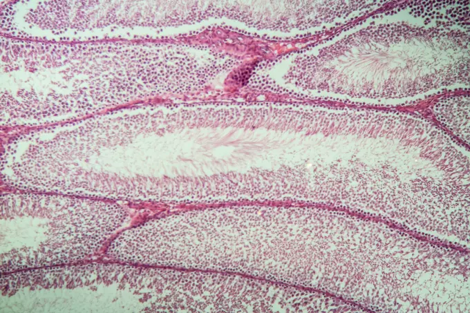
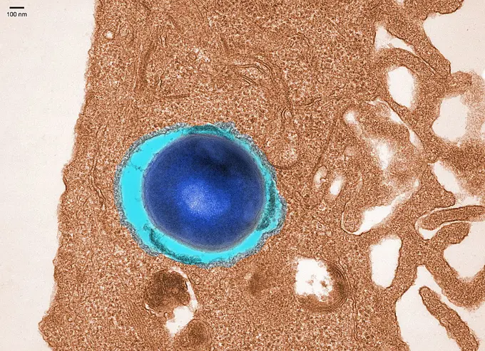
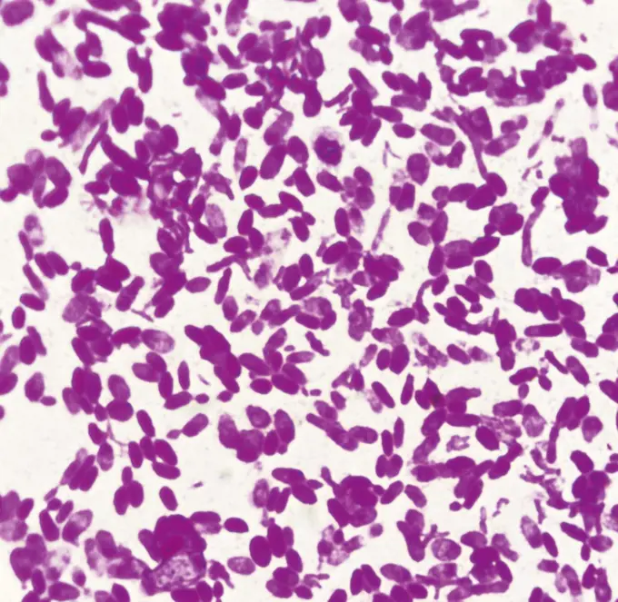
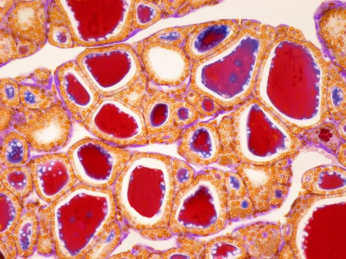
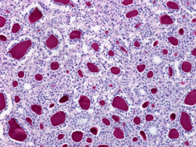
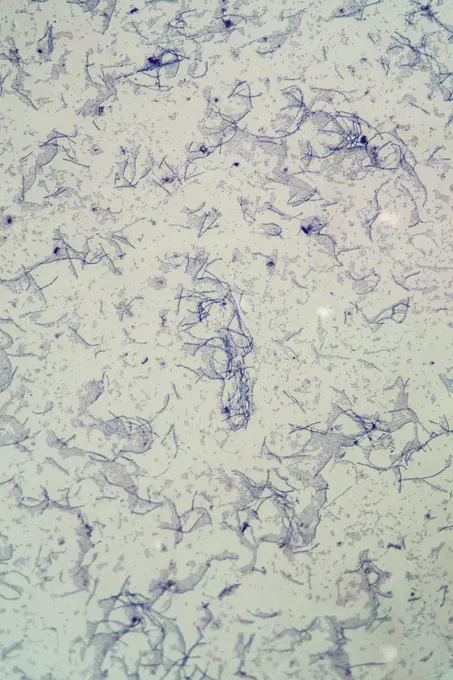
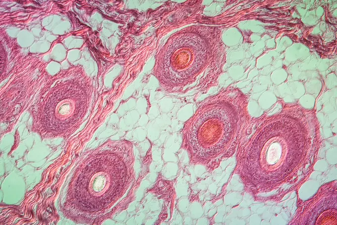
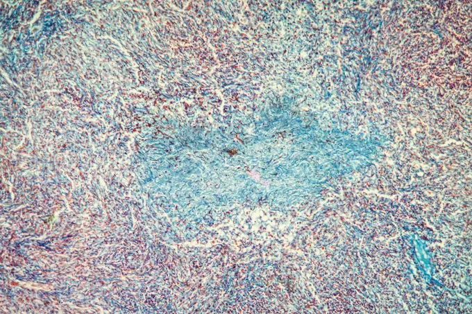
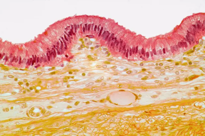
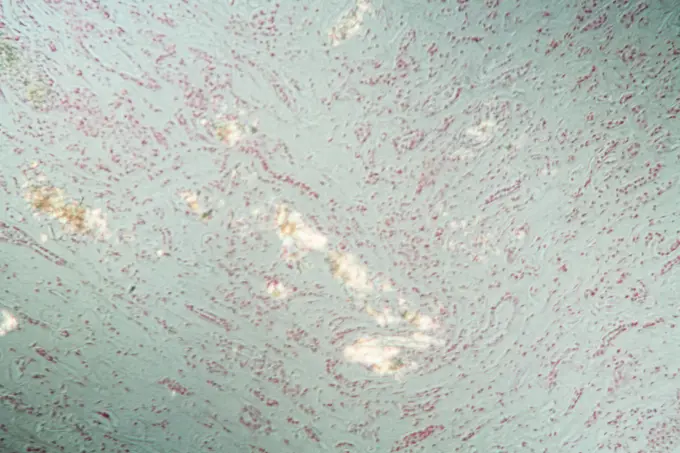
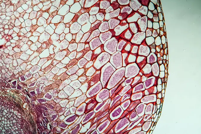
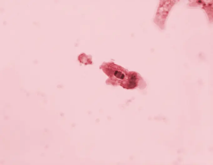
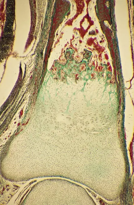
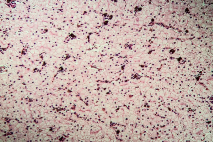
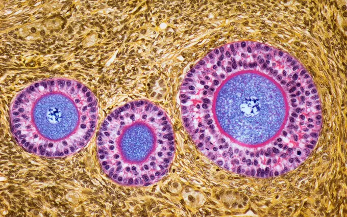
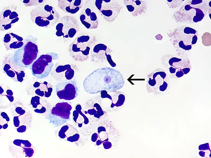
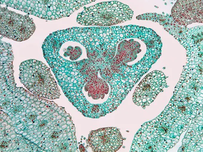
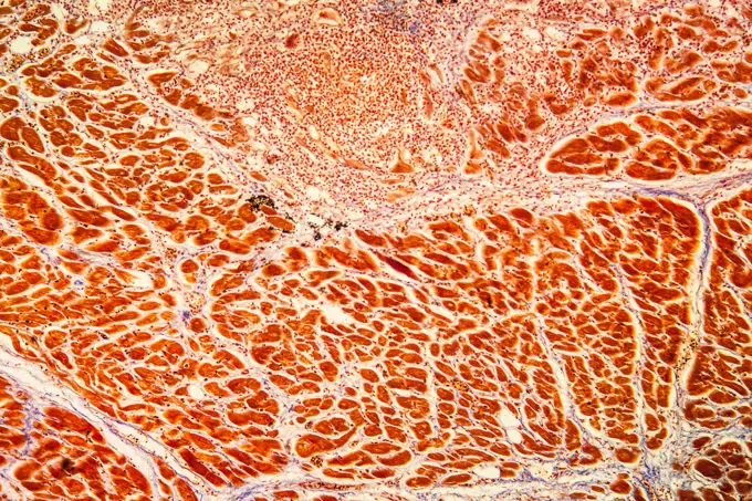
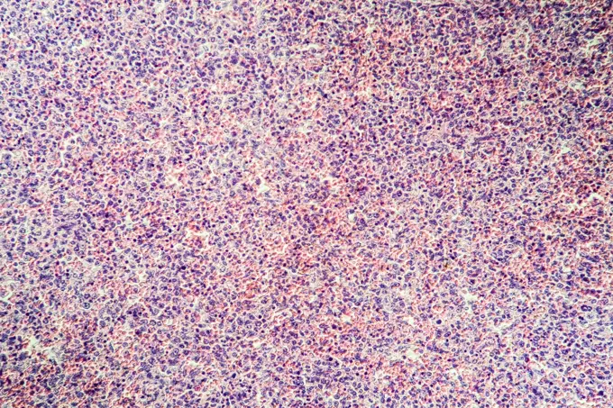
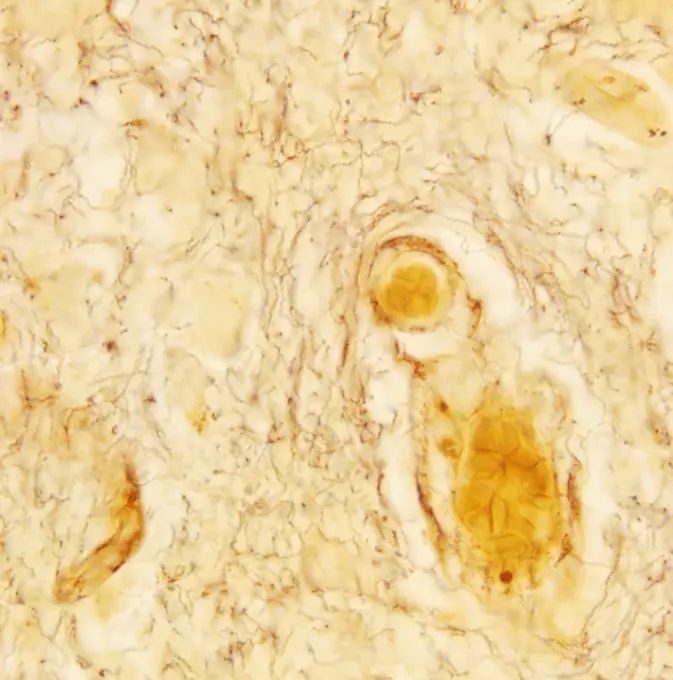
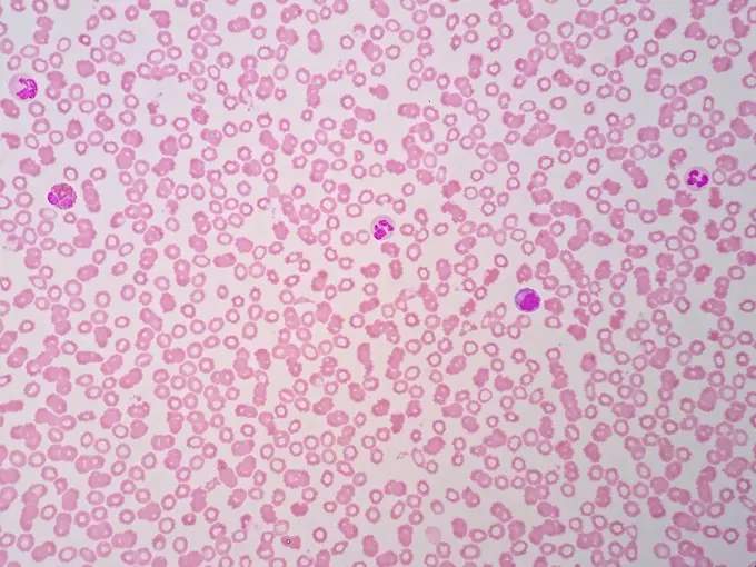
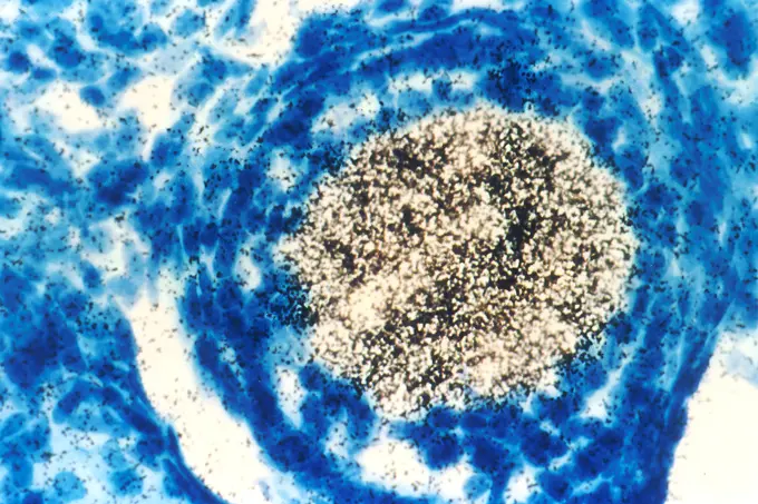
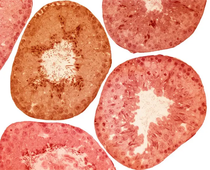
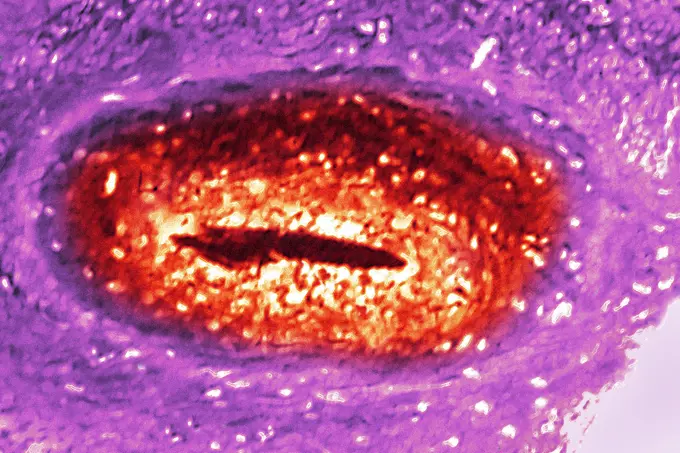
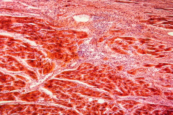
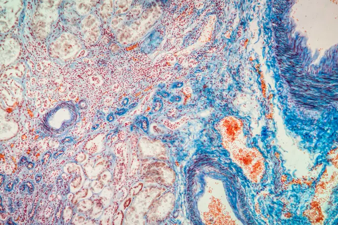
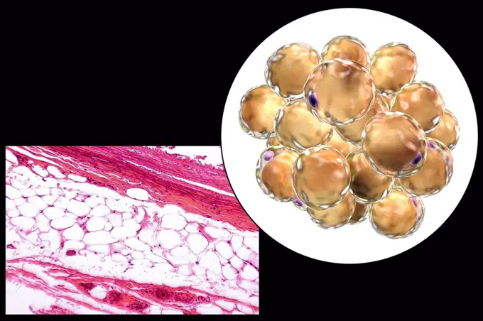
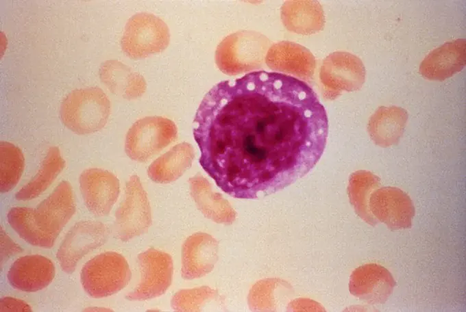
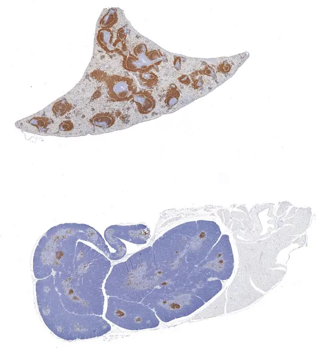
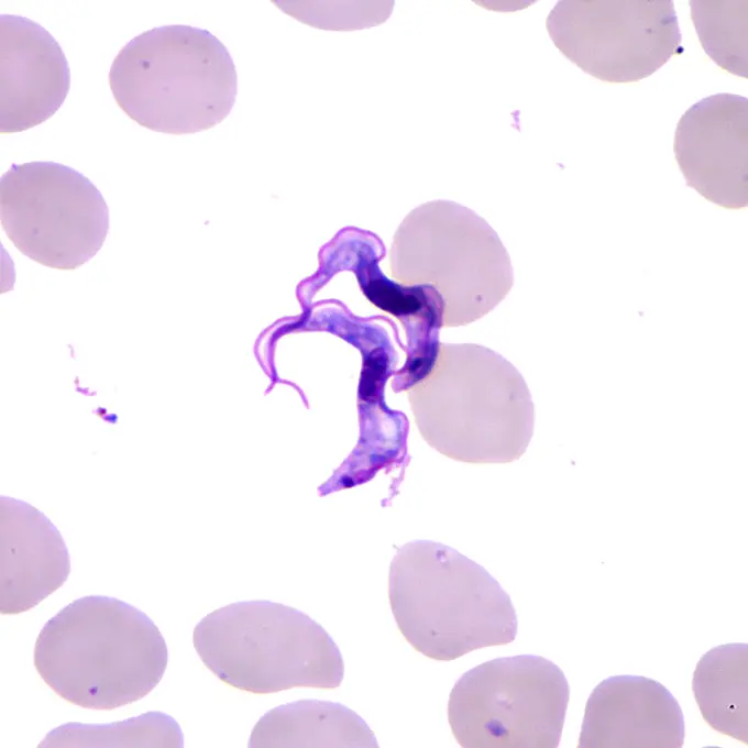
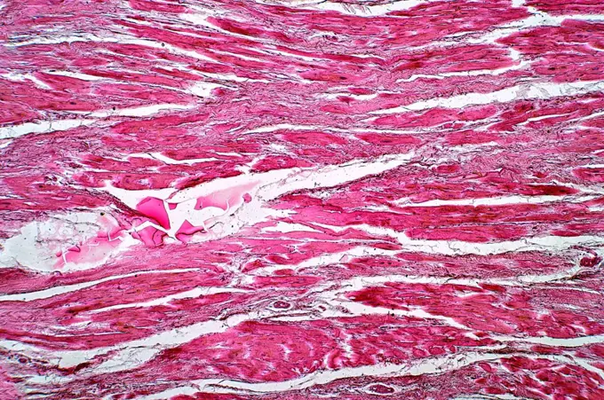
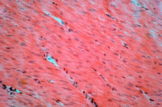
My Boards
View Pictures
  • My Boards

Microscopic Medical Images

Close-up views of various tissues under a microscope, illustrating medical conditions like tuberculosis, CMV infection, and prostate cancer.

Hods of the rat rat hotel cross -section 100x copyright: xzoonar.com/DR.XNORBERBERTXLANGEX 13712883
357 assets in this story
6188-55643760
Mouse brain mouse brain tissue under the microscope 100x copyright: xzoonar.com/DR.XNORBERBERTXLANGEX 13650388
1525-24460462
Transmission electron micrograph of Staphylococcus
4128-17810559
Mitosis, light micrograph
1788-111174298
Microphotograph of acetobacter bacteria.
4128R-13447242
Thyroid gland. Light micrograph (LM) of a thyroid gland showing the follicles. The follicles are lined by a single layer of cuboidal epithelial cells ...
4128R-10369
Thyroid, light micrograph
6188-55643642
Bacteria bacteria from the dental coat 200x copyright: xzoonar.com/DR.XNORBERBERTXLANGEX 13628428
6188-55643884
Kopfhaut Kopfhaut Haare quer 100x Copyright: xZoonar.com/Dr.xNorbertxLangex 13660697
6188-55643651
Gallenblasentumor Gallenblasentumor Gewebe unter dem Mikroskop 100x Copyright: xZoonar.com/Dr.xNorbertxLangex 13633403
1899-61461011
This photomicrograph revealed the presence of trichrome-stained parasitic Dientamoeba fragilis trophozoites in this specimen. CDC 1970
6188-55643669
Mixed tumor of the ears of the ears of the Mixture tumor of the ears of the ears of the ears of 100x copyright: xzoonar.com/dr.xnorbertxlangeex 13633532
6237-V70988061
4128-18929796
Gall bladder, LM
6188-55644412
Kidney tissue with gout kidney tissue with gout tissue under the microscope 100x copyright: xzoonar.com/dr.xnorberbertxlangeex 13722618
6188-55643629
We's We's We's Weighing
6188-55644165
Sarcocysteis beim Rind Sarcocysteis beim Rind Sporentiere im Muskel, 100x Copyright: xZoonar.com/Dr.xNorbertxLangex 13698991
4128R-22529
Bladder cancer. Light micrograph of a section through a transitional cell carcinoma TCC, cancer of the bladder wall. TCC is the most common form of bl...
1899-61460965
Under a magnification of 1750X, this photomicrograph of a nasal swab specimen, revealed the presence of a trophozoite-staged amoeba. The species was not indicated. This trophozoite was in the process of undergoing cell division, that would lead to the creation of two separate organisms. CDC
1439R-995036
Developing foetal bone
6188-55644359
Axolotl Axolotl Leberzellen 100x Copyright: xZoonar.com/Dr.xNorbertxLangex 13718007
4128R-23735
Spinal bone. Light micrograph of a section through developing bone from the vertebral column. Calcified bone is blue and uncalcified bone is red. Bone...
1788-20761693
Culture of cells of the colon.
1788-111173664
Microphotograph of a section of human endometrium during the intermenstrual phase.
1788-111173364
Microphotograph of a section of a hair root from the human scalp.
1788-111173939
Serum from peritoneal exudate cells, at x550 magnification.
4128R-13447178
Ovarian follicles. Light micrograph (LM) of a section through late primary follicles in an ovary. At centre is the egg's nucleus, surrounded by cuboid...
824-57657905
A fixed cerebrospinal fluid cytospin showing a Giemsa-Wright stained Naegleria fowleri trophozoite (arrow) amid polymorphonuclear leukocytes and a few lymphocytes. In the trophozoite, the nucleus and the nucleolus can be seen. Magnification: 1000x
4298-1047
Tradescantia reflexa (spiderwort) ovary cross section, magnification 100x
1525-22361446
Japanese paper
6188-55643699
chronische Myokarditis chronische Myokarditis Erkranktes Gewebe 100x Copyright: xZoonar.com/Dr.xNorbertxLangex 13644835
824-63194561
This colorized transmission electron micrograph (TEM) revealed the presence of a number of Novel H1N1 virus virions in this tissue culture sample. Novel H1N1 (referred to as swine flu early on) is a new influenza virus causing illness in people. This new virus was first detected in people in the United States in April 2009. This virus is spreading from person-to-person worldwide, probably in much the same way that regular seasonal influenza viruses spread. On June 11, 2009, the World Health Organization (WHO) reported that a pandemic of novel H1N1 flu was underway.
6188-55644563
Gas brandy of the liver gas fire of the liver fabric under the microscope 100x copyright: xzoonar.com/dr.xnorbertxlangex 13735503
1788-111174306
Microphotograph of treponema pallidum, causative agent of syphilis, example of spirochaetes seen with counterstaining, at x650 magnification.
4128-17810027
Blood smear, light micrograph
824-63227344
A technique called in situ hybridization shows whether a gene is expressed in cells and also provides clues about the function of the gene. This technique has helped identify activated oncogenes in cancer cells and their normal counterparts in normal cells, in many different species. In this photograph, a labeled segment of DNA (a known oncogene) has been placed into a mouse oocyte, a cell that develops into a mature egg. The labeled DNA paired with (or hybridized to) multiple copies of RNA in the mouse oocyte. The presence of this RNA (shown here as black dots inside the nucleus of the immature cell) shows that the normal cellular counterpart of the oncogene is active, suggesting that it is essential for normal cell development germline. Gene expression is manifested by the production of RNA transcripts in cells. Hybridization histochemistry (in situ hybridization) allows localization of these transcripts with cellular or higher resolution. Furthermore, the relative amounts of transcr
4128R-23740
Seminiferous tubules. Coloured light micrograph of a section through the testis, showing seminiferous tubules pink and lending cells yellow. This is t...
6145-29256932
Skin: cutaneous amoebiasis
4413-4201
Larvae of oyster under the microscope
1788-111173344
Section of Guinea pig cochlea (Cavia porcellus), Caviidae, seen under a microscope.
6188-55644324
Tongue of the ratte rat tongue with taste buds cross 200x copyright: xzoonar.com/dr.xnorberbertxlangex 13712896
4128-17928152
Venule, TEM
824-57656858
Arteritis of the lower limbs (disease affecting the arteries of the legs, it is almost always due to a deposit of cholesterol in the arteries, which limits blood circulation). microscopy on a histological section of a diseased artery.
4128-18929757
Spinal column. LM
6188-55643976
Cyst of the ovary cyst of the ovary fabric fabric 100x copyright: xzoonar.com/dr.xnorbertxlangex 13667640
1848-V64947727
4128R-9308
Tendon, light micrograph
6188-55643662
Atrophia granularis renis Atrophia granularis renis Gewebe unter dem Mikroskop 100x Copyright: xZoonar.com/Dr.xNorbertxLangex 13633483
4128R-13372878
Fat cells, light micrograph and computer illustration. White adipose tissue composed of adipocytes (fat cells). Adipocytes form adipose tissue, which ...
4297-1830
Micrograph of an atypical enlarged lymphocyte found in the blood smear from a patient with Henoch-Schoenlein purpura, HPS
1788-111174235
Smear of malarial blood with red blood cells invaded by Plasmodium malariae, Plasmodiidae, seen under a microscope.
1788-111172677
Enlarged and artificially coloured section of pea root tubercles affected by Rhizobium leguminosarum.
1746-21105990
CD79a: Immunoperoxidase staining of formalin-fixed, paraffin-embedded mouse spleen and thymus showing cell membrane staining in the B-cells
824-63194921
This Giemsa-stained light photomicrograph revealed the presence of two Trypanosoma brucei parasites, which were found in a blood smear. Humans are the main reservoir for Trypanosoma brucei gambiense, but this species can also be found in animals.
4128-18680650
Cardiac callosity, light micrograph
824-63227415
Brain sections from normal mice (left) and tauopathies (right). The dark purple lines in the left image represent the hippocampus, the area most responsible for learning and memory. This structure is almost completely absent in the image on the right. An NIH-funded mouse study identifies a possible therapeutic target for a family of neurodegenerative diseases.
4128-18680653
Human smooth muscle, light micrograph
6188-55644288
Tongue of the cat tongue of the cat tissue with flavor buds cross 200x copyright: xzoonar.com/dr.xnorberbertxlangex 13706821
PREVIOUS
of 4
NEXT
2401 S. Ervay, Suite 206
Dallas, Texas 75215
United States
Get Started
Free ResearchMy BoardsMy Cart
For Creators
How To License Your ContentContributor PortalFrame of Mind
Resources
API accessPricing
Contact
+1 866 236 0087help@viewpictures.co.uk Contact form
©2026 View Pictures. All Rights Reserved. -A
This site is protected by reCAPTCHA and Google Privacy Policy and Terms of Service apply.