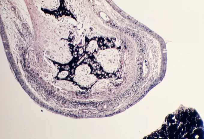
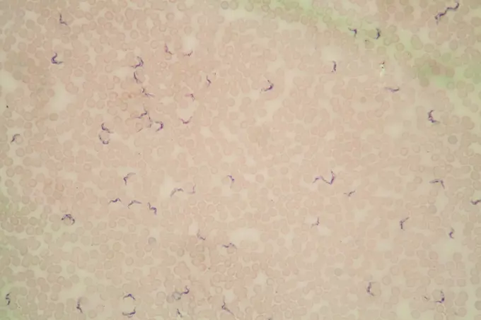
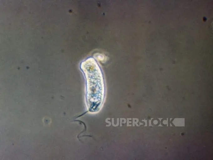
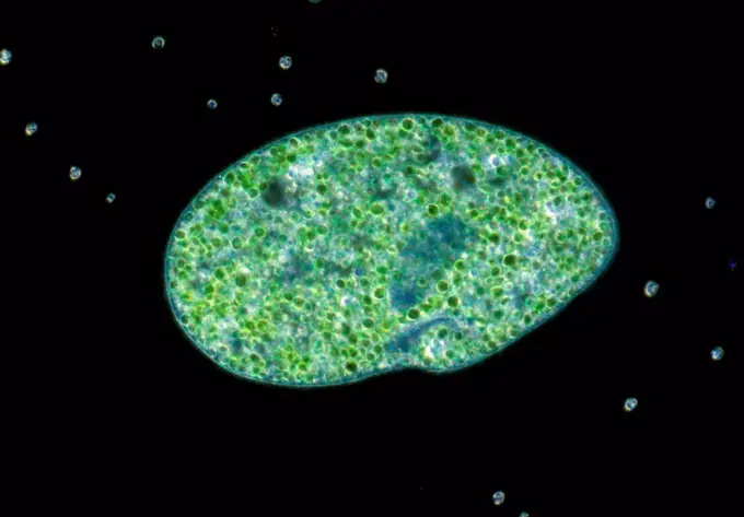
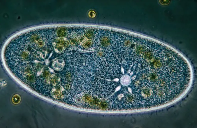
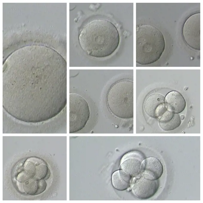
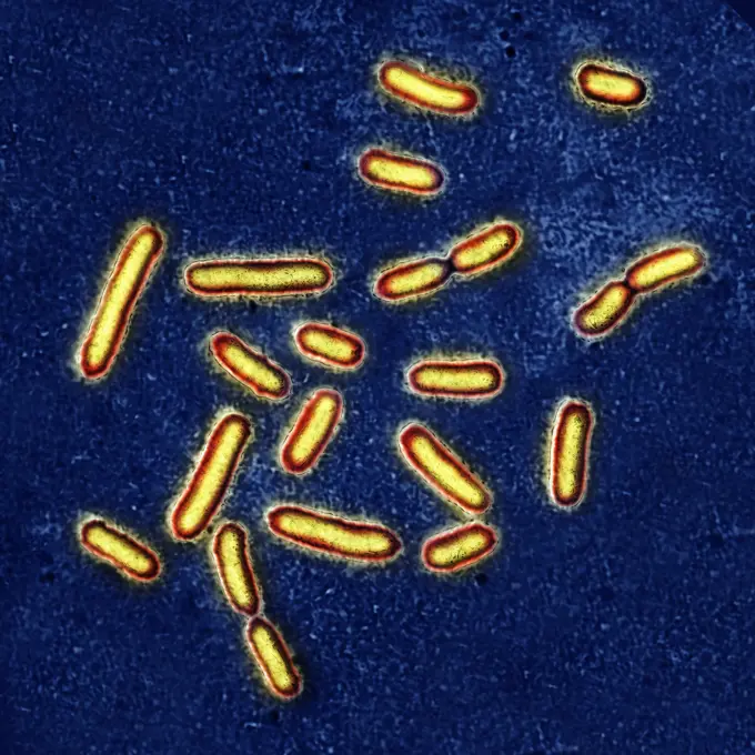
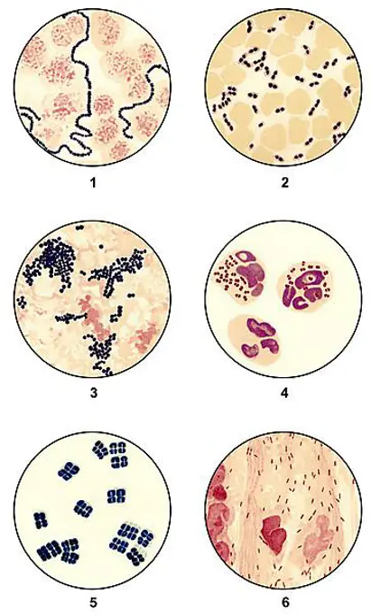
My Boards
View Pictures
  • My Boards

Microscopic Organisms and Tissue Types

Close-up images of microscopic organisms, including amoebae and cancerous tissue sections, showcasing cellular structures and colors.

Microphotograph of a Section of the respiratory mucosa of a dog, at x100 magnification.
238 assets in this story
4128-V58573367
824-63168547
Report at the Rouen University Hospital, PMA (Medically Assisted Procreation) service. Biology laboratory. IVF by ICSI.
4128-20044975
Cholera bacteria, illustration
4413-109880
Microscopic view of the chicken mite
1525-20846633
Swiss Cheese 02
1899-61461208
Meningococcus or meningococcal disease or Neisseria meningitidis, is a bacteria involved in meningitis. These bacteria are the cause of mild pharyngitis and only one person in approximately 400 becomes the victim of a serious meningococcal infection, most often in the form of acute purulent meningitis. Image produced from optical microscopy
6188-55561711
Ciliaten auf Futtersuche Ciliaten auf Futtersuche Copyright: xZoonar.com/Dr.xLangex 13174906
1525-22717524
bright jellyfish floating in the dark sea water
6188-55643647
Schlafkrankheit Schlafkrankheit Trypanosomen unter dem Mikroskop im Blut 400x Copyright: xZoonar.com/Dr.xNorbertxLangex 13630470
1788-20761710
Euglena unicellular alga viewed under a microscope, Euglenaceae.
4128R-13620000
Darkfield light micrograph of Paramecium bursaria, a ciliate protozoan, that contains endosymbiotic green algae (Chlorella sp.). Paramecium are found ...
6177-V53848781
1525-22717530
bright jellyfish floating in the dark sea water
4443-73161323
Animalcula, Infusoria, Fluvia, Tilia, Et, Marina, Hauniae, Typis, N. Mölleri, 1786, Infusoria, Animalcules, A meticulously illustrated plate showcasing various botanical structures, featuring a range of seeds and spores. The collection is systematically organized, with intricate details highlighting each specimen. Numbers accompany each illustration, indicating individual species or types. The diversity in shape and size is evident, with circular, oval, and elongated forms, capturing the intricacies of plant reproduction. This educational representation emphasizes the significance of each element in botanical studies, reflecting a historical approach to plant morphology and classification.
6145-52833945
A pencil sketch by Carl von Hasenauer depicting architectural design and details on paper.
824-57659491
This iodine-stained photomicrograph revealed ultrastructural morphology exhibited by a binucleate parasitic amoebic cyst of Entamoeba histolytica, in a concentrated wet mount specimen. Notice the chromatoid body with blunt and rounded ends, and that only two nuclei are visible in this focal plane. CDC/Dr Mae Melvin 1977
4421-25060
Plankton - Dinoflagellate (Peridinium depressum) x 43
1848-68970444
Screw algae
1848-49277173
Blue Liquid falling into Water against White Background
4201-66319
Torch Lily (Kniphofia sp) SEM close-up view of pollen at 1050x magnification
6145-44934449
Earth Observations taken by Expedition 34 crewmember.
6145-29256509
Nerve cells.
4128-20044978
Cholera bacteria, illustration
1788-111172842
Stages of the life cycle of Glenodinium sanguineum, Dinoflagellates. Drawing.
1788-111172770
Biflagellated gametes of Acetabularia, Polyphysaceae. Drawing.
4297-1457
Scanning electron micrograph (SEM) at an extremely high magnification of 44, 818X, of a single dividing Gram-negative Escherichia coli bacterium
1525-19842940
Frog egg under the microscope, background. (Frogg Egg Sec.) 100x

824-63128596
This scanning electron micrograph (SEM) depicts a flagellated Vibrio vulnificus bacterium, Mag. 13184x. Vibrio vulnificus is a bacterium in the same family as those that cause cholera. It normally lives in warm seawater and is part of a group of vibrios that are called halophilic because they require salt. V. vulnificus can cause disease in those who eat contaminated seafood or have an open wound that is exposed to contaminated seawater. Among healthy people, ingestion of V. vulnificus can cause vomiting, diarrhea, and abdominal pain. In immunocompromised persons, particularly those with chronic liver disease, V. vulnificus can infect the bloodstream, causing a severe and life-threatening illness characterized by fever and chills, decreased blood pressure (septic shock), and blistering skin lesions. V. vulnificus bloodstream infections are fatal about 50% of the time. See image n0831905 on the site.
824-63168551
Report at the Rouen University Hospital, PMA (Medically Assisted Procreation) service. Biology laboratory. IVF by ICSI.
4141-32736
slipper animalcule compressed paramecium aurelia magnification x125
1525-20337735
Human IVF
824-57660038
Pseudomonas aeruginosa or pyocyanin bacillus, pathogenic bacteria, very resistant and often responsible for nosocomial infections. It is one of the most difficult bacteria to eliminate clinically. The infections it causes concern: the eyes, burns, surgical wounds, urine, lungs, stomach, intestines, meninges, blood. Image taken from an X 1000 optical microscopy.
1848-61699854
Bacteria smears of germs for microbiological diagnosis, microscope slides, clinical microbiology, state of scientific research around 1915, 1: Streptococci, 2: Pneumococci, 3: Staphylococci, 4: Gonococci, 5: Sarcina, 6: Influenza virus of the influenza epidemic of 1892
1525-20334880
Human IVF
1899-20311106
 Rouen University Hospital, France. Reproductive biology laboratory.
824-63225798
Transmission electron microscope images of Bacillus subtilis, using ambient and cryo-preparative methods. Mid-log phase cultures were prepared by conventional chemical fixation (A), MW-assisted chemical fixation (B), traditional FS (C), or by rapid FS without (D) or with MW irradiation (E-F). Bars: 50 nm...
6145-52893972
Culpepper Compound Microscope Slide, 18th Century. From microscope storage box 1965T5099.1.. Purchased from Edward H Pinto, 1965.
4443-73206796
Hundt, Fr., Rutherford, Lewis M., Sonnenfleckengruppe in ihren Changeungen an sieben Tagen, photograph, foto, foto's, height mm, width mm, photographer, 1870-09 - 1870-09, third quarter 19th century, salted paper print, salt print
PREVIOUS
of 3
NEXT
2401 S. Ervay, Suite 206
Dallas, Texas 75215
United States
Get Started
Free ResearchMy BoardsMy Cart
For Creators
How To License Your ContentContributor PortalFrame of Mind
Resources
API accessPricing
Contact
+1 866 236 0087help@viewpictures.co.uk Contact form
©2026 View Pictures. All Rights Reserved. -A
This site is protected by reCAPTCHA and Google Privacy Policy and Terms of Service apply.