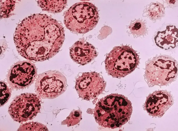
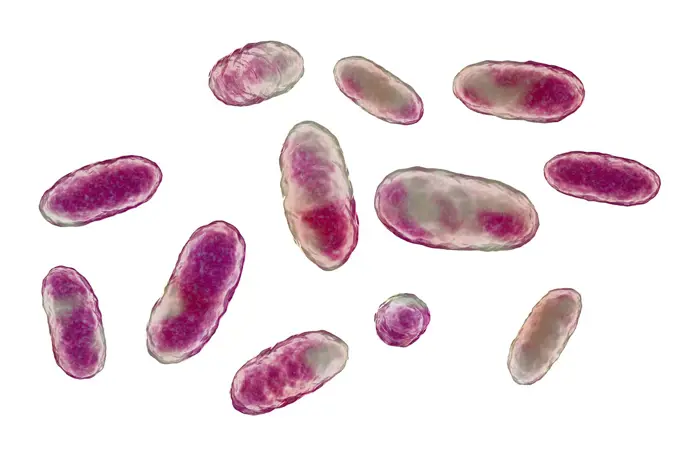
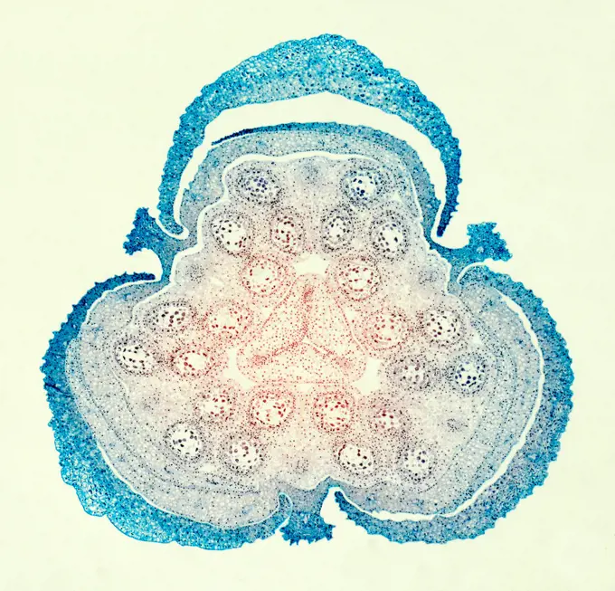
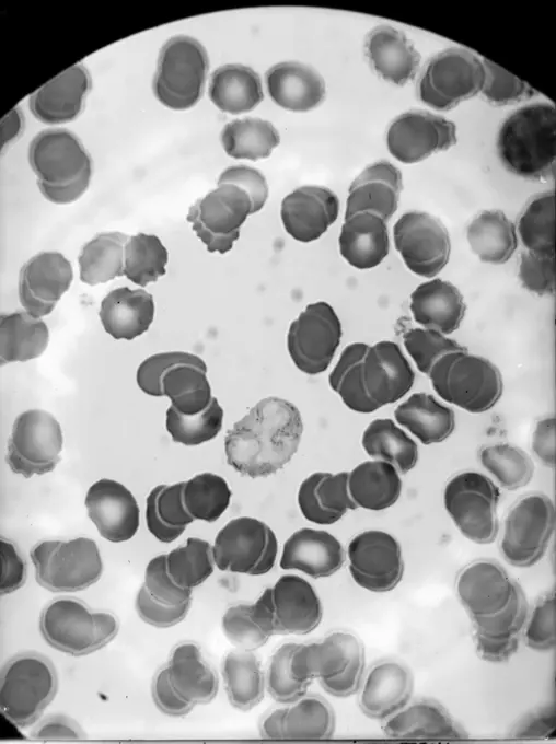
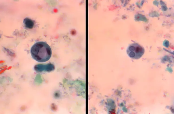
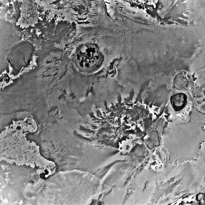
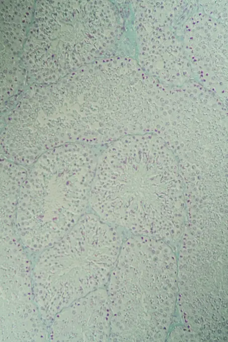
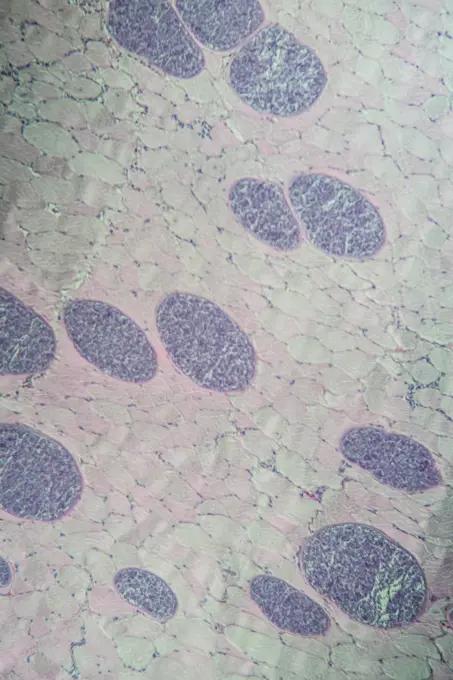
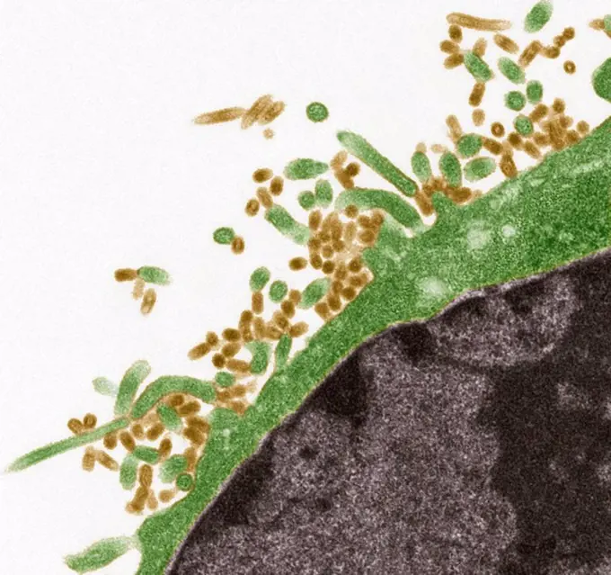
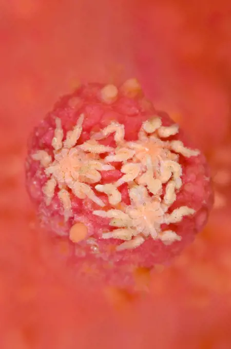
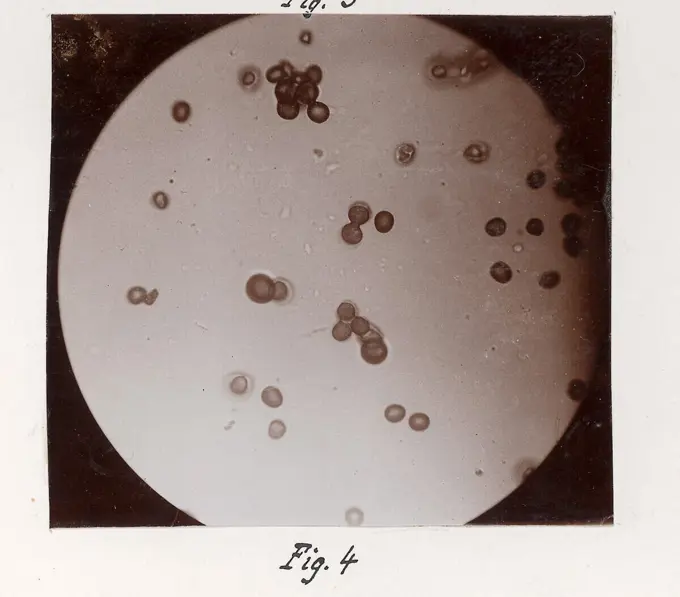
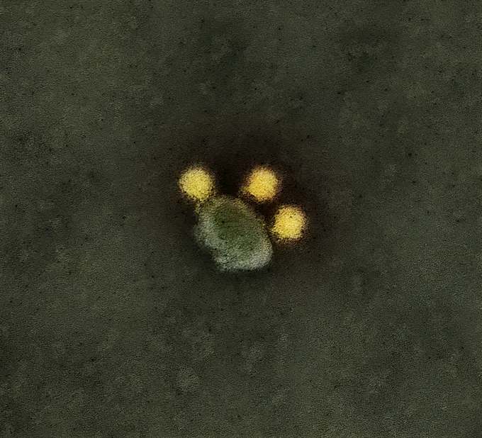
My Boards
View Pictures
  • My Boards

Microscopic Organisms Observed

Images showcasing various microscopic life forms, including plasmodium, protists, and amoebae, under different magnifications in scientific settings.

Colorized TEM image of human white blood cells
245 assets in this story
4128-28575319
Aggregatibacter bacteria, illustration
4141-111428933
Light micrograph cross section  through the young bud of a lily flower (Lilium species) showing the male and female reproductive organs. CS, UK
6188-55645428
Green algae in the water drop under the microscope 100x green algae in the water drop under the microscope 100x copyright: xzoonar.com/DR.XNORBERBERTXLANGEX 13856172
6188-55644466
Quergestreifter Muskel Quergestreifter Muskel Muskelgewebe 200x Copyright: xZoonar.com/Dr.xNorbertxLangex 13728189
6145-46770691
Glass negative, micrograph with medical motif.
1788-111175612
Larva of spiny lobster (Palinurus vulgaris), crustacean, seen under a microscope, at x25 magnification.
824-57659419
This photomicrograph shows two Entamoeba histolytica cysts. The cyst on the left contains a single nucleus and is therefore called a mononuclear cyst, while the cyst shown on the right contains two separate nuclei and is called a binuclear cyst. CDC/Dr Mae Melvin 1977
6145-44496958
Io
1899-53511520
Uterine cancer visualized by frontal MRI.
6188-55644214
Testicles guinea pig testicular cross -section under the microscope 200x copyright: xzoonar.com/dr.xnorbertxlangex 13702089
824-65831039
Colorized transmission electron micrograph of rabies virus particles (teal). Posted to commemorate World Neglected Tropical Diseases Day on January 30th. Rabies is one of the most neglected tropical diseases in underserved regions of the world. Per the World Health Organization, it causes an estimated 59,000 human deaths annually. It disproportionally affects rural poor populations and children with limited access to healthcare and veterinary services.
4128-17810048
Marine plankton, light micrograph
1788-20761828
Aspergillus clavatus viewed under the microscope, ascomycete fungi.
1850-47937
Lunaria annua, Honesty, Brown subject, White background.
6177-V54022777
6188-55644171
Sarcocysteis at the monkey Sarcocysteis at the monkey Spares in the muscle, 100x copyright: xzoonar.com/DR.XNORBERBERTXLANGEX 13698999
4409-66492
Skin diseases: Purple spots, Purpura simplex and P. haemorrhagica and rose rash, Exanthesis roseola. Handcoloured copperplate stipple engraving by John Pass from John Wilkes' "Encyclopedia Londinensis," J. Adlard, London, 1822.
4413-8317
Spores and sporangiums of a fern X40
6145-44719325
S73-15171 (4 Jan. 1973) --- These orange glass spheres and fragments are the finest particles ever brought back from the moon. Ranging in size from 20 to 45 microns (about 1/1000 of an inch) the particles are magnified 160 times in this photomicrograph made in the Lunar Receiving Laboratory at the Manned Spacecraft Center. The orange soil was brought back from the Taurus-Littrow landing site by the Apollo 17 crewmen. Scientist-astronaut Harrison H. Jack Schmitt discovered the orange soil at Shorty Crater during the second Apollo 17 extravehicular activity (EVA). This lunar material is being studied and analyzed by scientists in the LRL. The orange particles in this photomicrograph, which are intermixed with black and black-speckled grains, are about the same size as the particles that compose silt on Earth. Chemical analysis of the orange soil material has shown the sample to be similar to some of the samples brought back from the Apollo 11 (Sea of Tranquility) site several hundred mil
6176-55293824
BACTERIA SPIRILLUM SPIRILLUM VOLUTANS LARGEST KNOWN SPIRILLUM SHOWS THE FLAGELLA 1000X OIL STUDIO
1788-111174079
Protococcus viridis, Chlorophyceae. Drawing.
1525-26227457
Big sugar crystals macro closeup abstract background flatlay
1788-111174209
Microphotograph of Protozoan, genus Bodo, and two strands of seaweed.
4269-25050
Fertilized oocyte. 2 nuclei Light microscope
6145-29604696
Collection Dutch Heidematschappij. water moving /. undated
6145-29292853
Herpes simplex virus infection
6177-V53863604
6145-45231646
Biomedical research offers hope for a variety of medical problems, from diabetes to the replacement of damaged bone and tissues. Bioreactors, which are used to grow cells and tissue cultures, play a major role in such research and production efforts. Anchorage dependent cells on STS-95 will be grown on beads similar to these cells produced during previous investigations. Recombinant proteins may offer the possibility of reducing or eliminating transplant rejections. Research by Synthecon, Inc. using the BioDyn Bioreactor will focus on the preliminary process for growing a proprietary recombinant protein that can decrease rejection of transplanted tissue. The cells producing this protein are anchorage dependent, meaning that they must attach to something to grow. These cells will be cultured in the bioreactor in a medium containing polymer microbeads. Synthecon hopes that the data from this mission will lead to the development of a commercial protein that will aid in prevention of trans
824-57657952
Naegleria fowleri is an amoeba (one-celled living organism) that lives in soil and warm fresh water, such as lakes, rivers, and hot springs. It is commonly called the brain-eating amoeba because it can cause brain infection when water containing the amoeba rises in the nose. Only about three people in the United States are infected each year, but these infections are usually fatal.
4269-27856
Small intestine. Colored MRI (Magnetic Resonance Imaging) scan of the abdomen showing the small intestine (orange). Are also seen the liver at top (black), the stomach at right, part of the large intestine at left, and the baldder at bottom.
1899-61460921
This photomicrograph revealed the presence of both an Entamoeba hartmanni , and an Endolimax nana amoebic trophozoite. CDC 1970
4128-V58573223
4141-111428940
Light micrograph transverse section through the rachis of a Male fern (Dryopteris filix-mas). TS, UK. The rachis is the main axis of a compound structure.
4128R-8203
Influenza viruses, TEM
1788-20761588
Amoeba (Amoeba sp) viewed under a microscope, Protozoa.
4128R-23750
Blood vessel. Coloured transmission electron micrograph TEM of a section through a blood vessel, showing platelets blue and red blood cells red. Magni...
4070-V19746805
1848-61243026
Bacteria smears of germs for microbiological diagnosis, microscope slides, clinical microbiology, state of scientific research around 1915, 1: Diphtheria or Corynebacterium diphtheriae, 2: Diphtheria or Corynebacterium diphtheriae, 3: Yersinia pestis, 4 Salmonella Typhi, 5: Vibrio cholerae, 6: Spirillum
1788-111173991
Diagram of white blood cells. Drawing.
1899-61460888
This photomicrograph of an iron-hematoxylin-stained specimen, revealed the presence of two parasitic cysts, an Iodamoeba btschlii ( buetschlii ), and a Chilomastix mesnili . CDC/ Dr. Mae Melvin 1977
6145-52770929
Cyanotype photograph showing the dry side of a photo bar touched by a subject's fingertips and by heated or tempered rubber fingers, on a box format around 1897-1898.
4070-21285268
Three hairy sea spiders (Tanystylum grossifemorum) feeding on red soft coral (Gersemia rubiformis). Browning Pass, Port Hardy, Vancouver Island. British Columbia. Canada. North East Pacific Ocean.
6145-52771401
Microscopic photograph of F2 generation wheat and rye grains by Frana Jesenko on collodion paper mounted on a box box.
6145-44588700
Still photographs taken over 16 hours on Nov. 13, 2001, on the International Space Station have been condensed into a few seconds to show the de-mixing -- or phase separation -- process studied by the Experiment on Physics of Colloids in Space. Commanded from the ground, dozens of similar tests have been conducted since the experiment arrived on ISS in 2000. The sample is a mix of polymethylmethacrylate (PMMA or acrylic) colloids, polystyrene polymers and solvents. The circular area is 2 cm (0.8 in.) in diameter. The phase separation process occurs spontaneously after the sample is mechanically mixed. The evolving lighter regions are rich in colloid and have the structure of a liquid. The dark regions are poor in colloids and have the structure of a gas. This behavior carnot be observed on Earth because gravity causes the particles to fall out of solution faster than the phase separation can occur. While similar to a gas-liquid phase transition, the growth rate observed in this test is
824-65831083
Colorized transmission electron micrograph of negative-stained Zika virus particles (yellow).
PREVIOUS
of 3
NEXT
2401 S. Ervay, Suite 206
Dallas, Texas 75215
United States
Get Started
Free ResearchMy BoardsMy Cart
For Creators
How To License Your ContentContributor PortalFrame of Mind
Resources
API accessPricing
Contact
+1 866 236 0087help@viewpictures.co.uk Contact form
©2026 View Pictures. All Rights Reserved. -A
This site is protected by reCAPTCHA and Google Privacy Policy and Terms of Service apply.