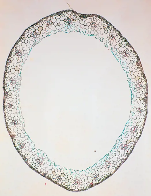
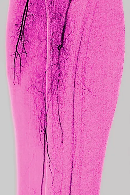
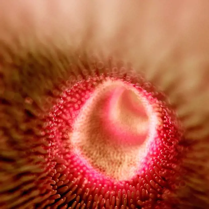
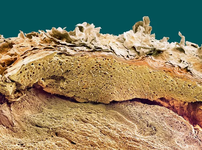
My Boards
View Pictures
  • My Boards

Microscopic Plant and Marine Textures

A collection of detailed microscopic images showcasing the intricate textures of plant tissue and marine life, featuring vibrant colors and patterns.

Candy Cane Sea Star (Fromia monilis) skin, Indonesia
260 assets in this story
4128R-3643
Pyoderma skin disease, coloured scanning electron micrograph SEM. Lymphocytes blue and red blood cells are present in the necrotic tissue on the surfa...
6145-29293003
Amoebiasis: colon
1788-111172818
Sea Sparkle, Noctiluca scintillans algae, Dinoflagellate .
4128R-1162
Tongue. Light micrograph of a section through the thick keratanised epithelium of a foliate papillae of the tongue. Magnification: x100 when printed a...
6145-54589486
Light print showing natural forms and objects from the natural kingdom by Martin Gerlach sen., produced between 1902 and 1904.
6145-46761703
Lumière autochrome. Medical photographs.O-blood cells not aggl. of A- or B serum. Immersion.
4128R-15290698
Aspergillus fumigatus fungus, illustration
4128R-10570
Developing pig eye, light micrograph
4128R-3517
Lung tissue, TEM
4141-111428959
Light micrograph cross section Brown algae (Fucus species) showing the monoecious conceptacles, UK
4141-74593
Commensal shrimp Periclemenes soror on oral face of sea star, Lembeh Strait, Northern Sulawesi, Indonesia
1525-28067569
Section through the rope and spinal cord of a young trout, vintage engraved illustration. From the Universe and Humanity, 1910.
1788-111176141
Microphotograph of the segmentation process of the fertilized egg of a frog, morula stage.
6188-55644708
Fush infection fungal infection false mildew on shepherd's paw herb 100x copyright: xzoonar.com/dr.xnorbertxlangex 13752009
7203-70648786
Bearded Fireworm (Hermodice carunculata), Underwater Fluorescence, Blue Heron Bridge, Intracoastal Waterway, West Palm Beach, Florida, USA
6188-55643571
Beach Hair Hair Leaf Cross -section 100x
4413-109843
Microscopic view of Sporangia Bryum Moss
4128-17811360
Mitosis, light micrograph
6145-44747508
Agricultural patterns are distinctly visible in this near-vertical false color infrared photography taken in November 1994. The area depicted on the photograph is south of Khartoum between the White and Blue Nile Rivers. By far the most important irrigation project in sub-Saharan Africa, both large and small scale agricultural enterprises have been developed using water transported from the perennial Nile Rivers. Hundreds of small rectangular fields and water-filled canals can be seen in this photograph.
1788-111176137
Microphotograph of the segmentation process of the fertilized egg of a frog, beginning of the fourth division of the egg.
6145-45240023
AS06-02-938 (6 April 1968) --- During the second revolution of the National Aeronautics and Space Administration's Apollo 6 spacecraft, this photograph of Senegal and Mauritania was taken at an altitude of 125 nautical miles. The predominant feature is the valley of the Senegal River (flowing to lower right). Lake Rkiz is to the left of the river in Mauritania. The Trarza Scrub Hills, a large area of elongated, fixed dunes, is in the lower left of the print. The land area seen in this photo is approximately 10,000 square miles or about the size of the state of Maryland. The photo was made one hour and fifty minutes after liftoff using a J.A. Maurer model 220G camera with Eastman Kodak SO-121 high resolution aerial Ektachrome film (exposure setting of f 5.6 at 1/500 sec.).
4128R-10365
Ovarian follicle, light micrograph
4269-27252
Plasmodium falciparum. Light micrograph of Plasmodium falciparum schizonts inside two erythrocytes, one of the early development stage of the parasite, agent of malaria. Magnification x1,500.
6188-55643021
Rogue leaves cross section 100x noise leaves cross section 100x copyright: xzoonar.com/dr.xlangeex 13397086
4128-48285231
Healthy bone tissue, illustration
1788-111173124
Cross section of a culm of wheat (genus Triticum), Gramineae, seen under a microscope.
1899-53511326
Arteritis in a lower limb (progressive occlusion of the arteries supplying the limbs and organs). Angiography of a lower limb.
4128-15979740
Intestinal villi, computer illustration.
4128R-5086
Skin, SEM
4128-V58557873
824-659
Herpes Virus
4128R-10371
Wart,, light micrograph
4201-22116850
Scarletina Bolete (Boletus luridiformis) detail of porous underside, Netherlands
6188-55644838
Giftefeu Giftefeu Blatt Querschnitt 100x Copyright: xZoonar.com/Dr.xNorbertxLangex 13769877
1788-111175876
Microphotograph of a silicotico nodule in the lung of a man suffering from silicosis.
1397R-86690
Footprint on green liquid
4128R-8349
Cut agate
1788-111174003
Isogamia sexual reproduction through similar but opposite-sex gametes of green algae. Drawing.
1899-61460372
Colorized transmission electron micrograph of monkeypox particles (pink) found within an infected cell (green), cultured in the laboratory
4141-111428918
Light micrograph cross section through the young branching root of a Broad bean (Viia faba). The image shows the lateral branching roots growing from the main root.
1890-110069
Light Micrograph LM of a transverse section of a stem of a Hedge Woundwort plant Stachys sylvatica, magnification x60
4128R-9674
Rapeseed pollen, SEM
4070-18049
Panther chameleon close-up of skin of tail Furcifer pardalis} green / blue colouration, Madagascar.
4384-138
This thin-section transmission electron micrograph (TEM) revealed some of the ultrastructural morphology found within the cyst-stage of a Giardia sp. protozoan.  The outer cyst wall is composed of filamentous and membranous portions, and is separated from the cytoplasm of the trophozoites contained within by the peritrophic space.  This cyst wall is approximately 0.25 microns thick. The protozoan Giardia  causes the diarrheal disease called giardiasis.  Giardia species exist as free-swimming (by
4128-28515227
Bacteria and fungi cultured on Petri dish
1788-111172948
Cross section of the thallus of sea lettuce (Ulva lactuca), Ulvaceae, seen under a microscope with counterstaining.
4413-20031496
Cross-section of a Wheat leaf. Dyes were used to distinguish the different parts of the leaf. Magnification of 20 in 24x36
4128-17857800
Maize root. Light micrograph (LM) of a section through the root of a maize plant (Zea mays) showing typical monocot arrangement of vascular bundles. T...
4128R-6321
Wart. Light micrograph of a section through a wart verruca vulgaris. The epidermis dark pink is irregularly thickened and covered by a layer of hyperk...
4413-109865
Detail of a spring diving beetle female
1899-65662465
Colorized transmission electron micrograph of mpox virus particles (pink) found within an infected cell (green), cultured in the laboratory.
824-65830963
Colorized transmission electron micrograph of mpox virus particles (pink) found within an infected cell (green), cultured in the laboratory.
6145-29269449
Ernst Haeckel, The evolution of man; embryos
7193-70583938
Australia, Nourlangie, Kakadu. Aboriginal paintings
1788-111173268
Wasp sting with venom gland seen under a microscope.
1788-20761871
Medicine - Human body - Heart attack phases, illustration
5514-53894988
HIV vaccine experiment, conducted by Swedish professor Eric Sanstram, in Dar es Salaam, Tanzania.
4128-16224590
Round cabbage leaf, X-ray
1788-111172650
Punctiform algae of the genus Nostoc, blue-green algae or cyanobacteria, seen under a microscope. Drawing.
4286-69830
Tongue models as an illustrative material for tongue diagnosis
PREVIOUS
of 3
NEXT
2401 S. Ervay, Suite 206
Dallas, Texas 75215
United States
Get Started
Free ResearchMy BoardsMy Cart
For Creators
How To License Your ContentContributor PortalFrame of Mind
Resources
API accessPricing
Contact
+1 866 236 0087help@viewpictures.co.uk Contact form
©2026 View Pictures. All Rights Reserved. -A
This site is protected by reCAPTCHA and Google Privacy Policy and Terms of Service apply.