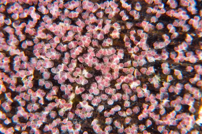
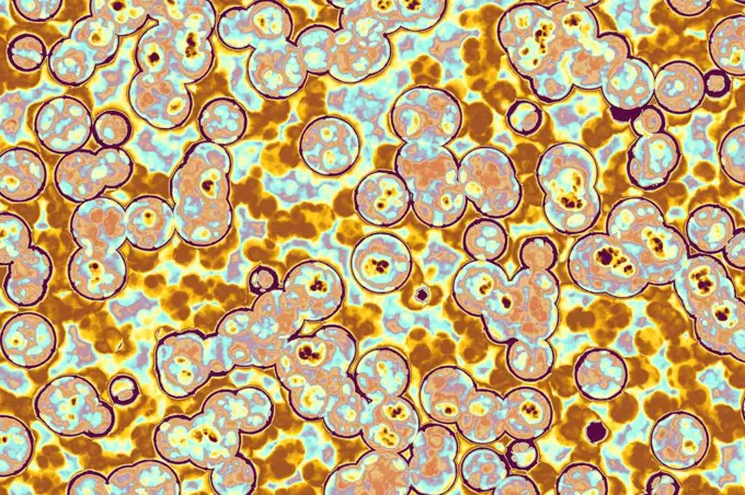
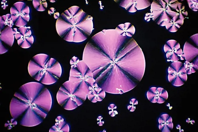
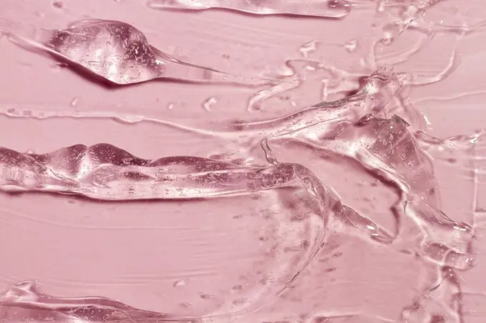
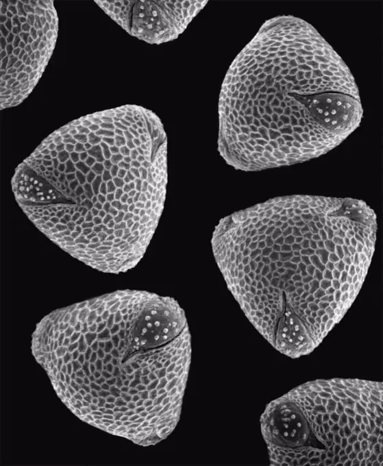
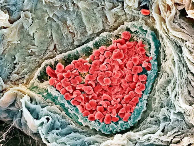
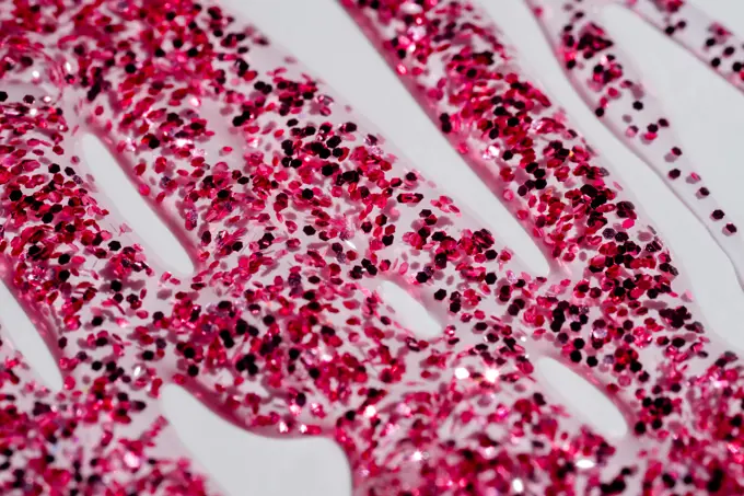
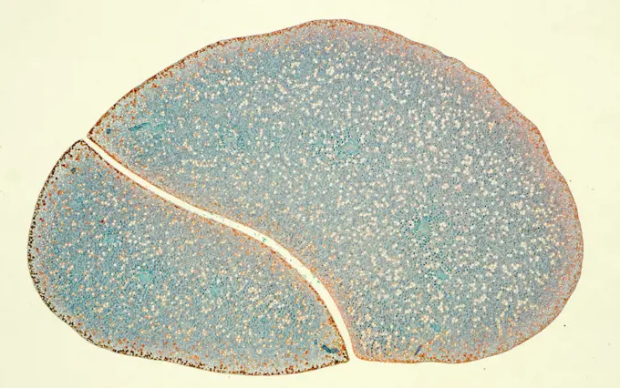
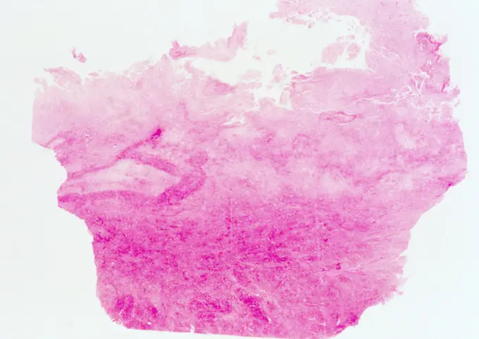
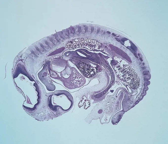
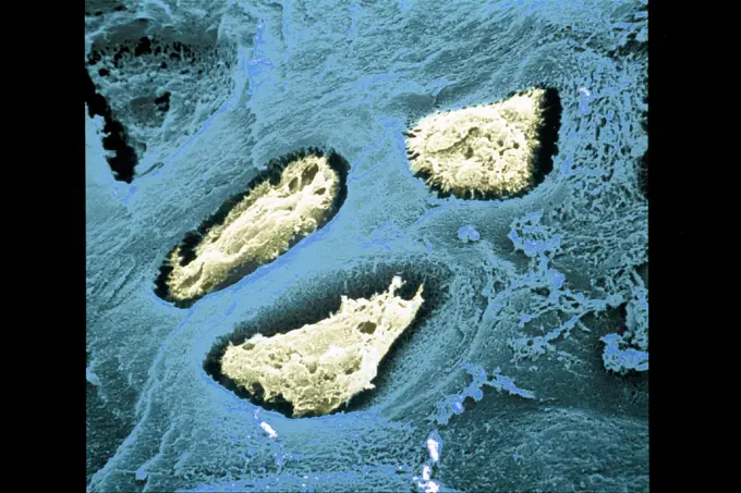
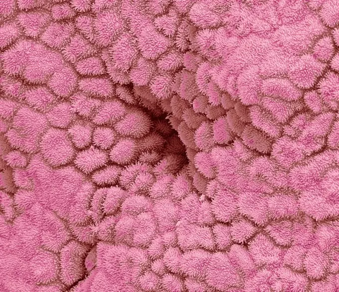
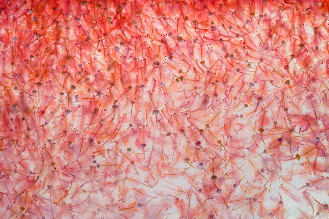
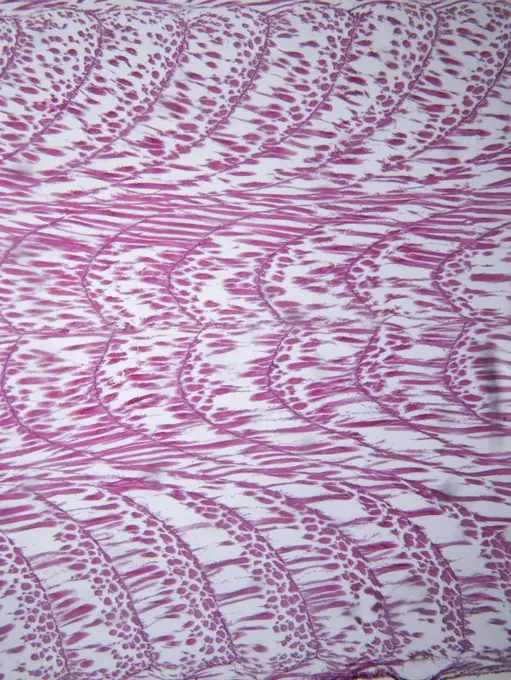
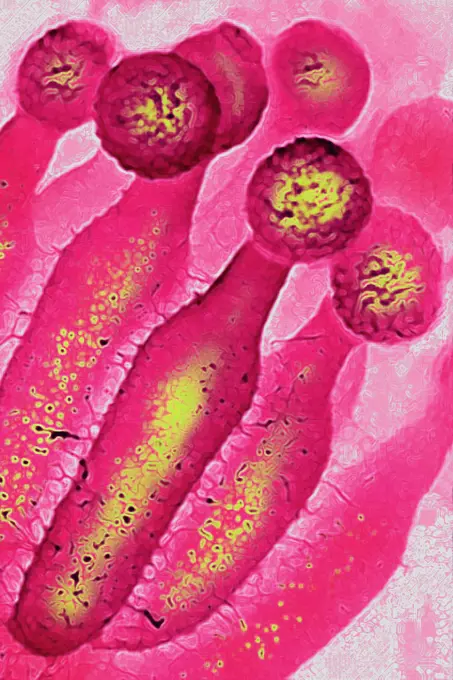
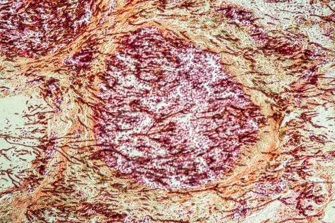
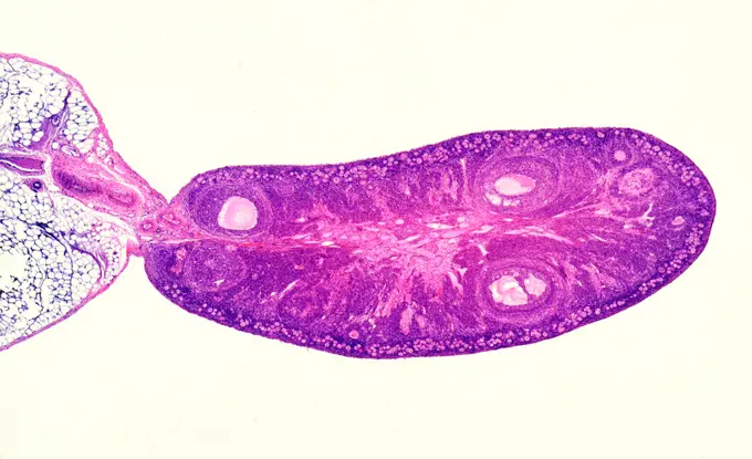
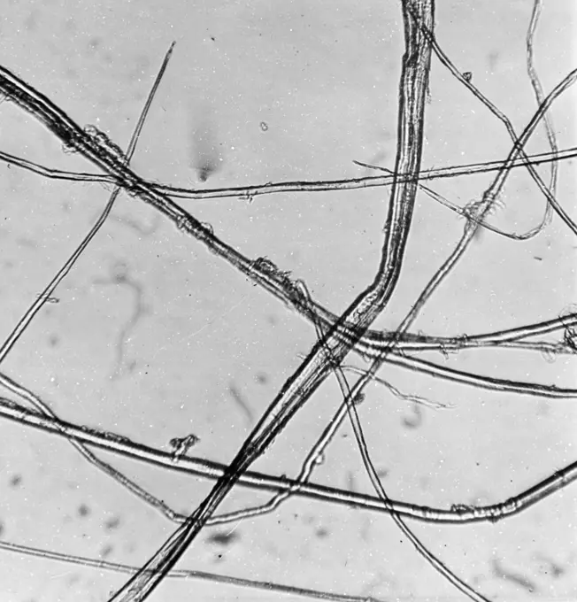
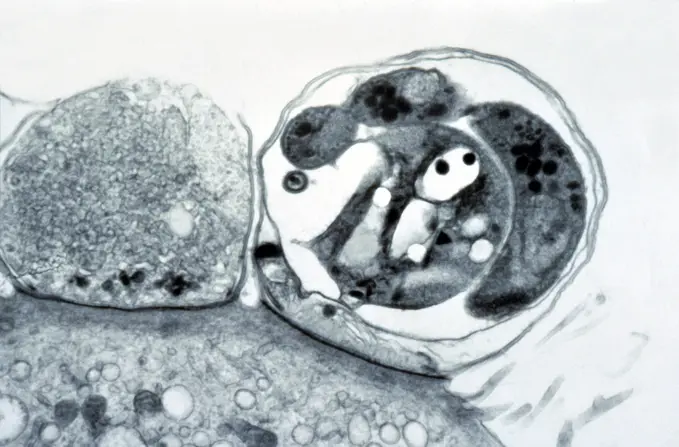
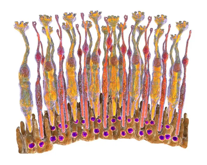
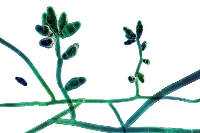
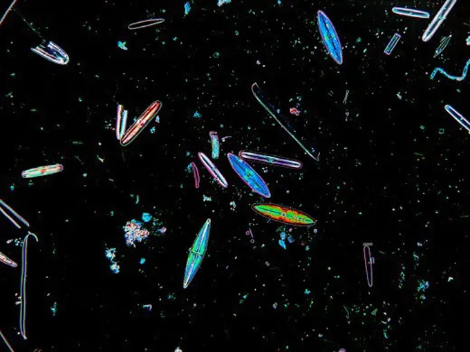
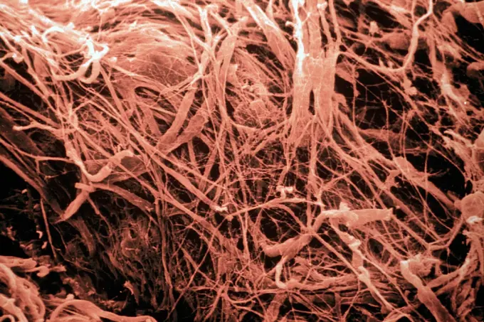
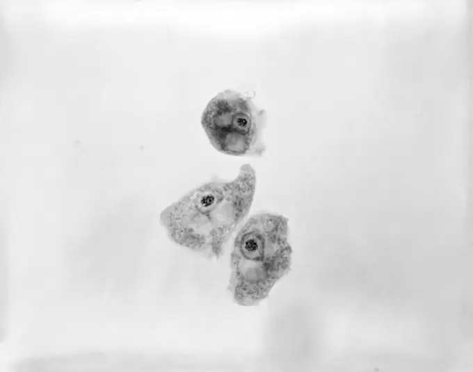
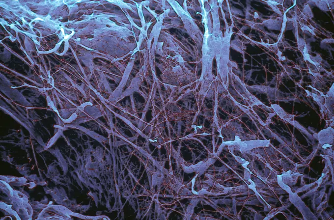
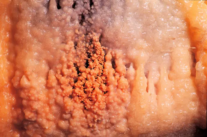
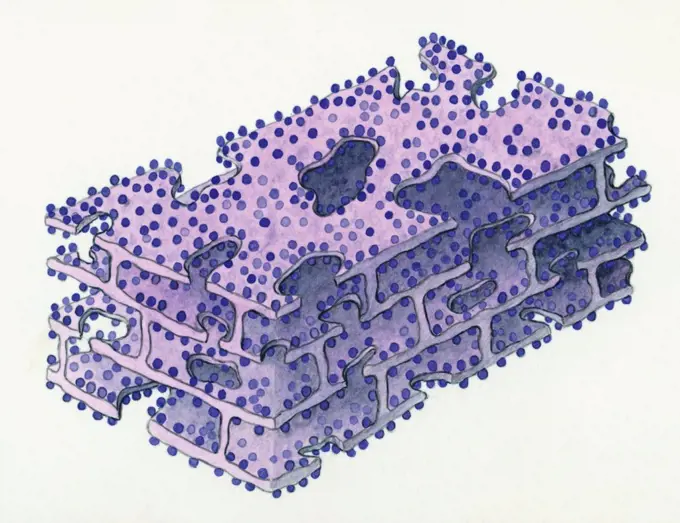
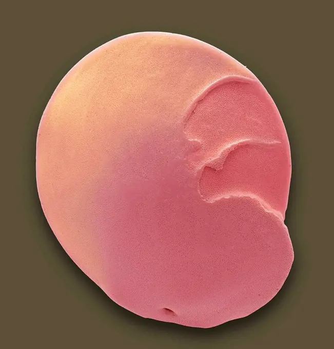
My Boards
View Pictures
  • My Boards

Microscopic Plant Structures

Detailed images of plant tissues viewed under a microscope, showcasing cell structures and colors, offering insights into botanical anatomy and morphology.

Swords swords cross 100x copyright: xzoonar.com/DR.XNORBERBERTXLANGEX 13688506
254 assets in this story
1889-48989848
Computer Generated Image
1788-111172626
Acrosymphyton purpuriferum, red algae, Florideophyceae.
4269-27741
Staphylococcus. Color enhanced scanning electron micrograph of a Staphylococcus biofilm on the inner surface of a needleless connector.
1525-21023822
Bubbles of dung in a creek
4293-1562
Indo-Pacific Sergeant, Abudefduf vaigiensis, egg brood, Mabul, Sabah, Malaysia, Borneo.
5514-16077616
Shape of bacterial cell: cocci, bacilli, spirilla bacteria
4128-111494396
Coloured scanning electron micrograph (SEM) of a freeze fracture of the vagina showing the stratified squamous epithelium and the underlying lamina pr...
4348-114
S-benzyl iso-thiouronium chloride, Crossed Polars, x60
1525-26183500
flat lay hydroalcoholic gel close up
4201-66259
Common Nasturtium (Tropaeolum majus) SEM close-up view of pollen grains at 1050x magnification
4128R-2140
Red blood cells, SEM
6188-66548581
Kosmetische schimmernde Peeling oder Maske smudge gel peeling swatch isoliert auf weiß Cosmetic shimmering scrub or mask smudge gel peeling swatch isolated on white Copyright: xZoonar.com/IrinaxKvyatkovskayax 22736403
4141-111428923
Light micrograph cross section through the fruit of a Loquat tree (Eriobotrya japonica), UK. This evergreen tree of shrub is also known as Japanese medlar or Japanese plum.
6188-55644784
Stechpalme Stechpalme Blatt Querschnitt 100x Copyright: xZoonar.com/Dr.xNorbertxLangex 13762695
6145-29255310
Amoebiasis: amoebic abscess in liver
1525-27799738
Germs of tuberculosis, vintage engraved illustration.
1788-111173220
Microphotograph of a small jellyfish, genus Obelia, Coelenterata, at x38 magnification.
7008-69757047
Rhodochrosite, Argentina
1788-111174365
Microphotograph of a longitudinal section of a pig embryo, at x6.3 magnification.
1525-19882751
Winter Jasmine leaf under a microscope (Leaf of Winter Jasmine C.S.) (Jasminum nudiflorum), 100x
4128-17928325
Liver fluke, light micrograph
4269-24801
Cartilage. Cartilage with 3 holes containing 3 cells ESM (Electron Scanning Microscope)
4128R-10583
Uterus lining, SEM
1525-25435435
Artemia plankton Brine shrimp
4128-V58557937
4128-17810021
Fish muscle tissue, light micrograph
1525-28067495
Pigeon blood cells, vintage engraved illustration. From the Universe and Humanity, 1910.
824-63208123
Penicillium chrysogenum, a penicillin-producing mold, visualized by scanning electron microscopy.
6188-55644590
Lungs of an aid patient lung of an aid patient lung tissue 200x copyright: xzoonar.com/dr.xnorberbertxlangex 13735558
4141-111428908
Light micrograph transverse section through the ovary of a rat showing a range of developing follicles. TS, UK
6145-46703944
Power cabinet image of flax. Uppmed Plant Fibergiberg enlarged 200 times.
6145-29256649
Gastrointestinal tract: cryptosporidiosis and HIV
4128R-11293533
The retina, inside the eye, contains a light-sensitive membranous layer of cells. These are specialized nerve cells: elongated rods (flower like shape...
824-63223205
Transmission electron micrograph of SARS-CoV-2 virus particles, isolated from a patient. Image captured and color-enhanced at the NIAID Integrated Research Facility (IRF) in Fort Detrick, Maryland. Credit: NIAID.
4128R-8493
Vaccinia virus particles, TEM
4128-15750426
Liver cancer, CT scan
6145-29270750
Six patches of skin showing types of skin disease. Coloured stipple etching by J. Pass, c. 1822.
6145-29288510
Liver: bacterial cholangitis and HIV
1788-111174095
Keratella square, Rotifers, seen under a microscope.
4128-19358308
Curvularia mould fungus, computer illustration. Curvularia is a group of about 35 species, most of which only infect plants. However, C. geniculata ca...
6145-29261177
The eye, as seen through a microscope: two figures. Colour lithograph, ca. 1862.
4298-1058
Diatoms found in pond water near Grant, NY. USA. Image enhanced to show structure.  Magnification 100x
4269-24793
Skin. Derm after action of collagen disease (connectivity) Elastic fibres after destruction of collagen ESM (Electron Scanning Microscope)
4306R-10163
Vinestain on a white table.
4409-17334043
CALCETIN DE LANA.
824-57659516
This 1973 photomicrograph depicts three Naegleria gruberi, free-living amoebae. CDC/Dr George Healy 1973
6145-29831058
Sample ca. 1830 French, Mulhausen. Sample 219981
4269-24642
Skin. Derm after action of collagenosis Deterioration of the elastic fibres further to destruction of collagen SEM (Scanning Electron Microscope)
4292-167190
Italy, Apulia, Castellana Caves, detail
1788-111173983
Three-dimensional pattern of a cell endoplasmic reticulum. Drawing.
1525-28244020
Bacteria cells square banners. Microorganism collection. Vector doodle style composition.
4128R-13022181
Foraminiferan. Coloured scanning electron micrograph (SEM) of a foraminiferan microfossil from maldives beach sand. Microfossils' are roughly 0.05 to ...
6145-29606374
Collection Dutch Heidematschappij. development. undated
6145-48895404
Favus. Dr. Selden I. Rainforth (American, 1879 - 1960)
PREVIOUS
of 3
NEXT
2401 S. Ervay, Suite 206
Dallas, Texas 75215
United States
Get Started
Free ResearchMy BoardsMy Cart
For Creators
How To License Your ContentContributor PortalFrame of Mind
Resources
API accessPricing
Contact
+1 866 236 0087help@viewpictures.co.uk Contact form
©2026 View Pictures. All Rights Reserved. -B
This site is protected by reCAPTCHA and Google Privacy Policy and Terms of Service apply.