My Boards
View Pictures
  • My Boards

Microscopic Protozoan Observations

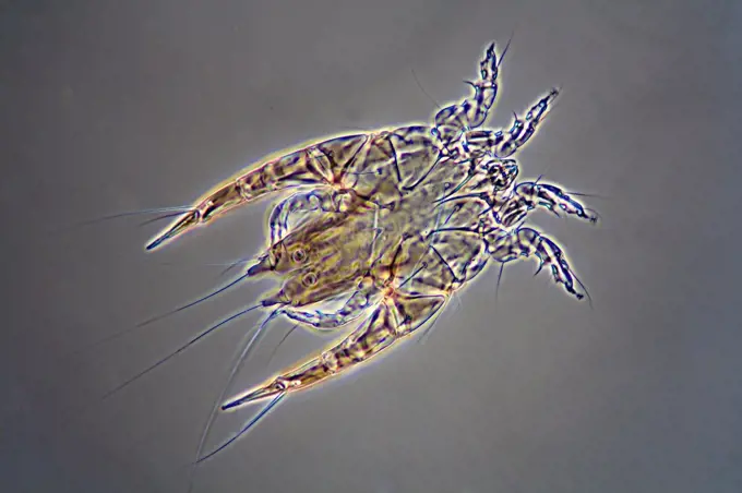
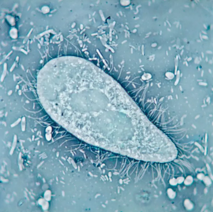
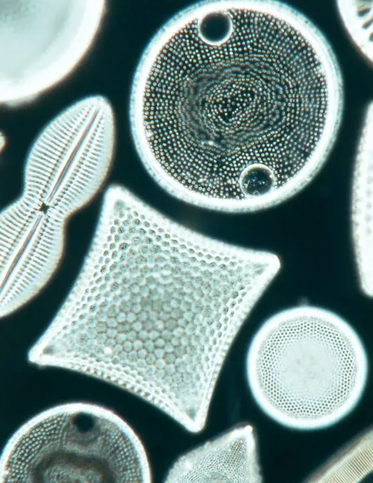
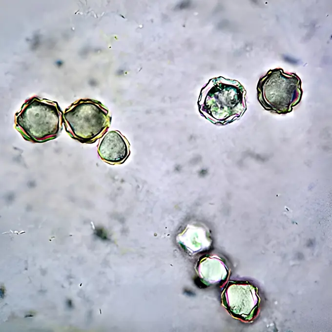
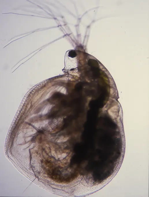
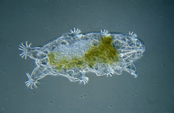
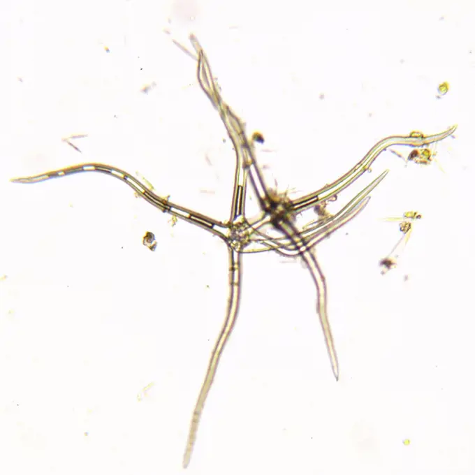
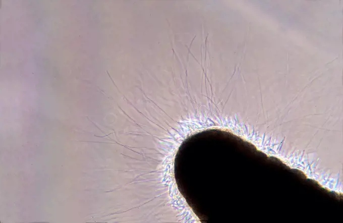
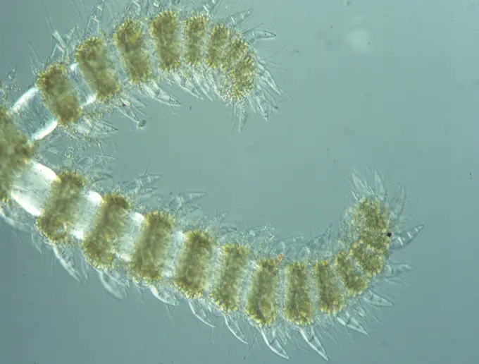
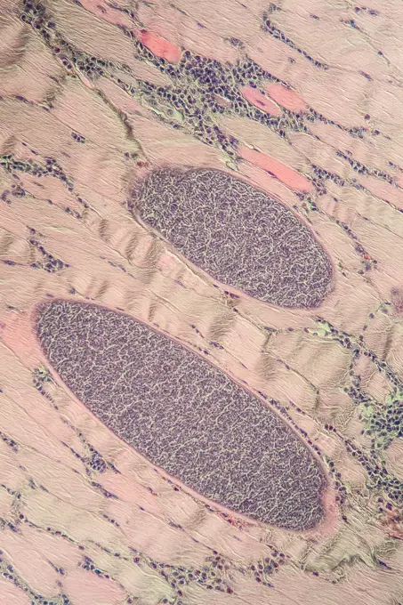
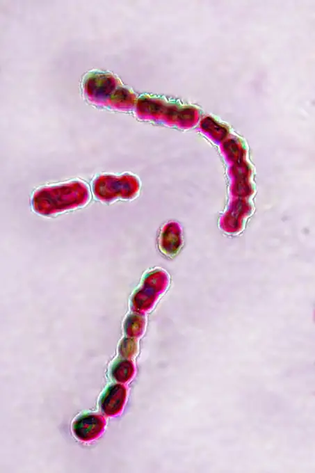
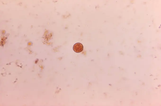
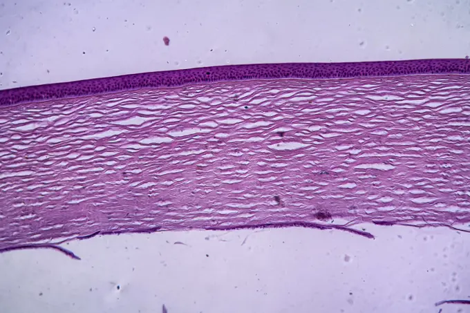
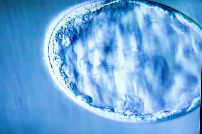
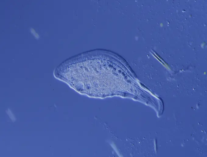
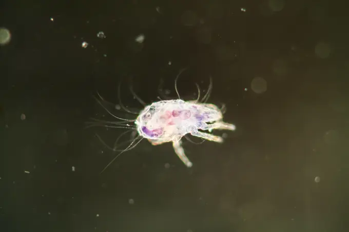
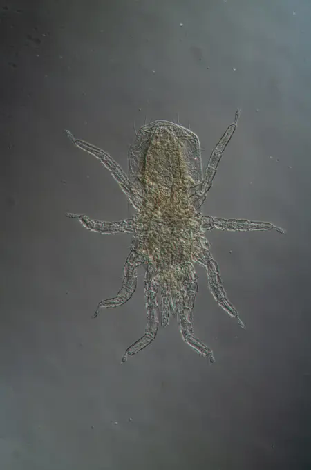
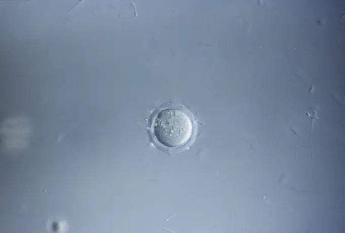
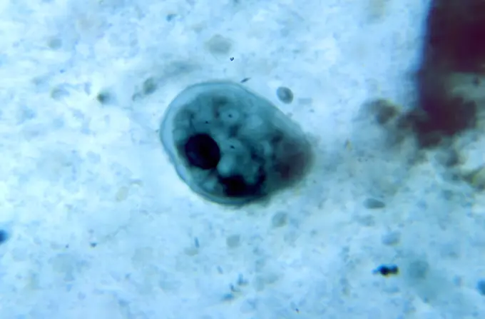
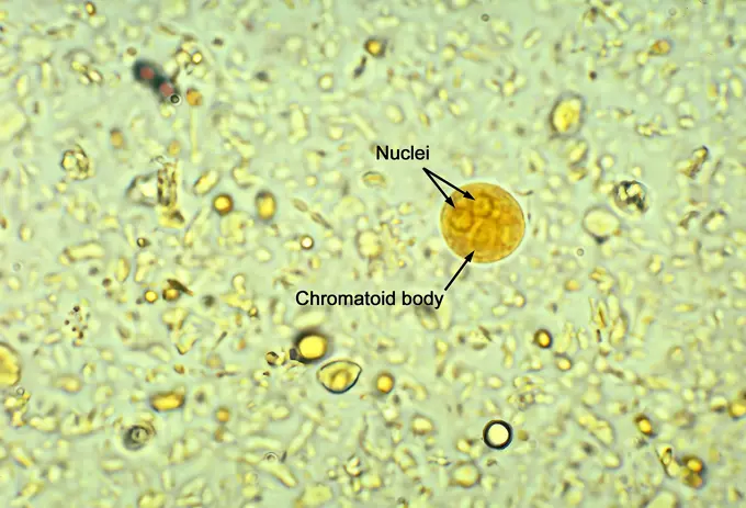
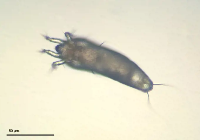
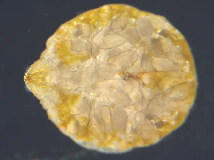
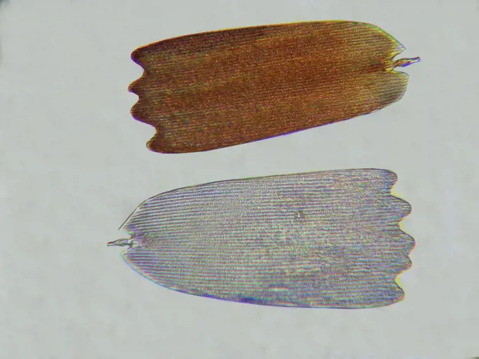
A series of photomicrographs depicting various protozoans like Naegleria gruberi and Paramecium, with diverse shapes and structures visible under high magnification.

Under 125X magnification, this photomicrograph of an agar medium revealed the presence of numerous non-pathogenic Naegleria gruberi amoebae. CDC 1971
166 assets in this story
6188-55645572
Zieralge unter dem Mikroskop 100x Zieralge unter dem Mikroskop 100x Copyright: xZoonar.com/Dr.xNorbertxLangex 13915964
824-63123145
View at the SEM.
4413-109878
Microscopic view of the chicken mite
1788-111174129
Ciliated infusorians seen under a microscope with counterstaining, at x320 magnification.
1788-111176733
Diatom algae with concentric lines and pinnate markings, seen under an optical microscope.
824-63123144
View at the SEM.
824-57659361
This photomicrograph of an iron-hematoxylin stained specimen revealed the presence of two spheroid parasitic cysts, Iodamoeba butschlii (buetschlii). The cyst on the right contained a clearly visible achromatoid granule in its cytoplasm (arrow). CDC/Dr Mae Melvin 1977
1899-61460539
Acanthamoeba is a microscopic, free-living amoeba, or amoeba (single-celled living organism), that can cause rare but serious eye, skin, and central nervous system infections. The amoeba is found worldwide in the environment in water and soil. The amoeba can spread to the eyes through the use of contact lenses, cuts or wounds in the skin, or by being inhaled into the lungs. Most people will be exposed to Acanthamoeba in their lifetime, but very few will become ill from this exposure
6188-55645380
Water flea under the microscope 100x water flea under the microscope 100x copyright: xzoonar.com/DR.XNORBERBERTXLANGEX 13847794
4201-40459
Tardigrade (Echiniscoides sigismundi) microscopic image, animal is less than one mm in length, can enter cryptobiosis to withstand temperature and moisture extremes, worldwide distribution
1850-41994
Algae
1788-20761837
Expanded melanophores chromatophores viewed under a microscope.
4413-1785
Discharge of cnidocystes from a Sea anemone tentacle
6188-55645426
Rotalgen unter dem Mikroskop 100x Rotalgen unter dem Mikroskop 100x Copyright: xZoonar.com/Dr.xNorbertxLangex 13856168
6188-55644182
Sarcocysteis at the monkey Sarcocysteis at the monkey Spares in the muscle, 200x copyright: xzoonar.com/DR.XNORBERBERTXLANGEX 13699021
824-63222957
Bacteria streptococcus pyogenes, responsible for infections, including angina, bronchopulmonary infections pneumonia, infection of wounds and skin. Optical microscopy view.
1788-111173796
Microphotograph of tecamebas, tropical Protozoan, at x400 magnification.
1899-61460933
Under 500X magnification, this photomicrograph revealed the presence of a parasitic cyst, Entamoeba histolytica. CDC/Dr LLC Moore, Jr. 1969
6188-55643761
Corn skin eye cornea eye tissue under the microscope 100x copyright: xzoonar.com/DR.XNORBERBERTXLANGEX 13650409
6176-67038967
Suckers on tentacles of Octopus Vulgaris, 20th century. Artist: CM Dixon.
4201-66206
Copepod nauplius larval stage of development at 120x magnification with light sensing organ visible in red, Spain
4128-V58573231
4128R-11288303
Blastocyst embryo, light micrograph. This embryo is five to six days old.
4269-27790
Flea infected with plague. A Thrassis bacci johnsoni flea 41 days after infection with Yersinia pestis, the bacterium that causes bubonic plague. Thrassis bacci, a flea-carried pathogen of rats, is one of the primary vectors of plague to humans.
1525-24951185
Goggles
6188-55645357
Ciliat im Wassertropfen unter dem Mikroskop 100x Ciliat im Wassertropfen unter dem Mikroskop 100x Copyright: xZoonar.com/Dr.xNorbertxLangex 13828581
4413-43515
Young Olive Psyllid
4297-1460
Under a magnification of 5000X, this colorized scanning photomicrograph shows numbers of clustered Gram-negative Salmonella typhimurium bacteria
4269-24902
In Vitro Fertilization. IVF (In Vitro Fertilization) Cellular division 2 cells stage Light microscope
1788-20761832
Meiosis of an egg cell in the ovary of an Ascaris, nematode worm, viewed under a microscope.
824-63128594
This scanning electron micrograph (SEM) depicts a Vibrio parahaemolyticus bacterium, Mag. 21851x. Vibrio parahaemolyticus is a bacterium in the same family as those that cause cholera. It lives in brackish saltwater, and causes gastrointestinal illness in humans. V. parahaemolyticus is a halophilic, or salt-requiring organism, naturally inhabiting coastal waters in the United States and Canada, and is present in higher concentrations during summer. When ingested, V. parahaemolyticus causes watery diarrhea often with abdominal cramping, nausea, vomiting fever and chills. Usually these symptoms occur within 24 hours of ingestion. Illness is usually self-limited and lasts 3 days. Severe disease is rare and occurs more commonly in persons with weakened immune systems. See image n1310105 on the site.
6188-66542753
Water flea in a drop of water. Water flea in a drop of water.
4269-27253
Plasmodium falciparum. Light micrograph of parasitic Plasmodium falciparum trophozoites in erythrocytes, agent of malaria.
4220-21334717
Light Micrograph using Nomarski Technique: Intestinal Roundworm - juvenile hatching from egg.     Date:
6188-54892429
two burnt holes two burnt holes in a paper ,model released, Symbolfoto Copyright: xZoonar.com/RuslanxNassyrovx 1979872 ,model released, Symbolfoto ,property released
6145-45231082
Along with hemoglobin the primary oxygen storage and transport proteins in all higher animals including humans. Important for medical reasons because they are primary blood proteins.
4201-24506
Rotifer (Rotaria rotatoria) a microscopically small animal with a length of 004 to 240 mm, waterborne and can inhabit highly acidic environments
1899-61460902
This photomicrograph of an iron-hematoxylin-stained specimen, revealed the presence of two amoebic organisms in their cystic stages of their development. In this focal plane, the smaller Entamoeba histolytica cyst (top), contained four nuclei, and two bluntly-tipped chromatoid bodies, while six nuclei could be detected in the larger Entamoeba coli cyst (bottom), which also contained two chromatoid bodies as well. CDC/ Dr. Mae Melvin 1977
4286-21418
Petri dish with a variety of bacterial and fungal colonies
1788-20759937
Limonitic oolite, thin section using parallel nicols, x100.
6188-55644968
House dust mite under the microscope house dust mite under the microscope 50x copyright: xzoonar.com/DR.XNORBERBERTXLANGEX 13778312
1525-27232783
Corona Virus2019-nCoV or Covid 19. Asian flu infection outbreak as pandemic risk around the world in medical concept. close up of microscope virus cells. 3d abstract illustration.
6188-55643128
Endoparasitische Hünermilbe 100x Endoparasitische Hünermilbe 100x Copyright: xZoonar.com/Dr.xNorbertxLangex 13445835
4269-27447
Escherichia coli. This scanning electron micrograph (SEM) of a Escherichia coli bacterium ; Magnification 12800x. E. coli are rod-shaped Gram-negative bacteria that normally colonize the digestive tract in humans and other animals. However, some strains can produce a toxin that leads to severe illness or even death.
4220-21334602
Mouse Ovum being fertilized.     Date:
1788-111173292
Microphotograph of chetognato, genus Sagitta in vivo, at x10 magnification.
4413-43406
Young larva of Oleander scale
6188-55645077
Radiolarien Radiolarien Schalen unter dem Mikroskop 100x Copyright: xZoonar.com/Dr.xNorbertxLangex 13785171
824-63184384
Hepatitis C virus (HCV). Image produced using high-dynamic-range imaging (HDRI) from an image taken with transmission electron microscopy. Viral diameter around 22 nm.
4220-20101211
Namibia - The balloon ground crew tries to follow the flight direction of the hot-air balloon, crossing a sandy plain at the edge of the Namib Desert in the NamibRand Nature Reserve. The so-called 'Fairy Circles' are circular patches without any vegetation which according to recent scientific studies are caused by the Harvester Termite (Hodotermes mossambicus). NamibRand Nature Reserve, Namibia.
4128R-13818843
Petri dish with bacteria.
1899-61460980
Under 900X magnification, this photomicrograph revealed some of the ultrastructural morphology exhibited by a trophozoite-stage Entamoeba coli microorganism, in a specimen prepared using the Kohn staining method. CDC/Dr Healy 1964
1899-61461004
This iodine-stained photomicrograph revealed ultrastructural morphology exhibited by a binucleate parasitic amoebic cyst of Entamoeba histolytica, in a concentrated wet mount specimen. Notice the chromatoid body with blunt and rounded ends, and that only two nuclei are visible in this focal plane. CDC/Dr Mae Melvin 1977
4128-28515225
Bacteria and fungi cultured on Petri dish
6145-44936643
Earth Observations taken by Expedition 34 crewmember.
824-57659357
This photomicrograph of an iron-hematoxylin-stained specimen, revealed the presence of two parasitic cysts, an Iodamoeba btschlii ( buetschlii ), and a Chilomastix mesnili . CDC/ Dr. Mae Melvin 1977
4413-43408
Grape erineum mite
4128-17928307
Liver fluke, light micrograph
4413-43404
Larva of Oleander scale
1848-51674324
Jellyfish, Common jellyfish (Aurelia aurita), Other animals, Cnidarians, Animals, Jellyfish, Common two, stranded on seashore
4128-V58573561
4128-15665011
Human sperm, illustration
1788-111172606
Thallus Spirogyra (genus Spirogyra), filamentous green algae, with visible strip of ribbonshaped chloroplasts, seen under a microscope.
1525-20334797
Human IVF
4413-42437
Scales of Camberwell Beauty wing enlarged five hundred times
824-63217276
Human immunodeficiency virus (HIV), the retrovirus responsible for AIDS. Image produced with transmission electron microscope (viral diameter ranges from around 110 to 125 nm).
PREVIOUS
of 2
NEXT
2401 S. Ervay, Suite 206
Dallas, Texas 75215
United States
Get Started
Free ResearchMy BoardsMy Cart
For Creators
How To License Your ContentContributor PortalFrame of Mind
Resources
API accessPricing
Contact
+1 866 236 0087help@viewpictures.co.uk Contact form
©2026 View Pictures. All Rights Reserved. -A
This site is protected by reCAPTCHA and Google Privacy Policy and Terms of Service apply.