My Boards
View Pictures
  • My Boards

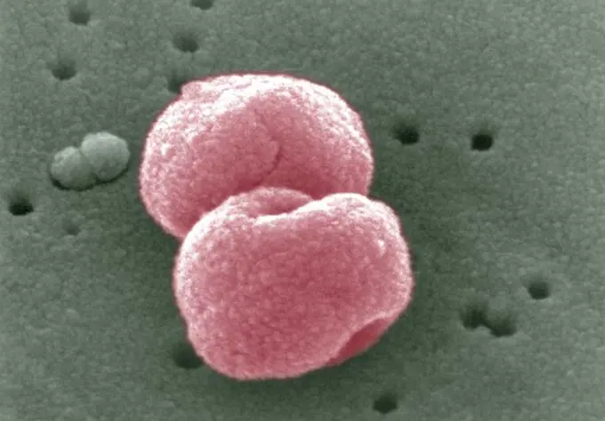
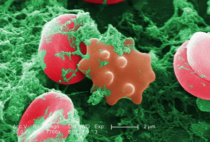
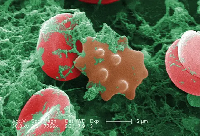
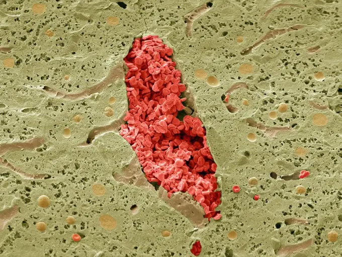
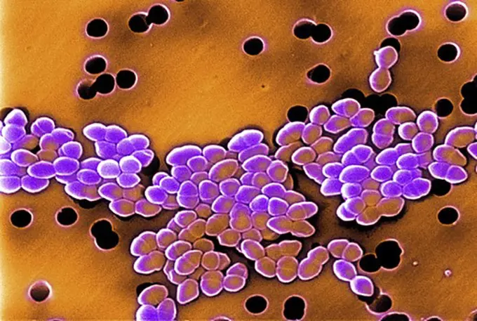
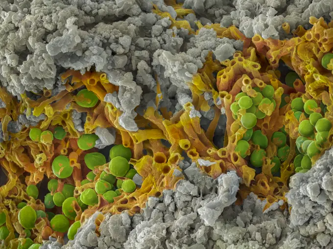
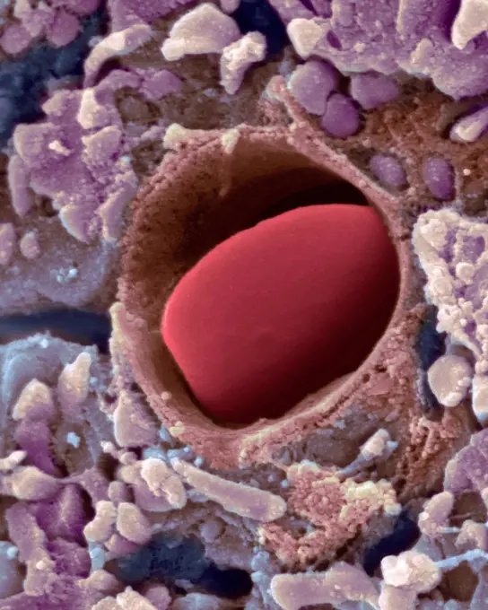
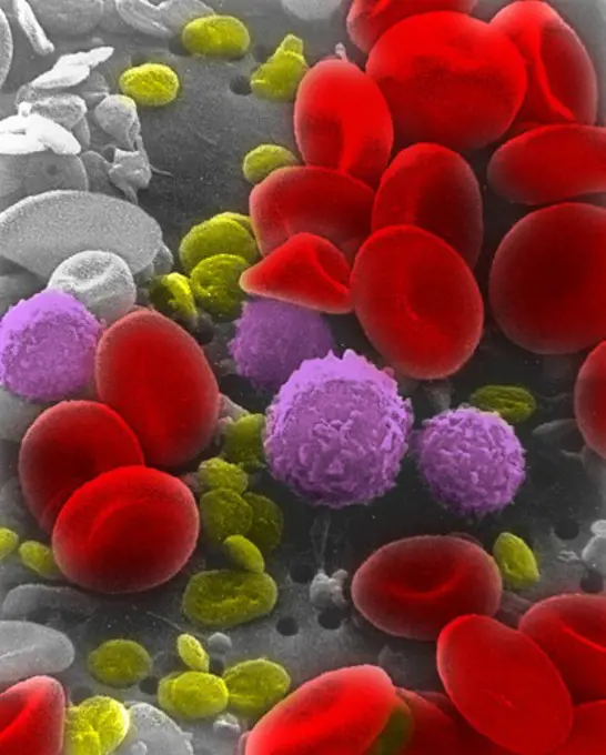
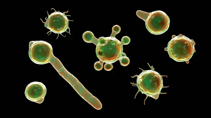
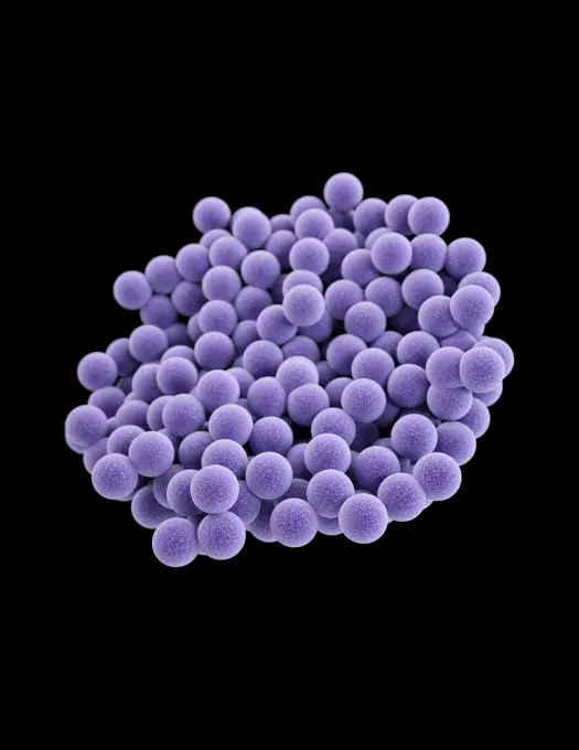
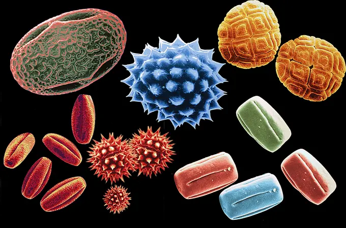
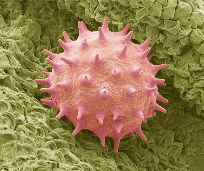
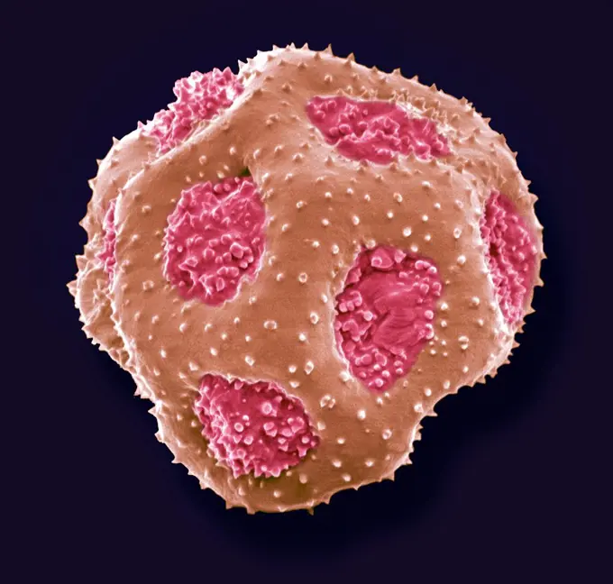
Microscopic Views of Bacteria

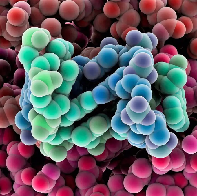
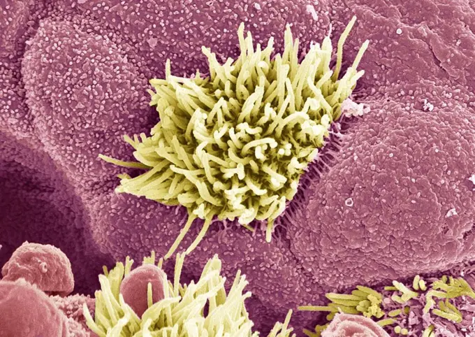
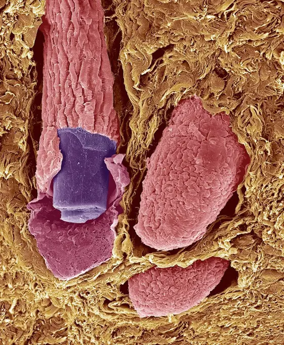
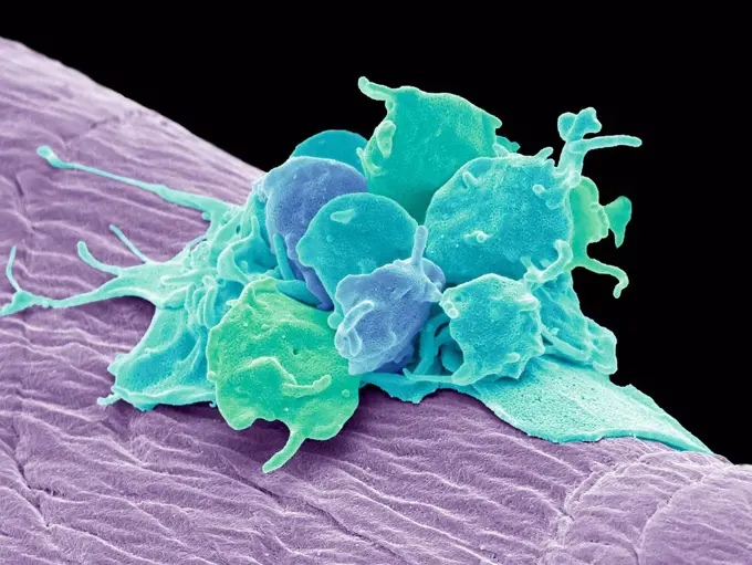
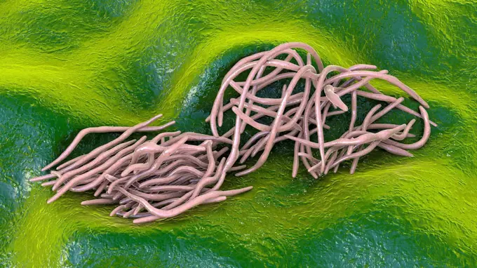
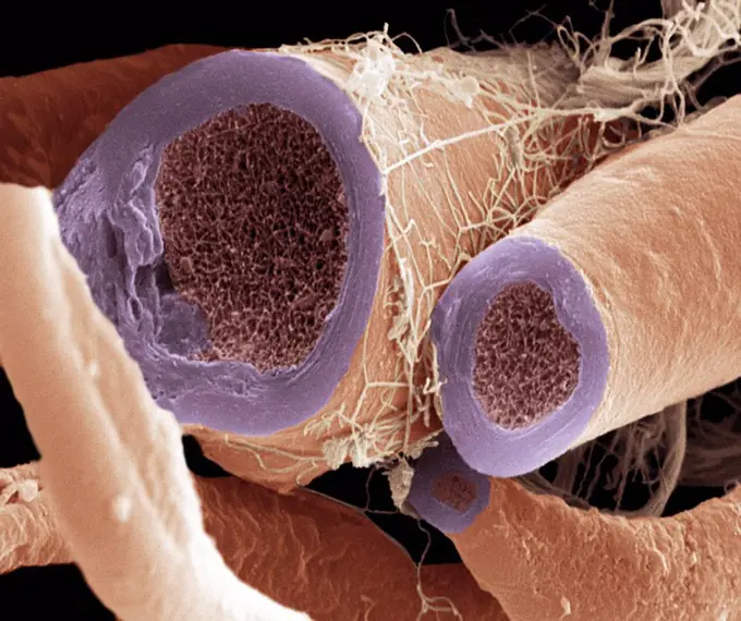
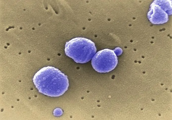
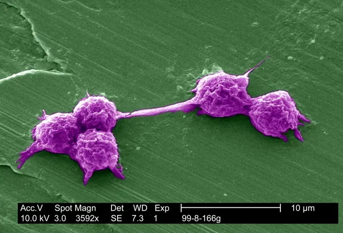
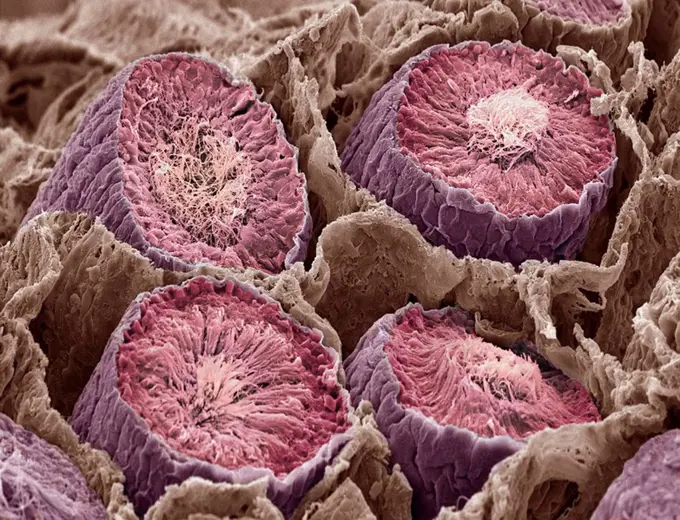
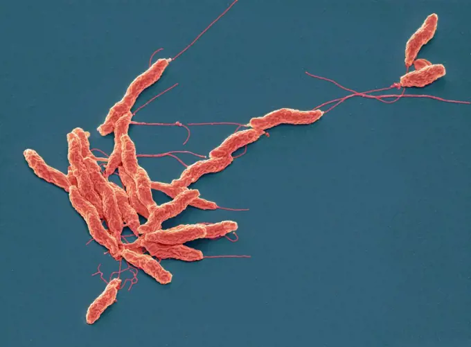
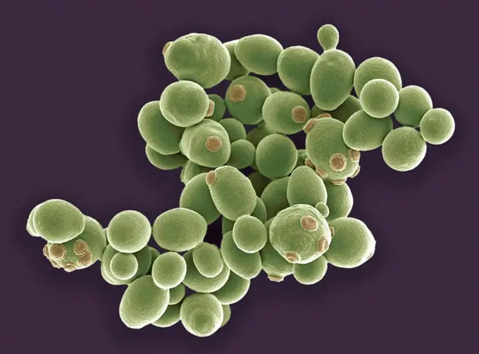
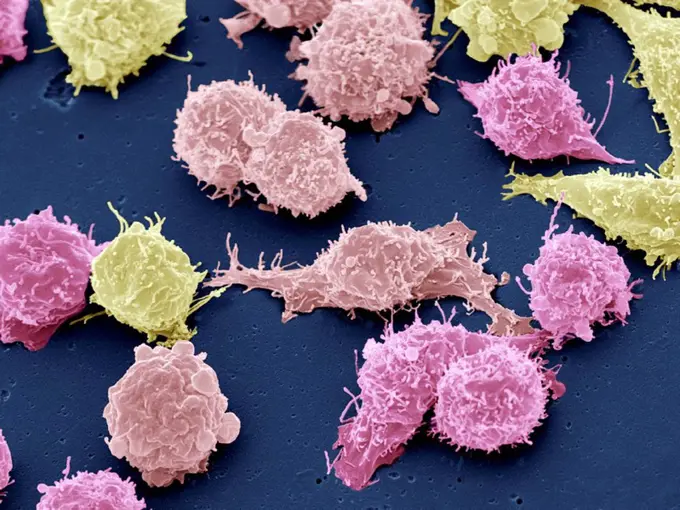
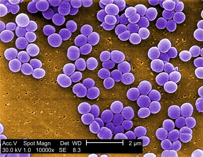
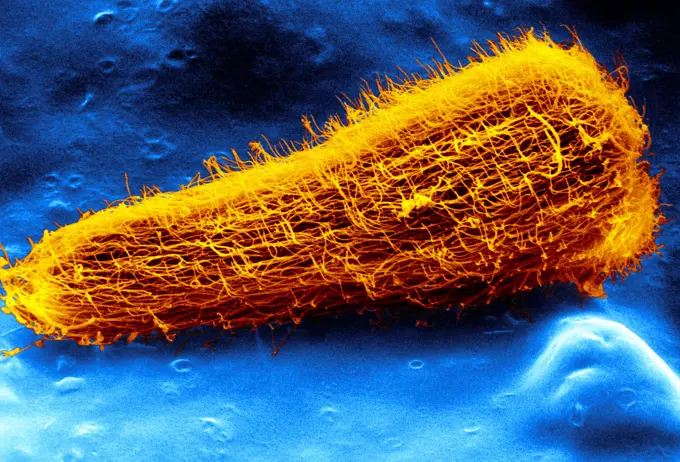
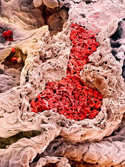
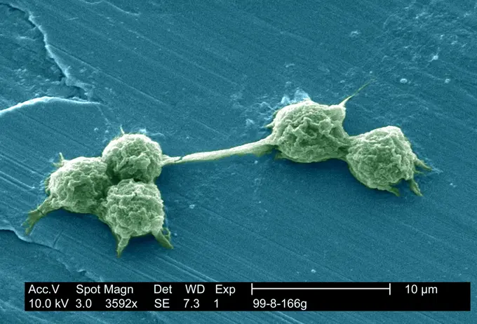
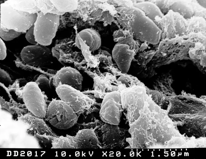
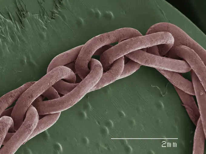
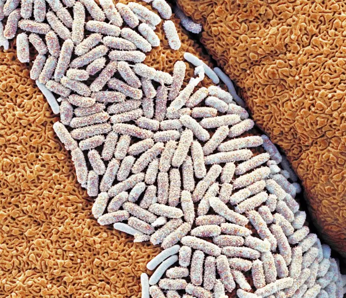
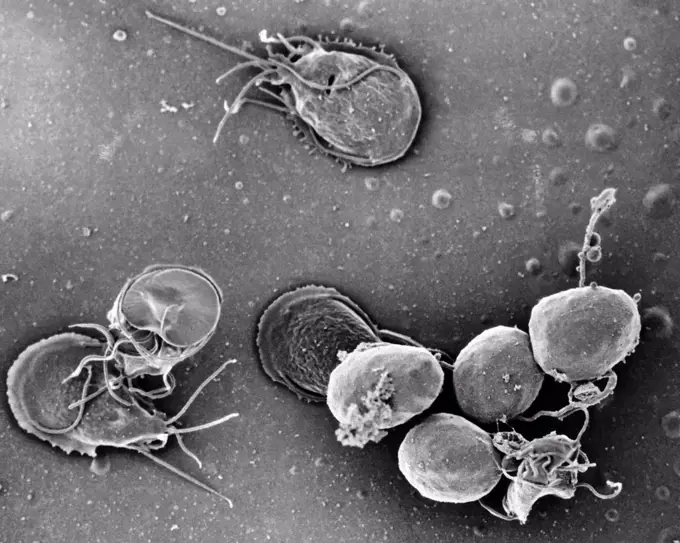
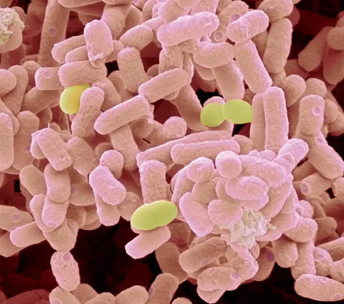
Colorized scanning electron micrographs showcasing various bacteria and human cells, emphasizing intricate details and scientific microscopy.

Escherichia coli bacteria pink, coloured scanning electron micrograph SEM. A few cocci bacteria yellow are also present. Magnification: x40,000 when p...
167 assets in this story
4128R-10582
Tongue bacteria, SEM
4128R-13619971
Used wax dental floss with cheek cells (purple) and bacteria (green) on dental floss fibres (blue), coloured scanning electron micrograph (SEM). Cheek...
4128R-11476199
Protein molecules, computer artwork.
4269-27389
Salmonella infantis. Scanning electron micrograph (SEM) of rod-shaped, motile, Gram-negative Salmonella infantis bacteria. Magnification 18875x. When ingested with spoiled food, the bacteria of the genus Salmonella are responsible for salmonellosis, a small intestine infection (gastroenteritis).
4269-24791
Collagen. Fibroplasts on external surface of intestine ESM (Electron Scanning Microscope)
824-63225910
Colorized scanning electron micrograph of a cell infected with a variant strain of SARS-CoV-2 virus particles (turquoise), isolated from a patient sample. Image captured at the NIAID Integrated Research Facility (IRF) in Fort Detrick, Maryland.
4128R-6971
Trachea lining, SEM
4128-111494390
Hair follicles. Coloured scanning electron micrograph (SEM). The outer layer of hair (the cuticle) has overlapping scales of keratin. These scales are...
4128R-15465093
Activated platelets, SEM
4297-1796
SEM image of Gram-positive Streptococcus pneumoniae bacteria
824-63225869
Colorized scanning electron micrograph of a cell (brown) infected with SARS-CoV-2 virus particles (blue), isolated from a patient sample. Image captured at the NIAID Integrated Research Facility (IRF) in Fort Detrick, Maryland.
824-63194910
This scanning electron micrograph (SEM) depicted a number of red blood cells found enmeshed in a fibrinous matrix on the luminal surface of an indwelling vascular catheter, Magnified 7766x. In this instance, the indwelling catheter was a tube that was left in place creating a patent portal directly into a blood vessel. The erythrocyte in the center had undergone the process of crenation, whereupon, it developed a number of cell wall projections, thereby, transforming it into what is termed an acanthocyte, or spur cell.
4297-1465
Scanning electron micrograph of red blood cells found enmeshed in a fibrinous matrix on the luminal surface of an indwelling vascular catheter
4384-172
Under a moderate magnification of 2000X, this digitally-colorized scanning electron micrograph (SEM) of an untreated water specimen extracted from a wild stream mainly used to control flooding during inclement weather, revealed the presence of unidentified organisms, which included bacteria, protozoa, and algae. In this particular view, a single copepod-like microorganism was seen occupying the field of view, which seemed to be encased in an outer shell of armour-like plates, or scales. Looking
4128R-4069
Liver vein, SEM
4297-1883
Scanning electron microscopic image of vancomycin-resistant enterococci bacteria
4128R-12573027
Red blood cells. Coloured scanning electron micrograph (SEM) of red blood cells (RBCs, erythrocytes). Red blood cells are biconcave, disc-shaped cells...
4269-24748
Sinusoid. Blood capillary between hepatic cells Red blood corpuscules (red corpuscles) and fibrine filaments SEM (Scanning Electron Microscope)
4297-1760
Colorized scanning electron micrograph of E.coli O157:H7 at a magnification of 3418x.Escherichia coli bacteria of the strain O157:H7.E.coli O157:H7 is one of hundreds of strains of this bacterium
4128-16171819
Bacteria from a human beard. Coloured scanning electron micrograph (SEM) of bacteria cultured from a human beard. A European study has found that the ...
4070-48287222
False-coloured scanning electron micrograph of a cleaved sample of a crustose lichen growing on grey limestone, the orange are fungal threads and the green shows algal cells. The fungal threads anchor the algae in place, close to the surface of the lichen to optimise their position for photosynthesis. Size in real life: 0.14mm. Derbyshire, UK. February.
4128-15659921
Heart capillary, SEM
4297-1793
Colorized scanning electron micrograph of human blood cells showing red blood cells (red) white blood cells (purple) and platelets (yellow)
4128-17857602
Round worms, computer illustration. Roundworms, or nematodes, include numerous free-living and pathogenic species. Nematodes that commonly parasitise ...
4128R-4520
Nerve fibres, SEM
4297-1794
SEM image of Gram-positive Streptococcus pneumoniae bacteria
824-63123769
Scanning electron micrograph of a laboratory-grown potable water biofilm, with the presence of Hartmanella vermiformis cysts. Aquatic bacteria were grown as biofilm on steel for one week. Hartmanella vermiformis protozoans were then added, which phagocytized the bacteria, that multiplied within vesicles and became cysts in which the bacteria will live until they rupture.
4128R-13620174
Coloured scanning electron micrograph (SEM) Pet dander: dog hair, cat hair and dead skin. Dog and cats produce human allergens. Pet dander is the mate...
4128-28767191
Conidiobolus coronatus microscopic fungi, illustration.
824-63223144
Scanning electron micrograph of filamentous Ebola virus particles attached and budding from chronically infected VERO E6 cells (15,000x magnification). Image captured at the NIAID Integrated Research Facility in Ft. Detrick, Maryland. Credit: NIAID.
824-63194381
Scanning electron micrograph of Ebola virus budding from the surface of a Vero cell (African green monkey kidney epithelial cell line. Produced by the National Institute of Allergy and Infectious Diseases (NIAID), under a very-high magnification, this digitally-colorized scanning electron micrograph (SEM) depicts a number of filamentous Ebola virus particles (red) that had budded from the surface of a VERO cell (blue-gray) of the African green monkey kidney epithelial cell line. Ebola hemorrhagic fever (Ebola HF) is one of Viral Hemorrhagic Fevers. It is a severe, often fatal disease in humans and nonhuman primates (such as monkeys, gorillas, and chimpanzees). Ebola HF is caused by infection with a virus of the family Filoviridae, genus Ebolavirus. When infection occurs, symptoms usually begin abruptly. The first Ebolavirus species was discovered in 1976 in what is now the Democratic Republic of the Congo near the Ebola River. Since then, outbreaks have appeared sporadically.
824-63227420
This image shows how the mouth design makes ticks generally difficult to remove once they attach to feed.
4384-248
Scanning Electron Micrograph of Untreated Water Sample
4128-V58562062
824-63227404
Scanning electron microscopy image illustrates tiny extracellular links between stereocilia of an inner ear hair cell. One type of link, the tip link, rests the top of the shorter stereocilia to the side of a longer stereocilia and is crucial for hearing. The yellow dots in the inset are the immuno-OR particles that label specific proteins comprising the spike binding.
4128R-7346
Sperm production, SEM
4128R-13022176
Campylobacter jejuni bacteria. Coloured scanning electron micrograph (SEM) of a Campylobacter jejuni bacteria. Campylobacter jejuni is a Gram-negative...
824-63225853
Colorized scanning electron micrograph of a cell (teal) infected with SARS-CoV-2 virus particles (orange), isolated from a patient sample. Image captured at the NIAID Integrated Research Facility (IRF) in Fort Detrick, Maryland.
824-63223146
Colorized scanning electron micrograph of Ebola virus particles (green) in extracellular space between infected African green monkey kidney cells. Image captured and color-enhanced at the NIAID Integrated Research Facility in Ft. Detrick, Maryland. Credit: NIAID.
4297-1451
Under a moderately-high magnification of 8000X, this scanning electron micrograph (SEM) depicted a grouping of Gram-negative Legionella pneumophila bacteria
4128R-5413
Yeast cells, SEM
4128R-5657
Cancer cells, SEM
4128-15982693
Migrating sarcoma cancer cells. Coloured scanning electron micrograph (SEM) of cancer cells migrating down pores in the filter on which they have been...
824-63194417
Under a high magnification of 10,000x, this scanning electron micrograph (SEM) shows a strain of Staphylococcus aureus bacteria taken from a vancomycin intermediate resistant culture (VISA). Under SEM, one can not tell the difference between bacteria that are susceptible, or multidrug resistant, but with transmission electron microscopy (TEM), at least with VISA isolates one can see a thickening in the cell wall that may attribute to their reduced susceptibility to vancomycin. VISA and VRSA are specific types of antimicrobial-resistant staph bacteria. While most staph bacteria are susceptible to the antimicrobial agent vancomycin some have developed resistance. VISA and VRSA cannot be successfully treated with vancomycin because these organisms are no longer susceptibile to vancomycin. However, to date, all VISA and VRSA isolates have been susceptible to other Food and Drug Administration (FDA) approved drugs.
824-63128388
Miracidium larva of the liver fluke (fasciola hepatica) viewed in SEM.
4128R-2008
Lung blood vessel, SEM
4128R-13620207
Coloured scanning electron micrograph (SEM) Cat hair (Felis silvestris catus). Cats have four types of hair. There are short fluffy hairs called secon...
824-63191194
This illustration depicts a three-dimensional (3D) computer-generated image of a group of methicillin-resistant Staphylococcus aureus (MRSA) bacteria, which were arranged in a cluster. The artistic recreation was based upon scanning electron micrographic imagery. Methicillin-resistant Staphylococcus aureus (MRSA) causes a range of illnesses, from skin and wound infections to pneumonia and bloodstream infections that can cause sepsis and death. Staph bacteria, including MRSA, are one of the most common causes of healthcare-associated infections. Resistance to methicillin and related antibiotics (e.g., nafcillin, oxacillin) and resistance to cephalosporins are of concern. CDC estimates 80,461 invasive MRSA infections and 11,285 related deaths occurred in 2011. An unknown but much higher number of less severe infections occurred in both the community and in healthcare settings.
4128R-23746
Dental plaque. Coloured scanning electron micrograph SEM of dental plaque. Plaque consists of a film of bacteria embedded in a glycoprotein matrix. Th...
4128R-11288975
Dust mite, computer artwork.
4269-1216
Scanning electron microscope view of different pollens, responsible for respiratory allergies.
4128R-7144
Chinese hibiscus pollen, SEM
4128R-9673
Primula pollen, SEM
824-63123770
Scanning electron micrograph of a laboratory-grown potable water biofilm, with the presence of Hartmanella vermiformis cysts. Aquatic bacteria were grown as biofilm on steel for one week. Hartmanella vermiformis protozoans were then added, which phagocytized the bacteria, that multiplied within vesicles and became cysts in which the bacteria will live until they rupture.
824-63223275
Scanning electron micrograph depicting a mass of Yersinia pestis bacteria (the cause of bubonic plague) in the foregut of the flea vector. Credit: NIAID.
4128R-11286536
Flu virus particle, computer artwork. In the virus' envelope are two types of protein spike, haemagglutinin (H, pink) and neuraminidase (N, orange), w...
4384-251
Scanning Electron Micrograph of Untreated Water Sample
1439-57940152
A closeup view of a chain link under a microscope displaying intricate textures
824-63191234
Scanning electron micrograph of just-divided HeLa cells. Zeiss Merlin HR-SEM.
824-63191235
Scanning electron micrograph of just-divided HeLa cells. Zeiss Merlin HR-SEM.
4128R-13477
E. coli bacteria. Coloured scanning electron micrograph SEM of Escherichia coli bacteria white on a gecko´s tongue. Magnification: x8000 when printed ...
4384-141
This is a  scanning electron micrograph (SEM) of an in vitro  Giardia lamblia culture, which had been cultivated in bile-free TYI-S-33 medium for 48 hrs, then incubated 24 hrs with 10 mg/ml bovine bile in order to stimulate cyst formation.  This photograph contains both trophozoites, and a cluster of maturing cysts (bottom right).  At far left, the two trophozoites-staged organisms are positionally situated opposite to one another, with the farthest left  G.  lamblia  displaying its dorsal, or u
4128-V58562138
4128R-11474477
Neurone. Scanning electron micrograph (SEM) of a PC12 neurone in culture.The PC12 cell line, developed from a pheochromocytoma tumor of the rat adrena...
4128-V58562082
4128R-15466816
Bacteria found on mobile phone, SEM
4128-111453067
Ovarian cancer cells, sem
PREVIOUS
of 2
NEXT
2401 S. Ervay, Suite 206
Dallas, Texas 75215
United States
Get Started
Free ResearchMy BoardsMy Cart
For Creators
How To License Your ContentContributor PortalFrame of Mind
Resources
API accessPricing
Contact
+1 866 236 0087help@viewpictures.co.uk Contact form
©2026 View Pictures. All Rights Reserved. -A
This site is protected by reCAPTCHA and Google Privacy Policy and Terms of Service apply.