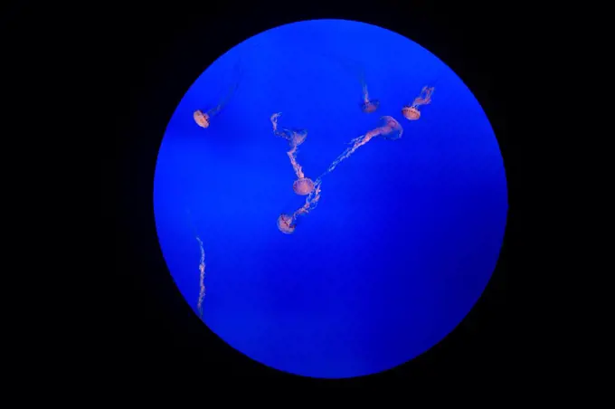
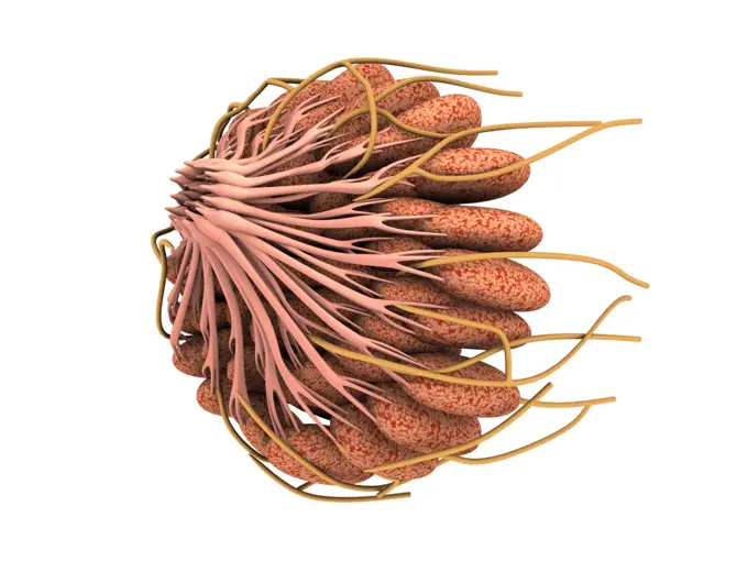
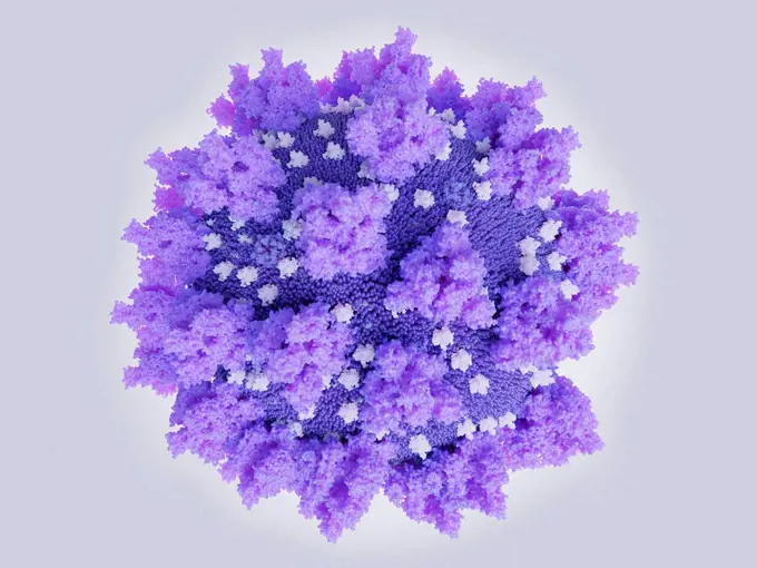
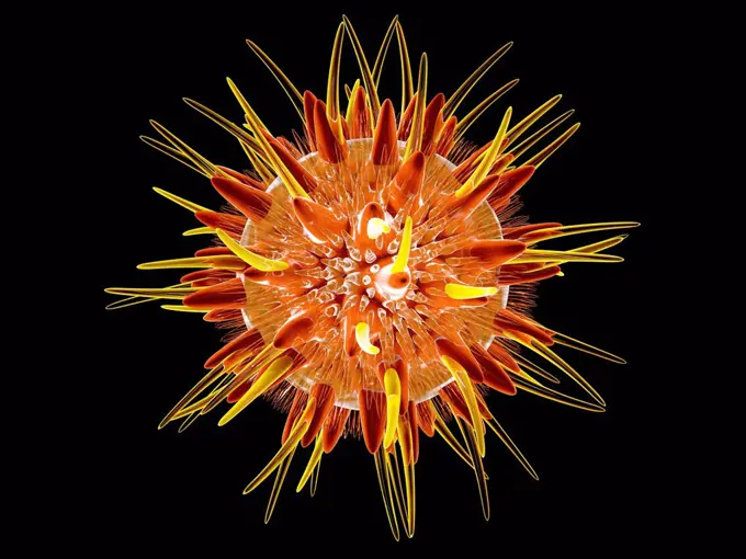
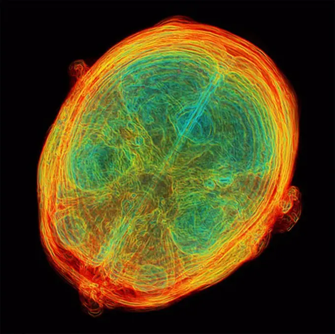
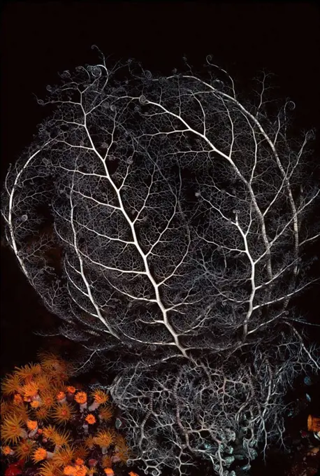
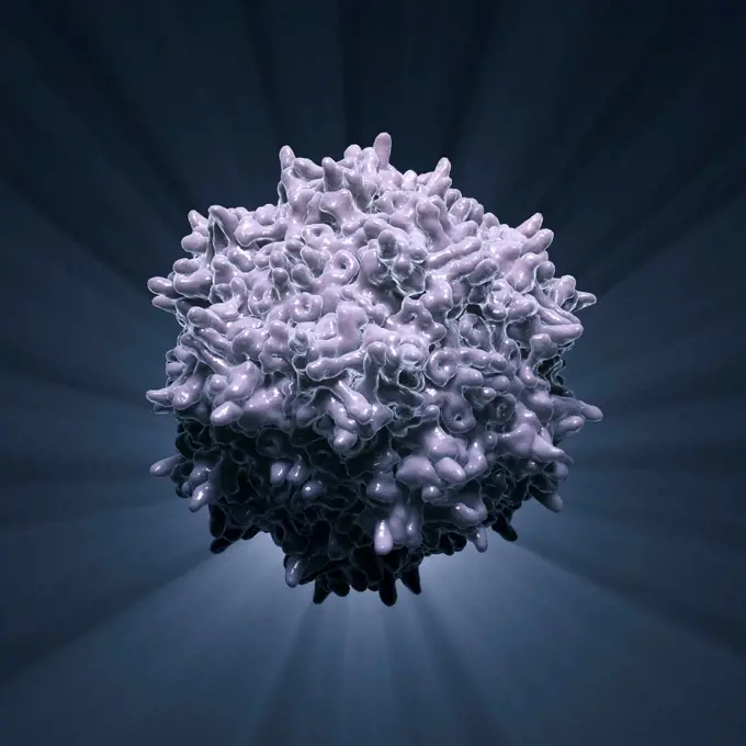
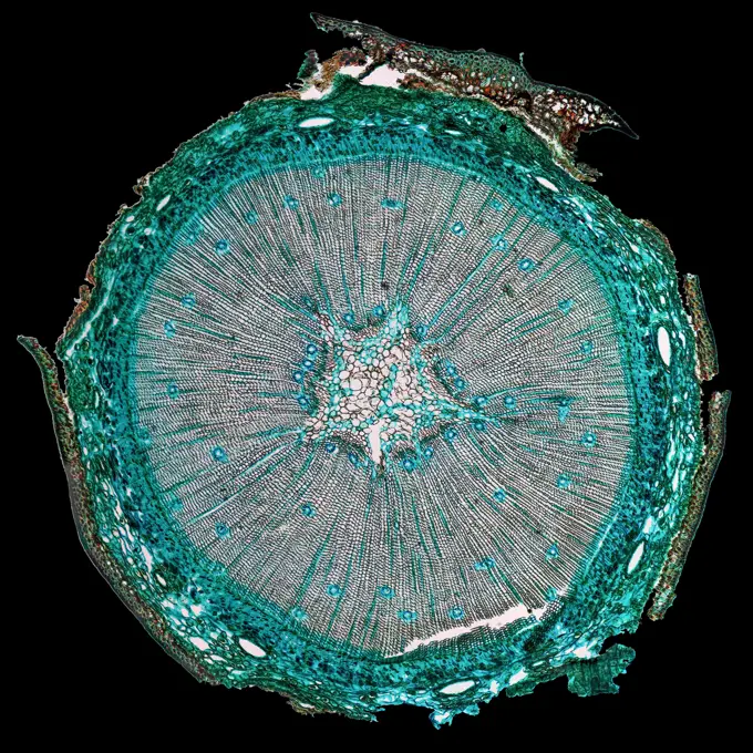
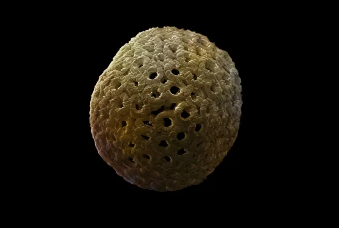
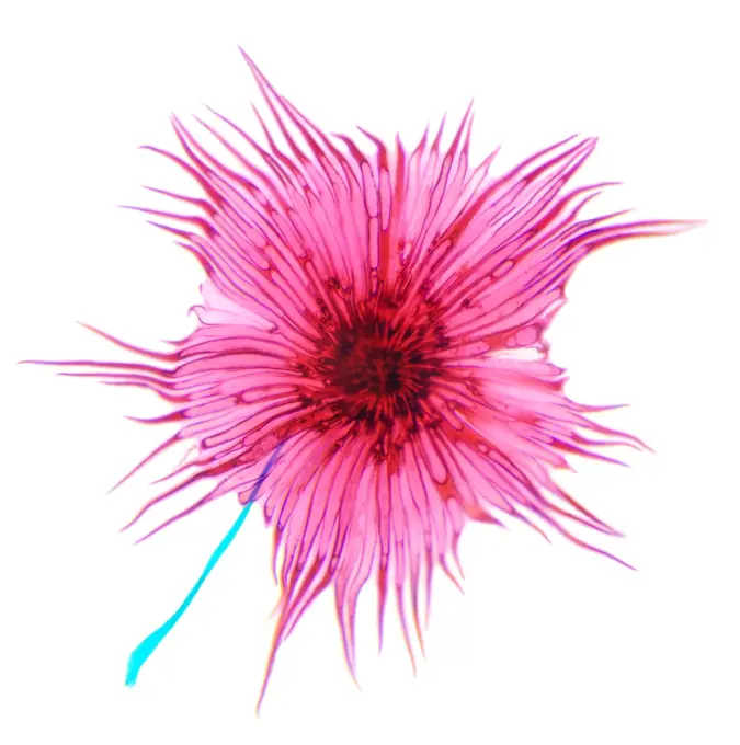
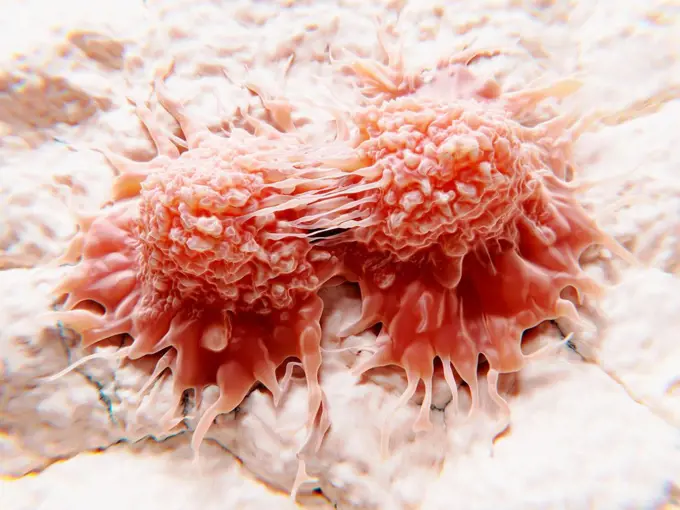
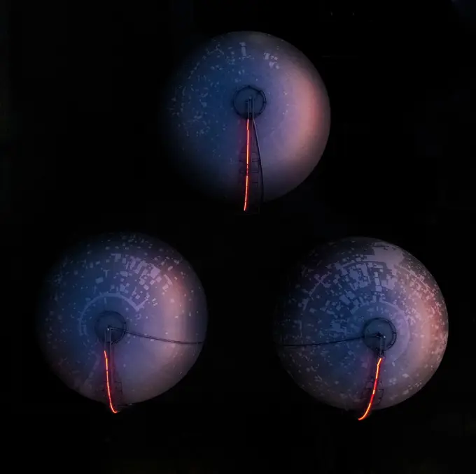
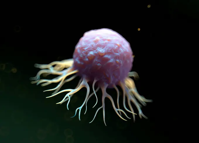
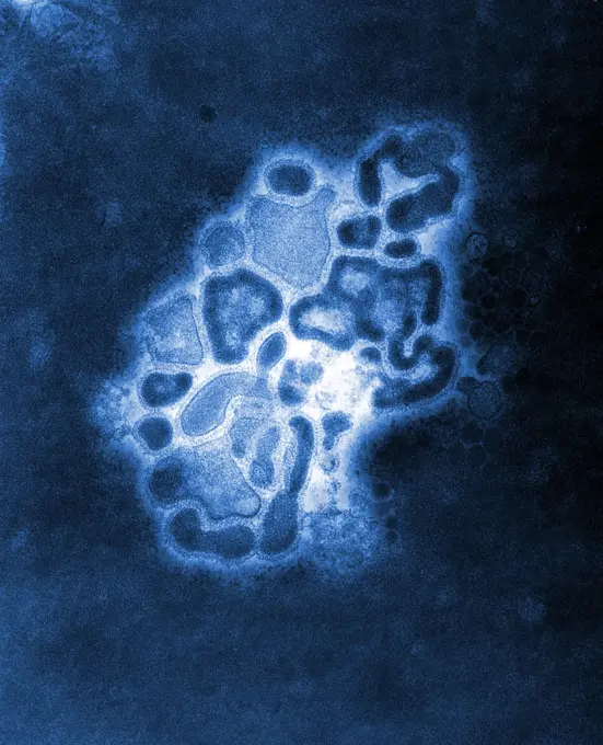
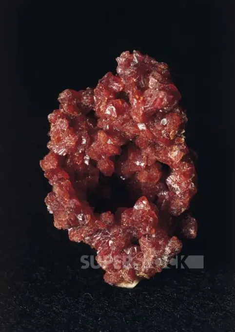
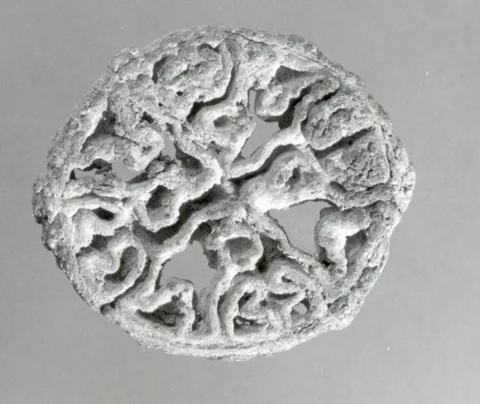
My Boards
View Pictures
  • My Boards

Molecular Structures and Diseases

Artistic representations of various molecular structures, including viruses and dendrimers, emphasizing the complexity of biological entities.

Virus, illustration
Virus, illustration
134 assets in this story
4413-53203
Jellyfish Monterey Bay Aquarium CaliforniaUSA
4239R-20483476
Conceptual image of female breast anatomy.
4128-V58576255
4128-15814059
Illustration of a SARS-CoV-2 coronavirus (previously 2019-nCoV) that was first identified in Wuhan, China, in December 2019. It is an enveloped RNA (r...
4128R-11287681
Diverticulitis. Coloured X-ray of the colon of a patient with diverticulitis. A diverticulum is a small pouch-like structure that protrudes out of wea...
4443-75471415
Ectocarpus brachiatus, 1844., 1844., Halstead Place, Sevenoaks, England. Publisher, Anna Atkins. Plants in art, Algae, Great Britain, Photography, Cyanotypes., 1 photograph
4201-78503
Ecuadorian Mantid (Pseudocanthops sp) matches the color of the lichen from which it hangs
1815R-13385689
3D Illustration of a virus
1848-54746145
Dendritic cell of the immune system detecting a virus, illustration
1532R-36076
A celeriac root
4128R-6390
Brain scans. 3D_magnetic resonance imaging MRI scans of a healthy brain. Axial horizontal sections through the brain from the top of the brain towards...
1760-7755
Indonesia, basket seastar with extended feeding arms
4378-3063
Atomic structure of Adeno-Associated Virus (PDB 1LP3), a vector for human gene therapy.
1848-49565933
Rhodochrosite, Catamarca Province, Argentina Rhodochrosite is a manganese carbonate mineral that ranges in color from light pink to bright red
6188-55638585
Melone Melonenscheibe Copyright: xZoonar.com/MikexRichterx 1457449
4128R-13386224
Bacteria, illustration.
6188-55585071
Pine Wood micrograph High resolution light photomicrograph of pine tree wood cross section seen through a microscope Copyright: xZoonar.com/ClaudioxDiviziax 7886942
6188-55645662
Zieralge Micrasterias unter dem Mikroskop Zieralge Micrasterias unter dem Mikroskop Copyright: xZoonar.com/Dr.xNorbertxLangex 14094967
4220-21334521
Scanning Electron Micrograph (SEM): Coccolithophore (Calcareous Nannoplankton), Umbilicosphaera sibogae.     Date:
4128-16224500
Broccoli (Brassica oleracea), X-ray
4070-21284413
Aerial view of Grand Prismatic Springs Midway Geyser Basin. Yellowstone National Park, Wyoming, July 2011.
1525-19842580
Cactus pollen under a microscope, (Stellato-Pilosus W.M.). 400x
4128-19357661
Illustration of a human immunoglobulin G1 (IgG1) antibody. This is a combination of the ribbon structure and molecular surface representations of the ...
4128-19497234
Dividing cancer cells, illustration
4070-28602150
Slime mould (Lamproderma scintillans) super close up of 1mm tall sporangia, Buckinghamshire, England, UK, March Focus Stacked.
6145-43966013
Calcite. minerals. North America; USA; New Jersey; Sussex County; Ogdensburg; Sterling Hill Mine
824-63218211
Fracture of the skull, seen on a radial section scanner.
824-63225810
Colorized scanning electron micrograph of macrophages.
6162-49120128
Aerial view of three reservoir gas tanks.
4128-48286024
Cancer cell, illustration
824-63194485
Under a plate magnification of 37,800X, this colorized transmission electron micrograph (TEM) depicted a strain of swine flu, the A/New Jersey/76 (Hsw1N1) virus, while in the virus' first developmental passage through a chicken egg. Swine Influenza (swine flu) is a respiratory disease of pigs caused by type A influenza that regularly cause outbreaks of influenza among pigs. Swine flu viruses cause high levels of illness and low death rates among pigs. Swine influenza viruses may circulate in swine throughout the year, but most outbreaks among swine herds occur during the late fall and winter months similar to humans. The classical swine flu virus (an influenza type A H1N1 virus) was first isolated from a pig in 1930.
1788-20759856
Minerals: Botryogen
6145-29758805
Compartmented stamp seal ca. late 3rd-early 2nd millennium B.C. Bactria-Margiana Archaeological Complex. Compartmented stamp seal 326792
4128R-14606953
Medical nanoparticles, conceptual illustration
PREVIOUS
of 2
NEXT
2401 S. Ervay, Suite 206
Dallas, Texas 75215
United States
Get Started
Free ResearchMy BoardsMy Cart
For Creators
How To License Your ContentContributor PortalFrame of Mind
Resources
API accessPricing
Contact
+1 866 236 0087help@viewpictures.co.uk Contact form
©2026 View Pictures. All Rights Reserved. -A
This site is protected by reCAPTCHA and Google Privacy Policy and Terms of Service apply.