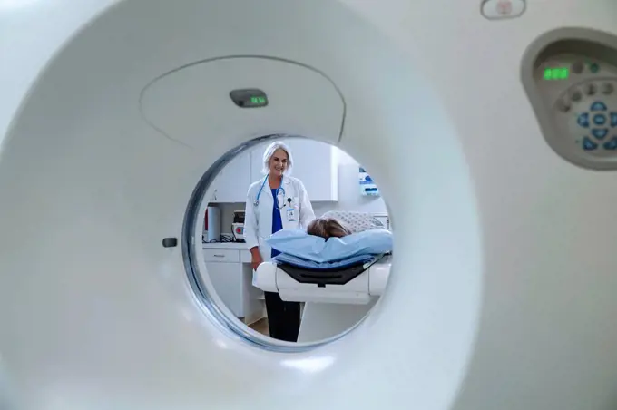
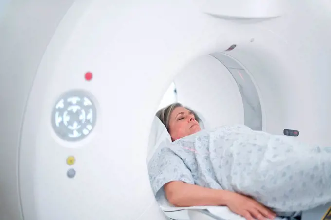
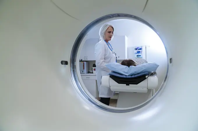
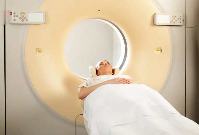
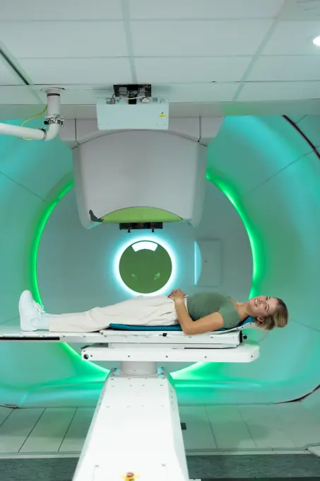
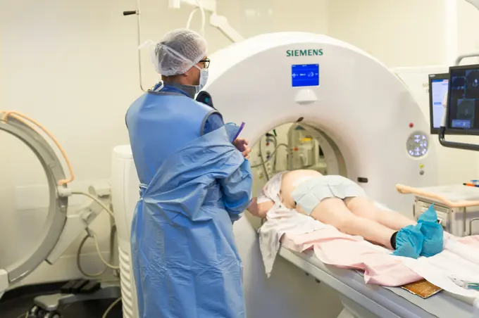
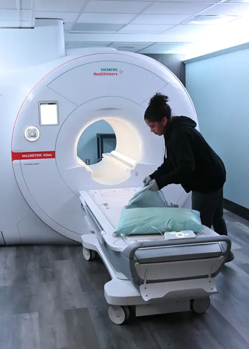
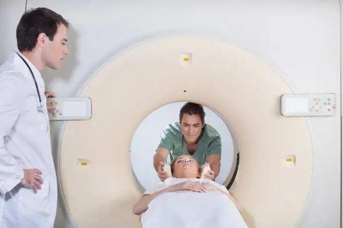
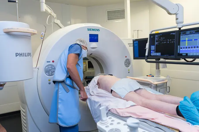
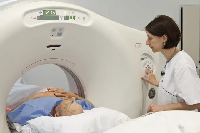
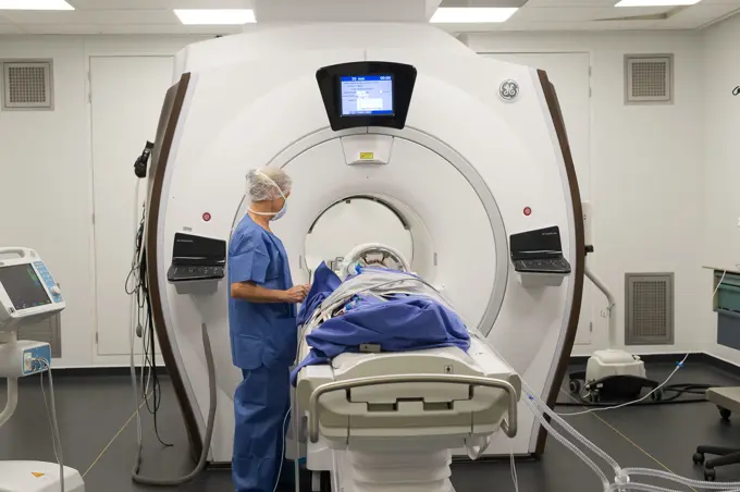
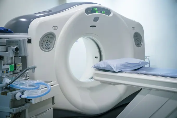
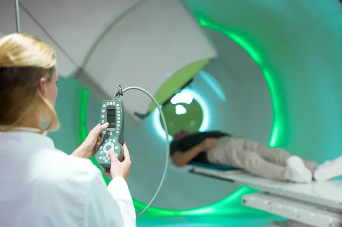
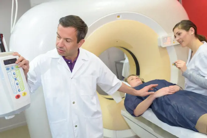
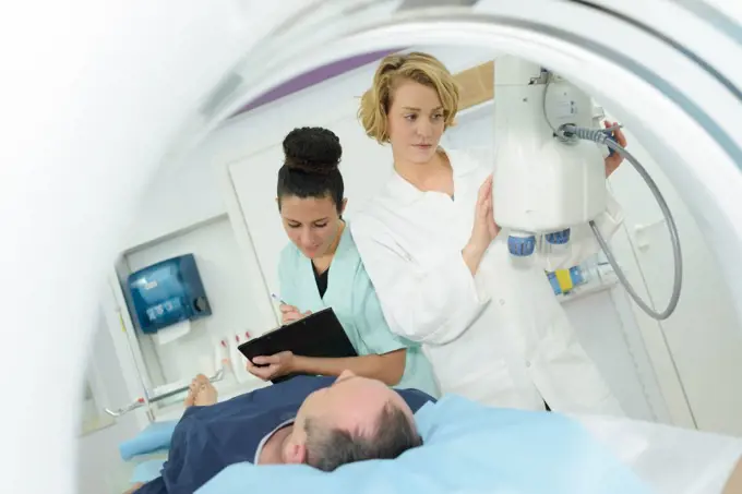
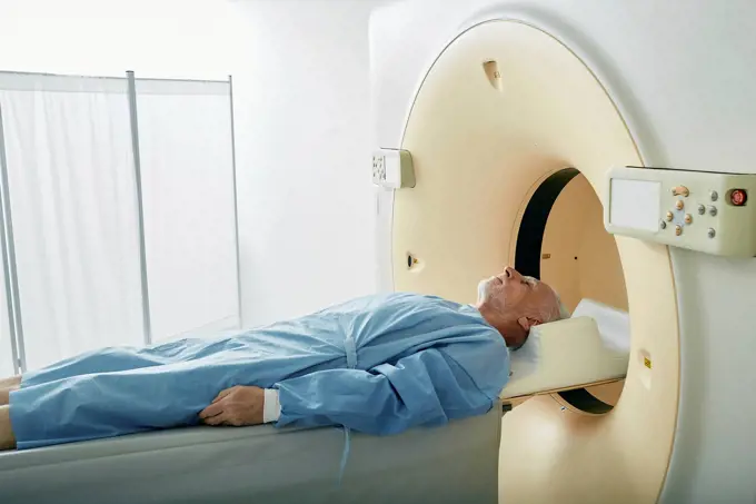
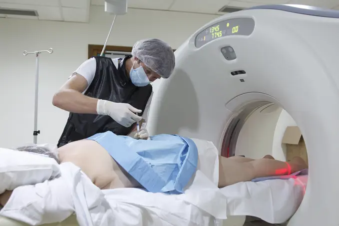
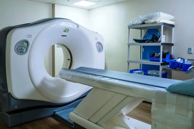
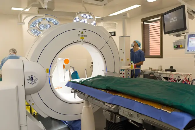
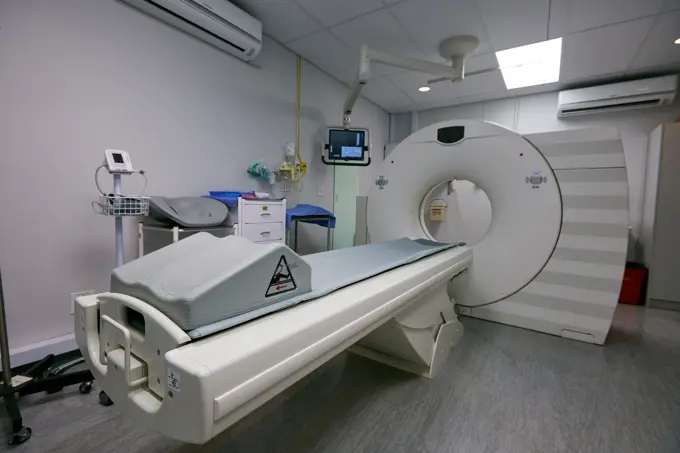
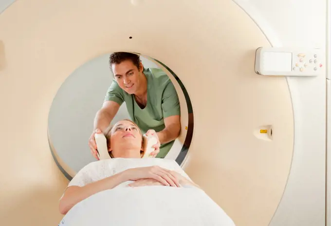
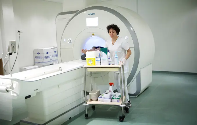
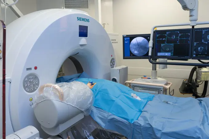
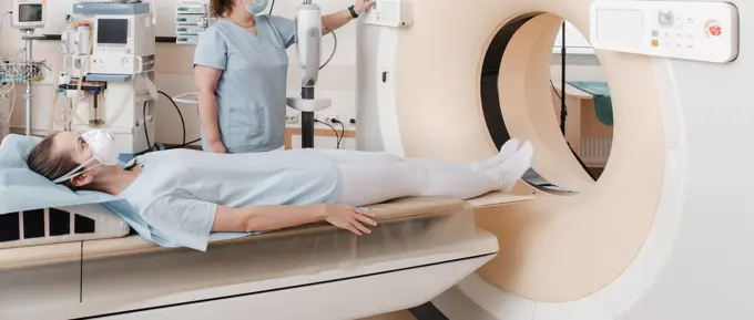
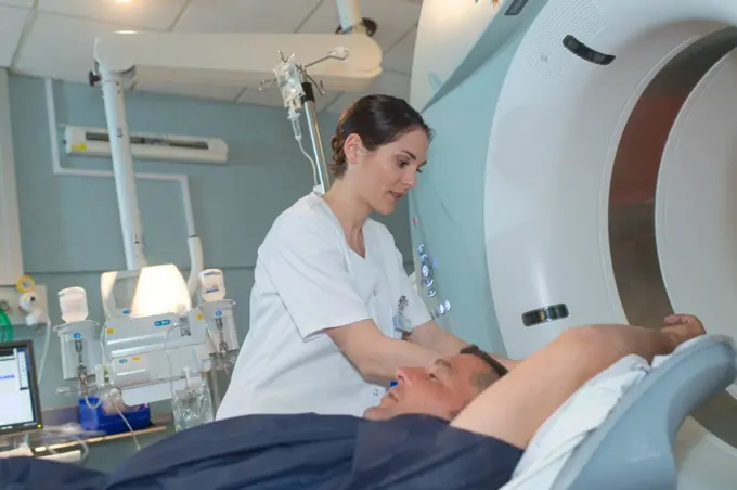
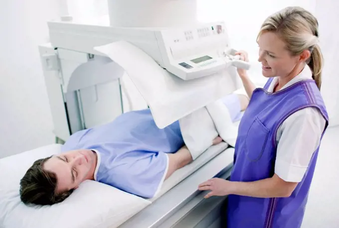
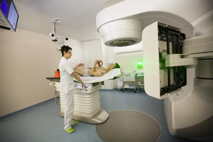
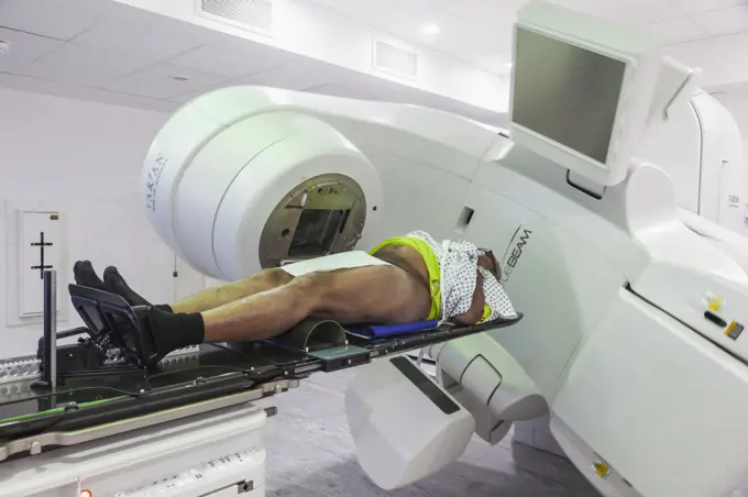
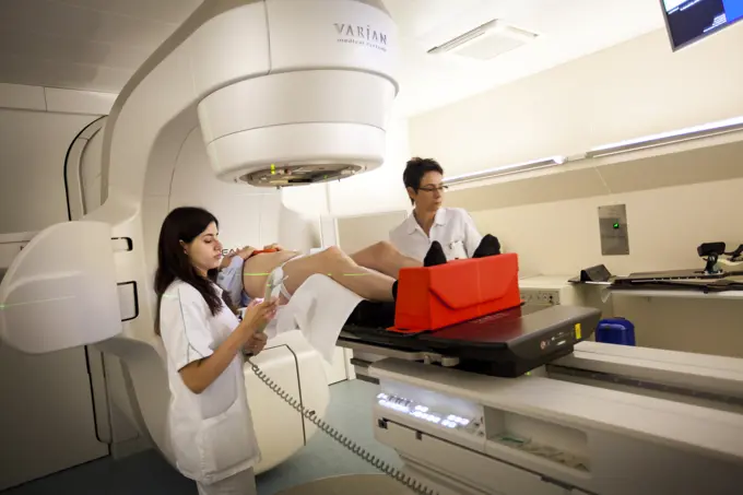
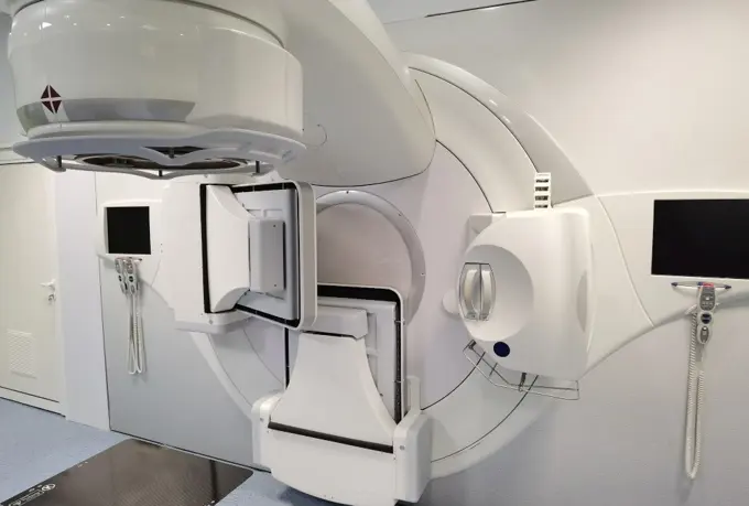
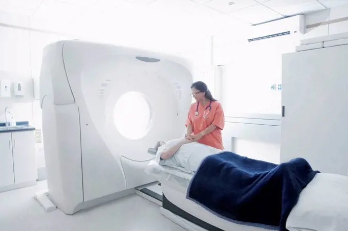
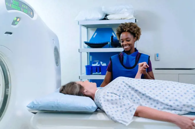
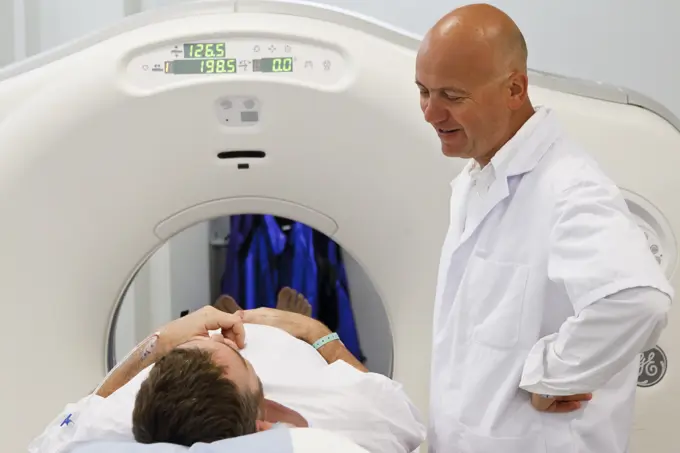
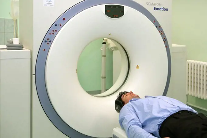
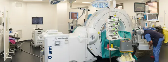
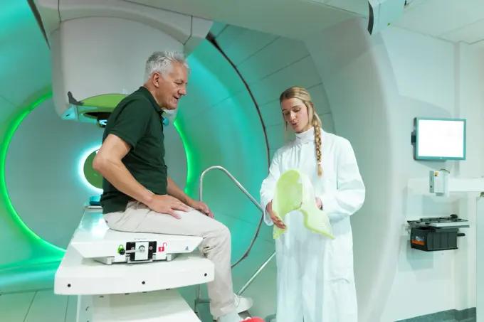
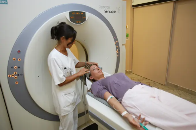
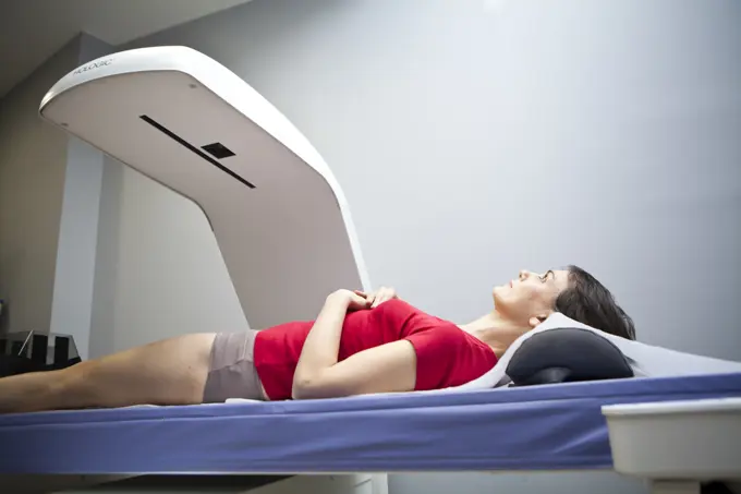
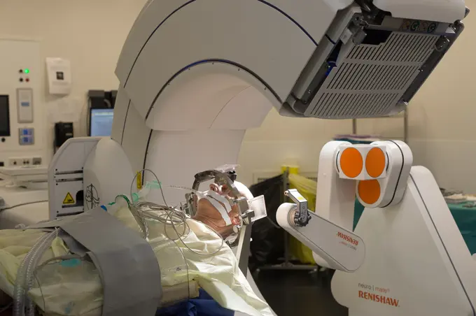
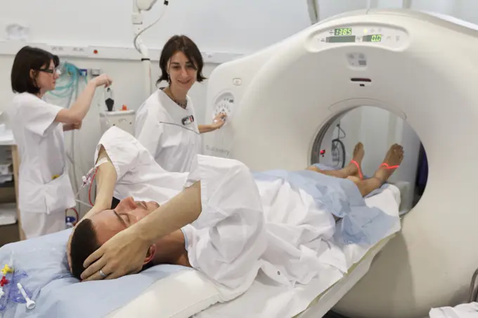
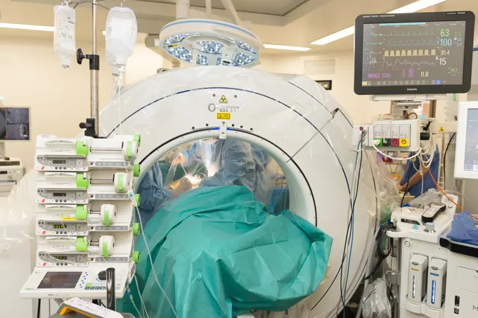
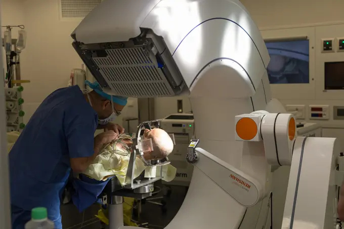
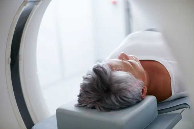
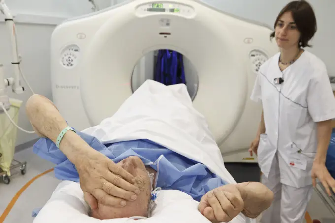
My Boards
View Pictures
  • My Boards

MRI And CT Imaging

Images capturing medical imaging processes, including MRI and CT scans with patients being assisted by healthcare professionals.

Caucasian doctor comforting patient near scanner
189 assets in this story
824-63181512
Digital medical imaging centre in Paris, France. Skull MRI.
824-63161113
MRI or magnetic resonance imaging of the skull.
4128R-14588495
Female patient in MRI scanner
1589R-14623142
Caucasian doctor talking to patient near scanner
4269-40469
Female patient undergoing a CT scan, Croix Saint Simon hospital, Paris,
824-63161120
MRI or magnetic resonance imaging of a patient's hips.
1589R-14623146
Caucasian doctor comforting patient near scanner
4269-33759
Radiotherapy radiographer with a male patient treated for a lung cancer with respiration-gated radiotherapy. Latest generation particle accelerator. Radiotherapy center pole of Oncology Hospital of Bordeaux. Hospital Haut-Leveque.
1525-25072313
A woman having a CT Scan taken
1525-57461855
doctor and nurse preparing patient for mri scan in hospital
1815-69224380
Patient undergoing proton therapy in a modern medical facility
1589R-14623170
Caucasian patient laying on scanner table
824-63214239
Reportage in the interventional scanner service in Pasteur 2 Hospital, Nice, France. Vertebroplasty- cementoplasty operation to treat a vertebral facture. The technique consists of injecting, under image guidance, a biocompatible cement mixture into the weakened vertebra to consolidate it.
1848-53914847
Computer tomography, CT, in a hospital
1525-25072365
Doctor instructing technician while preparing patient for MRI scan
6145-58911708
A radiologic technologist prepares a magnetic resonance imaging scanner for a patient at Kimbrough Ambulatory Care Center on Fort Meade, Maryland, Feb. 12. (Defense Health Agency
1525-25072364
Male nurse preparing patient for CT scan
867-1626
Patient having an MRI scan
824-63214243
Reportage in the interventional scanner service in Pasteur 2 Hospital, Nice, France. Vertebroplasty- cementoplasty operation to treat a vertebral facture. The technique consists of injecting, under image guidance, a biocompatible cement mixture into the weakened vertebra to consolidate it. The radiologist wears a protective X-ray apron to protect against the rays.
824-63211808
Report from the radiotherapy department of a hospital in Savoie. Radiotherapy session to treat brain metastases. A mask is applied to the patient to prevent movement during the treatment. The treatment is carried out with the Truebeam.
824-63173367
Ct scanner examination
1899-54027503
Stereotactic Neurosurgery operation, Pasteur 2 Hospital, Nice, France, Treating Parkinsons disease through deep brain stimulation, by implanting electrodes in brain, modulating abnormal cerebral electrical activity, Carrying out the pre-op MRI scan. The scan will enable electrode targets and trajectories to be established. (Photo by: BSIP/UIG via Getty Images)
1589R-14623255
Scanner at hospital
1815-69224370
Medical professional operating proton therapy equipment for cancer treatment in a clinical setting
4269-20404636
MRI scanner, Paris, France.
1525-25970714
professional with patient and doctor using cat scan
824-63187696
Reportage in a hospital. MRI scan.
1525-23688839
patient on mri machine while two female doctors operating it
824-63206535
Reportage in a radiology service in a hospital in Haute-Savoie, France. MRI scan.
1525-24502014
male patient undergoing test at ct scanner machine
4128-24794753
Patient undergoing CT scan
824-63173380
Lumbar vertebra, infiltration
1589R-14623133
Scanner in hospital
824-63181505
Digital medical imaging centre in Paris, France. Skull MRI.
824-63214493
Reportage in the stereotactic neurosurgery operating theatre in Pasteur 2 Hospital, Nice, France. Treating Parkinsons disease through deep brain stimulation. This technique consists of implanting electrodes in the brain, which modulate abnormal cerebral electrical activity. The room is equipped with a pre-op O-arm scanner and NeuroMate, a stereotactic robot. The robot enables surgeons to optimize and increase reliability of their operating technique, and the O-arm enables real time 3D images to be obtained to improve the precision, safety and efficiency of the operation. Preparing the O-arm scanner.
1848-54953289
Computer tomography, CT or cat-scan, computer-assisted tomography, hospital, Germany, Europe
4197-73070576
Healthcare, mri and machine in room at hospital for treatment, cancer or radiology for diagnosis. Ct scan, medical ward and equipment for oncology testing with consultation or checkup in empty clinic
1525-25072327
A CT Scan Technician preparing a patient for scanning
4269-5309
Lens Hospital, France. Patient undergoing MRI scan in the first high field ultra short bore MRI. The high height of the bore of the device makes it accessible for obese patients (weight up to 250 kg), pregnant women or claustrophobic patients.
824-63206522
Reportage in a radiology service in a hospital in Haute-Savoie, France. MRI scan.
824-63179446
Reportage in the A&E department of Robert Ballanger general hospital, France. Chest x-ray in the trauma centre.
824-63210925
Report on PET imaging. Positron Emission Tomography, or PET Scan, is used for the diagnosis and monitoring of cancer patients. This method makes it possible to detect a tumor thanks to a radioactive product which accumulates in an excessive way in the cells presenting a pathological hypermetabolism.
4269-40482
Male patient undergoing a CT scan, Croix Saint Simon hospital, Paris, France.
6188-60030443
Medical CT or MRI Scan in the modern hospital laboratory. Interior of radiography department. Technologically advanced equipment in white room. Magnetic resonance diagnostics machine Medical CT or MRI Scan in the modern hospital laboratory. Interior of radiography department. Technologically advanced equipment in white room. Magnetic resonance diagnostics machine. Copyright: xZoonar.com/MariyaxLvovax 17877496
4269-14040
A patient with Parkinson's disease is treated with thalamic stimulation : electrods are inserted into the brain and fed with a thalamic pacemaker implanted in the thorax. This neurosurgery is performed with the O-ARM Imaging System. This motorized intra-operative multi-dimensional surgical imaging platform provides real-time high resolution fluoroscopy and 3D CT scan imaging, and allows high precision and safety surgical procedures. Here, the stereotaxic frame is placed on patient's head. Pr Jea
1848-53791489
Computed tomography, CT scanner, Germany, Europe
824-63210924
Report on PET imaging. Positron Emission Tomography, or PET Scan, is used for the diagnosis and monitoring of cancer patients. This method makes it possible to detect a tumor thanks to a radioactive product which accumulates in an excessive way in the cells presenting a pathological hypermetabolism.
1899-54027384
Interventional scanner service, Pasteur 2 Hospital, Nice, France, Vertebroplasty, cementoplasty operation to treat a vertebral facture, technique consists of injecting a biocompatible cement mixture into the weakened vertebra to consolidate it. (Photo by: BSIP/UIG via Getty Images)
4128R-14324243
CT scan
824-63196925
Reportage in a radiation oncology service in Switzerland. The service is equipped with the latest linear accelerator, the Truebeam. This machine carries out intensity modulation radiation therapy with one or mutiple arcs (RapidArc), adapted to all areas and types of cancer. It also allows stereotactic radiation therapy. A scanner known as the Cone Beam CT has been integrated into the TrueBeam in order to optimise treatment. It allows images to be captured during treatment in order to follow the movement of the tumor during irradiation, and to adapt the treatment in real time. Thanks to the recordings of breathing movement, the TrueBeam allows the most mobile organs and tumors to be treated while limiting uncertainties on the volume to be covered during treatment.
6188-60030244
Doctor or nurse and patient with tomography CT or MRI Scan in hospital. Interior of radiography department. Technologically advanced equipment in white room. Magnetic resonance diagnostics machine Doctor or nurse and patient with tomography CT or MRI Scan in hospital. Interior of radiography department. Technologically advanced equipment in white room. Magnetic resonance diagnostics machine. ,model released, Symbolfoto Copyright: xZoonar.com/MaksimxPavlovx 17856792 ,model released, Symbolfoto ,property released
442-21712477
England, London, Male Cancer Patient Receiving Radiotherapy Treatment
1525-57373524
nurse taking care of patient entering into mri machine
824-63196931
Reportage in a radiation oncology service in Switzerland. The service is equipped with the latest linear accelerator, the Truebeam. Technicians set a patient up for prostate cancer treatment.
6188-64821591
Medical equipment in oncology department at hospital Medical equipment in oncology department at hospital Copyright: xZoonar.com/ALEXxTIHONOVx 4141474
4128R-12912
Abdominal X_ray.
1589R-12919215
Hispanic nurse examining patient in MRI machine
824-63196939
Reportage in a radiation oncology service in Switzerland. The service is equipped with the latest linear accelerator, the Truebeam. A technician sets a patient up for prostate cancer treatment.
442-21746274
England, London, Afro-Caribbean Man Receiving Radiotherapy Treatment for Prostate Cancer
824-63214220
Reportage in the interventional scanner service in Pasteur 2 Hospital, Nice, France. Vertebroplasty- cementoplasty operation to treat a vertebral facture. The technique consists of injecting, under image guidance, a biocompatible cement mixture into the weakened vertebra to consolidate it.
4286-35640
Patient lying on table and doctor using imaging machine with contrast to perform angioplasty and spiral CT scan.
824-63187694
Reportage in a hospital. CT scanner.
824-63196932
Reportage in a radiation oncology service in Switzerland. The service is equipped with the latest linear accelerator, the Truebeam. Technicians set a patient up for prostate cancer treatment.
1525-23830919
Medical advanced linear accelerator in oncological cancer therapy in a modern hospital.
824-63210939
Report on PET imaging. Positron Emission Tomography, or PET Scan, is used for the diagnosis and monitoring of cancer patients. This method makes it possible to detect a tumor thanks to a radioactive product which accumulates in an excessive way in the cells presenting a pathological hypermetabolism.
824-63181516
Digital medical imaging centre in Paris, France. Skull MRI.
1589R-13542571
Hispanic nurse comforting patient at scanner
1589R-14623191
Technician holding hand of patient at scanner
824-63187691
Reportage in a hospital. CT scanner.
1801-62846680
CT scanner. LycaHealth Centre Westferry Circus, London, United Kingdom. Architect: James Park Associates, 2015.
824-63173348
Ct scanner examination
6177-V54158641
4286-69310
Computertomograph, CAT scan machine in hospital
1848-61547679
C-arm, Angiology, doctor, x-ray, hospital, Germany, Europe
824-63214730
Reportage in the orthopedic surgery operating theatre in Pasteur 2 Hospital, Nice, France. Medullary liberation and arthrodesis in a patient suffering from lumbar stenosis caused by arthritis. The operation consists of freeing boney elements from the vertebral canal that are compressing it, and consolidating the spine with an arthrodesis. This operation is carried out using the pre-op 3D imagery O-Arm system.
824-63196929
Reportage in a radiation oncology service in Switzerland. The service is equipped with the latest linear accelerator, the Truebeam. This machine carries out intensity modulation radiation therapy with one or mutiple arcs (RapidArc), adapted to all areas and types of cancer. It also allows stereotactic radiation therapy. A scanner known as the Cone Beam CT has been integrated into the TrueBeam in order to optimise treatment. It allows images to be captured during treatment in order to follow the movement of the tumor during irradiation, and to adapt the treatment in real time. Thanks to the recordings of breathing movement, the TrueBeam allows the most mobile organs and tumors to be treated while limiting uncertainties on the volume to be covered during treatment.
1815-69224391
Doctor and patient in a proton therapy room discussing treatment options
824-63211794
Report from the radiotherapy department of a hospital in Savoie. Two technicians set up a patient for the radiotherapy session, to treat cervical and dorsal adenocarcinoma.
824-63181635
Digital medical imaging centre in Paris, France. The mammography room.
4269-1643
Patient undergoing a Computed Tomography (CT) scan of the brain.
4269-26523
PET scan. Positron Emission Tomography (PET) scan. A positon emission tomography exam allows the study of metabolic function of organs. It detects the presence of radioactive tracer molecules that have been injected into the patient's bloodstream and absorbed into the tissues.
824-63181685
Digital medical imaging centre in Paris, France. Bone densitometry.
824-63181506
Digital medical imaging centre in Paris, France. Skull MRI.
4269-25510
3D CT scan. Patient undergoing a 3D Computed Tomography (CT) scan.
824-63214500
Reportage in the stereotactic neurosurgery operating theatre in Pasteur 2 Hospital, Nice, France. Treating Parkinsons disease through deep brain stimulation. This technique consists of implanting electrodes in the brain, which modulate abnormal cerebral electrical activity. The room is equipped with a pre-op O-arm scanner and NeuroMate, a stereotactic robot. The robot enables surgeons to optimize and increase reliability of their operating technique, and the O-arm enables real time 3D images to be obtained to improve the precision, safety and efficiency of the operation. The start of the operation. The surgeon and his team prepare locations using the O-arm.
4269-6307
Patient undergoing heart 3D CT scan, Department of Medical Imaging, Centre Cardiologique du Nord, St Denis, France. The Lightspeed VCT 64-Slice CT Scanner produces 64 slices per rotation, and reduces radiation exposure by 70%.
4269-33765
Radiotherapy radiographer with a male patient treated for a throat cancer with radiotherapy. Latest generation particle accelerator. Radiotherapy center pole of Oncology Hospital of Bordeaux. Hospital Haut-Leveque.
824-63173354
Ct scanner examination
824-63197465
Reportage in the Rinecker Proton Therapy Centre in Munich, Germany. The centre is equipped with the latest protontherapy treatment technology. Protontherapy consists of irradiating cancer cells by concentrating a beam of protons on the core of the tumour, while preserving healthy neighbouring tissue. To treat localised tumours on organs that are likely to be moving due to breathing, the patient is placed under artificial apnea.
824-63209105
Reportage at the New Vision clinic in Nice, France. This clinic is one of the main centres for refractive surgery in France, with cutting-edge technology for all eye laser operations. Eye operation using the SMILE (Small Incision Lenticule Extraction) laser technique. It is a new generation of treatment which enables short-sightedness to be corrected without removing the superficial layer of the cornea, nor opening it. The SMILE technique involves producing with the Femtosecond laser, a lenticule (thin slice) in 3D in the thickness of the cornea, and removing it with a microincision in the shape of a smile. The worse the short-sightedness, the thicker the lenticule.
824-63188329
Reportage in the radiotherapy centre at the Baie walk-in clinic in Avranches, France. Breast cancer treatment.
4269-20589676
Surgical treatment of Parkinson's disease by deep brain stimulation with Rosa ® robot, Amiens hospital, France.
824-63214731
Reportage in the orthopedic surgery operating theatre in Pasteur 2 Hospital, Nice, France. Medullary liberation and arthrodesis in a patient suffering from lumbar stenosis caused by arthritis. The operation consists of freeing boney elements from the vertebral canal that are compressing it, and consolidating the spine with an arthrodesis. This operation is carried out using the pre-op 3D imagery O-Arm system.
824-63214501
Reportage in the stereotactic neurosurgery operating theatre in Pasteur 2 Hospital, Nice, France. Treating Parkinsons disease through deep brain stimulation. This technique consists of implanting electrodes in the brain, which modulate abnormal cerebral electrical activity. The room is equipped with a pre-op O-arm scanner and NeuroMate, a stereotactic robot. The robot enables surgeons to optimize and increase reliability of their operating technique, and the O-arm enables real time 3D images to be obtained to improve the precision, safety and efficiency of the operation. The start of the operation. The surgeon and his team prepare locations using the O-arm.
4197-73070582
Healthcare, mri machine or senior woman in clinic for medical procedure, radiology or ct scan for diagnosis. Patient, skull assessment or tech for tumor screening, cancer detection or monitor growth
824-63197802
Reportage in the National Oncology Particle Therapy Centre in Milan, Italy (The CNAO - Centro Nazionale di Adroterapia Oncologica). Particle therapy is a state-of-the-art technique used to treat cancer. It consists of using hadrons, protons (proton therapy) and cabon ions (carbon-ion treatment) to irradiate tumours. It has two therapeutic purposes, a high ballistic accuracy which allows healthy tissue surrounding the tumour to be spared, and a greater efficiency at treating certain cancers. The CNAO is the only centre to own carbon-ion treatment technology in Europe. An x-ray is carried out to check that the patient is in exactly the right position.
1525-57384936
doctor ready to screen male patient at xray machine
824-63173366
Ct scanner examination
1848-53791509
Patient lying in a linear particle accelerator for radiation treatment of the foot, Germany, Europe
4269-33761
Radiotherapy radiographer with a male patient treated for a brain cancer with radiotherapy. Latest generation particle accelerator. Radiotherapy center pole of Oncology Hospital of Bordeaux. Hospital Haut-Leveque.
PREVIOUS
of 2
NEXT
2401 S. Ervay, Suite 206
Dallas, Texas 75215
United States
Get Started
Free ResearchMy BoardsMy Cart
For Creators
How To License Your ContentContributor PortalFrame of Mind
Resources
API accessPricing
Contact
+1 866 236 0087help@viewpictures.co.uk Contact form
©2026 View Pictures. All Rights Reserved. -A
This site is protected by reCAPTCHA and Google Privacy Policy and Terms of Service apply.