My Boards
View Pictures
  • My Boards

MRI Scanning Procedures

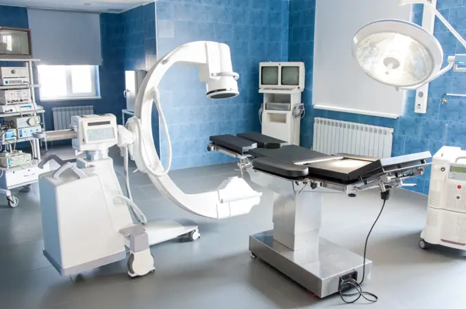
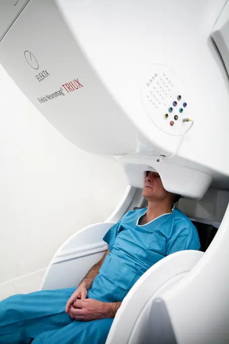
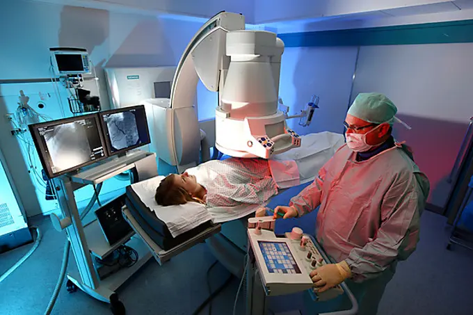
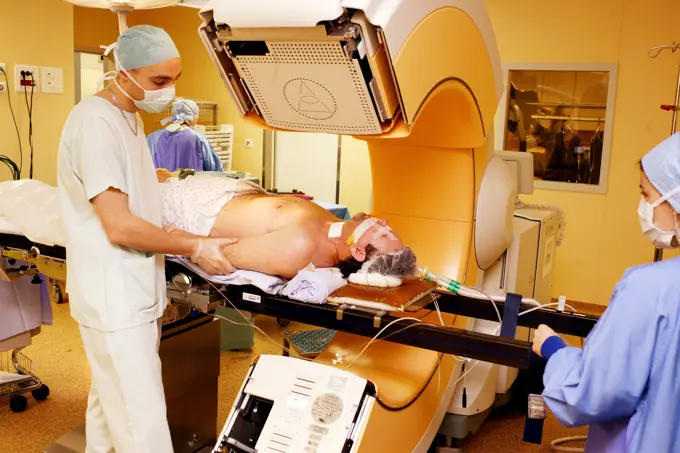
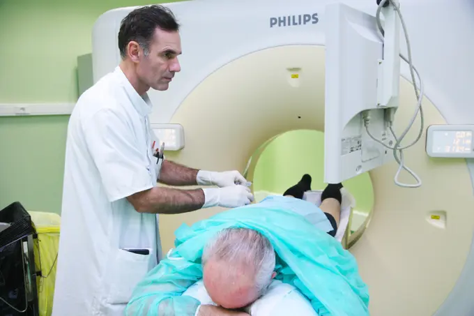
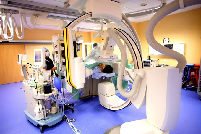
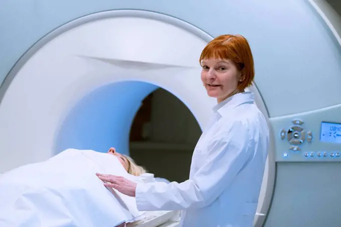
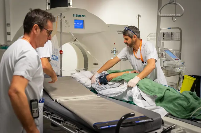
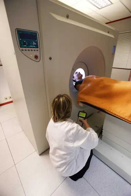
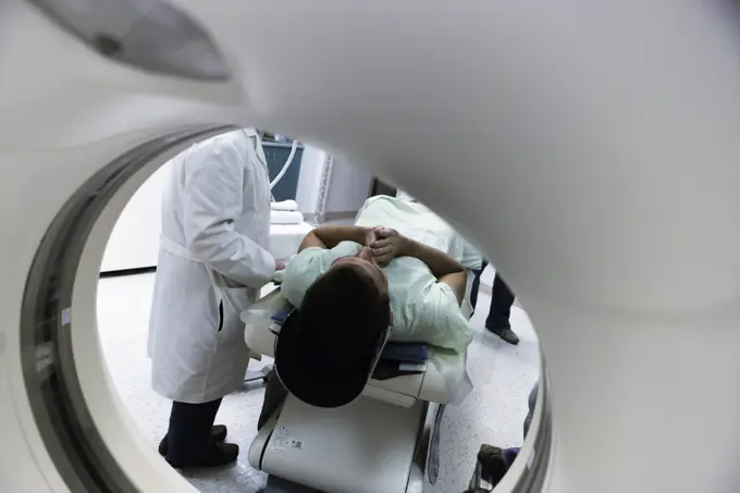
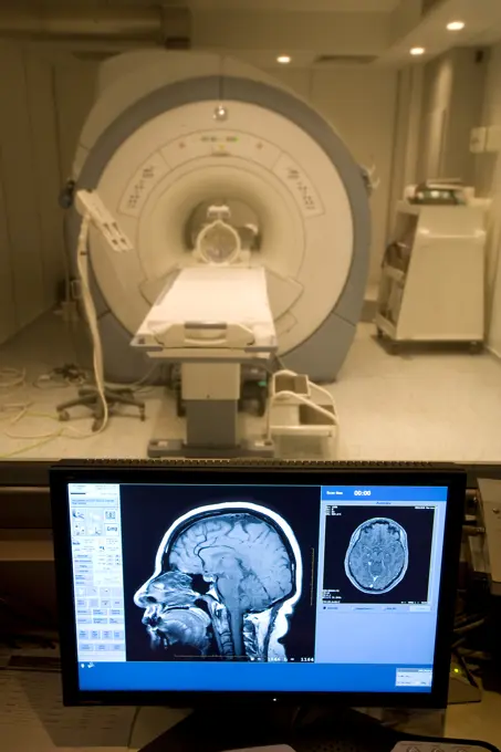
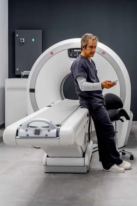
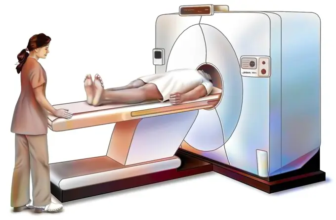
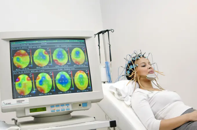
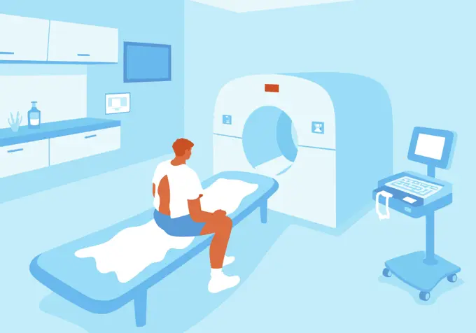
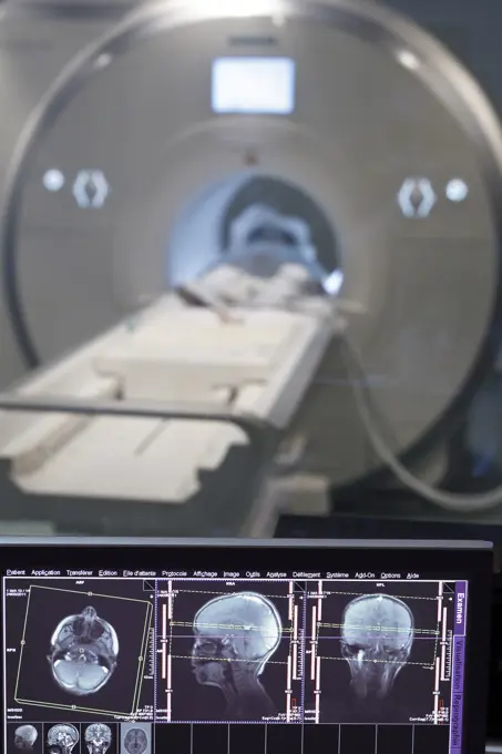
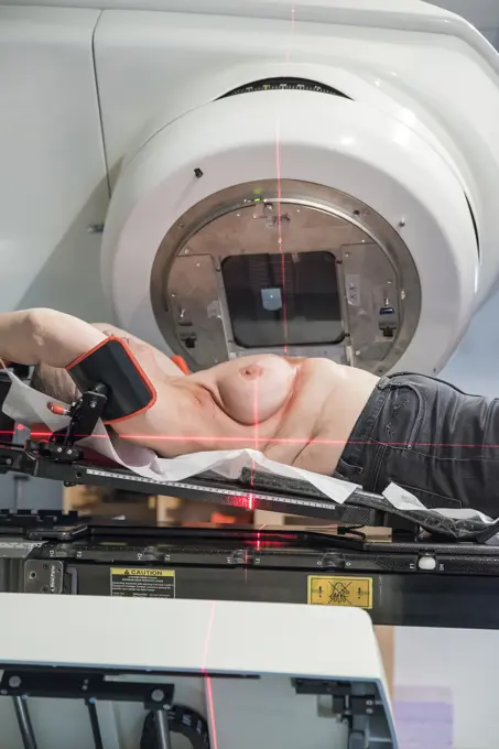
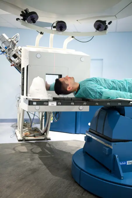
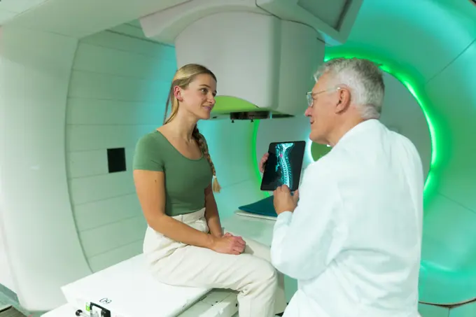
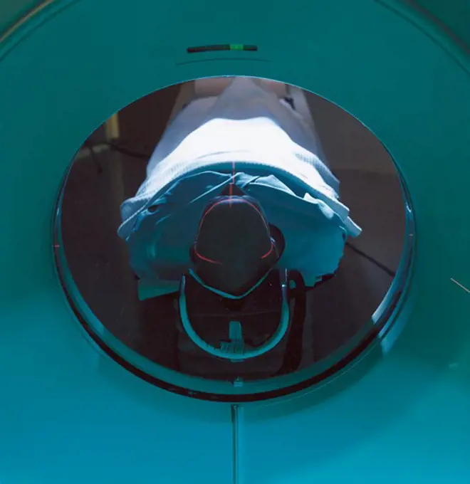
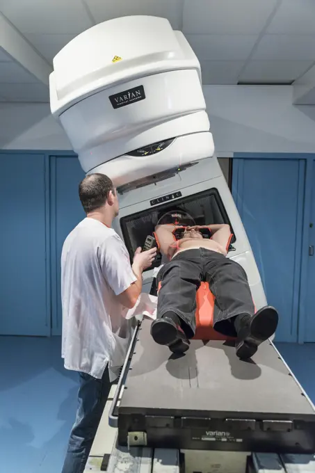
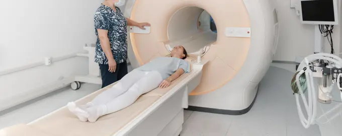
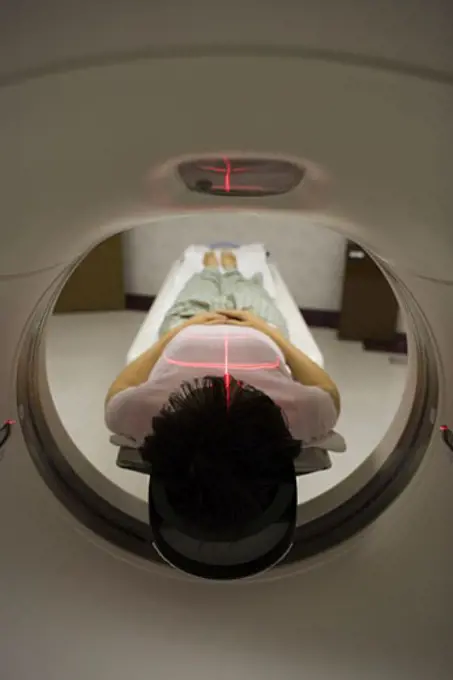
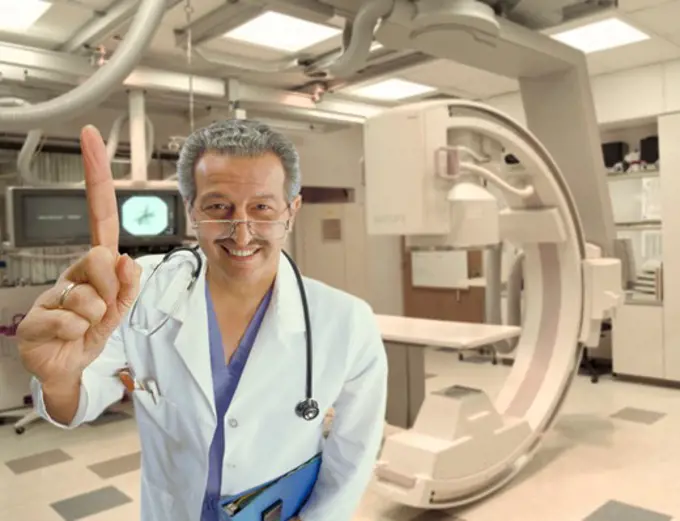
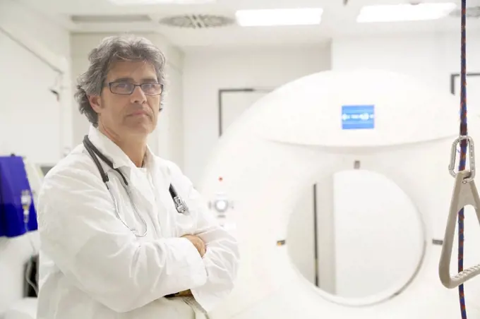
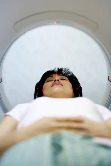
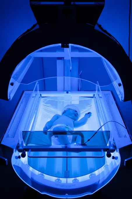
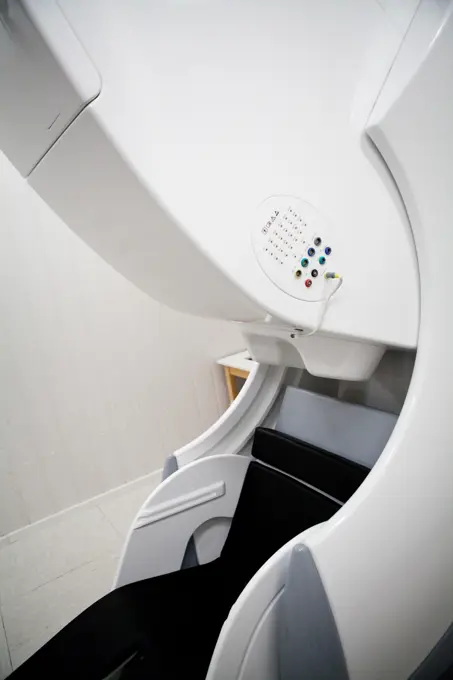
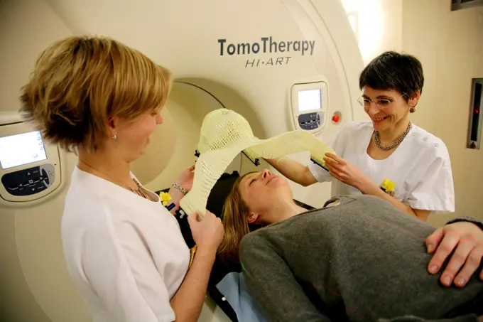
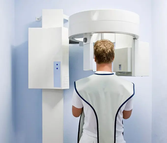
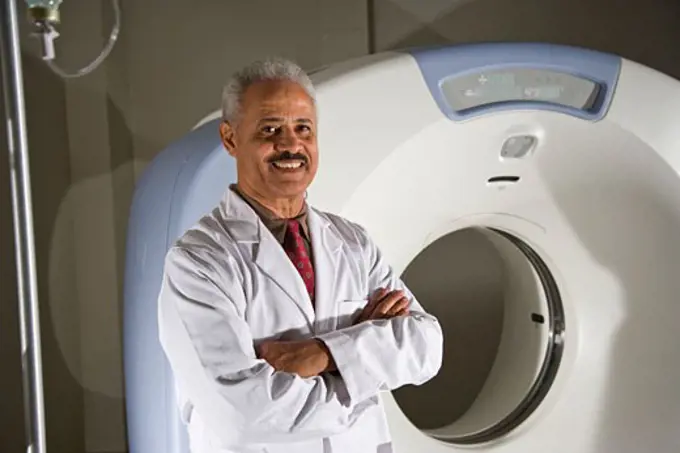
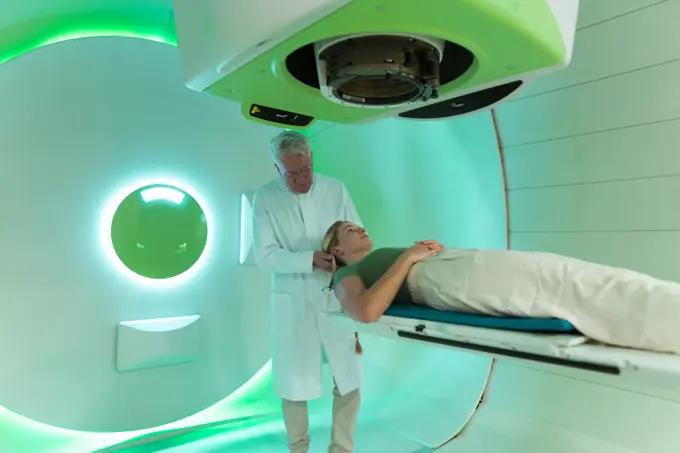
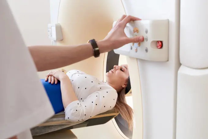
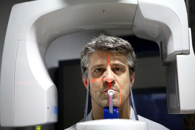
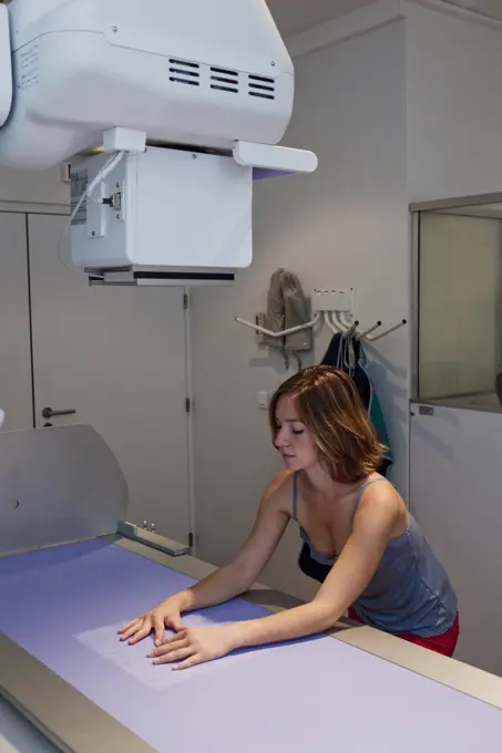
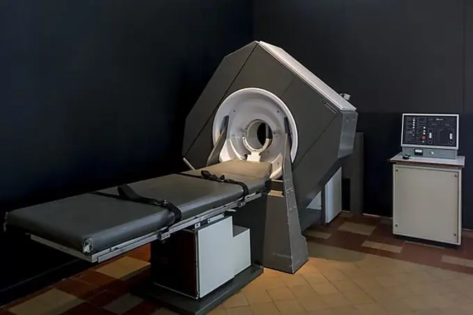
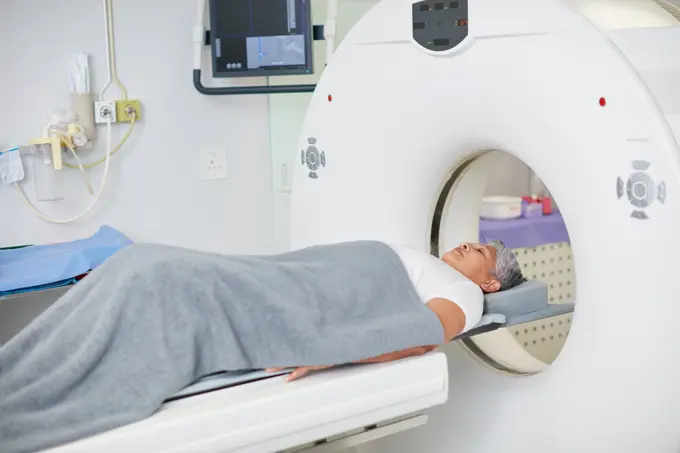
Images of patients undergoing MRI scans in clinical settings. Focus on the patients in machines, showcasing medical assessments and technology.

Appointment, healthcare and MRI with senior patient in hospital for assessment or brain anatomy. CT scan, diagnosis and medical with old woman in machine at clinic for neurological exam or function
179 assets in this story
6145-48920003
A view of the CT scanner in the new Fort Belvoir Community Hospital. The hospital, still under construction, is a 2005 Base Realignment and Closure project, and is scheduled to open later this year.
1525-21754323
operating room with X-ray medical scan.
1848-54953291
Computer tomography, CT or cat-scan, computer-assisted tomography, hospital, Germany, Europe
824-63179543
Reportage at the Neuroimaging research centre in Pitié Salpêtrière hospital in Paris, France. Magnetoencephalography (MEG) platform. MEG detects variations in the brain's magnetic field during various types of cerebral activity. It studies normal and abnormal brain function. Recording the magnetic field produced by neuronal currents requires ultra sensitive sensors called SQUIDs (Superconducting Quantum Interference Device). The 306 sensors spread over 102 areas allow both near and far magnetic fields to be measured, and the brain's deep structures to be 'seen'.
6177-V54158663
1848-54953283
Diagnostic and interventional radiology, angiography, examination and treatment for vascular diseases, hospital, Germany, Europe
824-63218509
An 81-year-old patient undergoes an injected brain scanner to detect a stroke.
1660R-60604
USA, Utah, Payson, Young woman during Magnetic Resonance Imaging
824-63197780
Reportage in the National Oncology Particle Therapy Centre in Milan, Italy (The CNAO - Centro Nazionale di Adroterapia Oncologica). Particle therapy is a state-of-the-art technique used to treat cancer. It consists of using hadrons, protons (proton therapy) and cabon ions (carbon-ion treatment) to irradiate tumours. It has two therapeutic purposes, a high ballistic accuracy which allows healthy tissue surrounding the tumour to be spared, and a greater efficiency at treating certain cancers. The CNAO is the only centre to own carbon-ion treatment technology in Europe.
4269-13811
Patient with degenerative arthosis receiving intervertebral disk prosthesis to treat herniated cervical disc. This cervical rachis surgery is performed with the O-ARM Imaging System. This motorized intra-operative multi-dimensional surgical imaging platform provides real-time high resolution fluoroscopy and 3D CT scan imaging, and allows high precision and safety surgical procedures. Surgery performed by Pr Jean-Jacques Moreau, neurosurgery department, Dupuytren hospital, Limoges, France.
4269-4760
Department of Pr Frija, Saint-Louis hospital, Paris, France. Pr de Kerviler, radiologist, set up of procedure of biopsy of peripheral lymph nodes assisted by medical imaging for lymphoma diagnostic. Here, doctor performing CT scan-assisted biopsy.
824-63161348
Device for radiotherapeutic treatment allowing stereotaxic radiotherapy and monitoring of the tumor during irradiation.
1815-69224389
Radiologist in a proton therapy room smiling confidently in a medical facility
4269-2923
High technology digital vascular angiography unit, medical imagery center of Poitiers University Hospital, France. This scanner, combining the two highly evolved biplane and flat detector technologies, generates high quality images for diagnostic purposes and optimised therapeutic procedures. Here, patient undergoing interventional neuroradiology treatment for a cerebral aneurysm.
1815R-13825407
Female doctor preparing patient for magnetic resonance imaging
6177-V53478321
824-63218508
An 81-year-old patient undergoes an injected brain scanner to detect a stroke.
824-63205903
Reportage at the Rinecker Proton Therapy Center in Munich, Germany. The centre has the latest equipment for proton therapy treatment. Proton therapy involves irradiating cancer cells by concentrating a proton beam on the heart of the tumour, while preserving the healthy surrounding tissue.
1589R-12401631
Older Hispanic man laying in MRI scanner
4269-4487
Patient undergoing Positrons Emission Tomography scan (PET scan) of the brain.
4269-39809
Female patient undergoing a CT scan, Angoulême hospital, France .
5514-58777438
Portrait of a young man about to have a MRI scan, rear view.
4294R-1468
Doctor with patient at CT scanner
4269-21307736
Bone scintigraphy, nuclear medicine department.
4269-33805
Elderly male patient talking to a radiotherapy radiographer during preparatory step to cancer treatment by radiotherapy. Radiotherapy center, pole of Oncology, Hospital of Bordeaux. Hospital Haut-Leveque.
6145-52086984
A member of the Army Health Services Command treats a cancer patient with radiation therapy. Country: Unknown
1525-22269527
Healthcare
4269-8943
Closed geometry Magnetic Resonance Imaging (MRI) unit, 3 tesla with a brain MRI scan on the control screen.
824-63197466
Reportage in the Rinecker Proton Therapy Centre in Munich, Germany. The centre is equipped with the latest protontherapy treatment technology. Protontherapy consists of irradiating cancer cells by concentrating a beam of protons on the core of the tumour, while preserving healthy neighbouring tissue. To treat localised tumours on organs that are likely to be moving due to breathing, the patient is placed under artificial apnea.
1815-15863293
Veterinarian using cell phone in clinic
824-63181504
Digital medical imaging centre in Paris, France. MRI knee.
1785-18842
Doctor holding test results next to CAT scan machine
1899-61460076
Report at Nice University Hospital, Pasteur Hospital, for a radical prostatectomy with lymph node dissection on a 66-year-old patient suffering from high-risk prostate cancer according to the D'Amico classification. Here, surgeon operating the Da Vinci robot. FRANCE
824-63169170
Report on blood donation at the EFS of Saint Louis Hospital in Paris. Apheresis device.
824-63161124
MRI or magnetic resonance imaging based on the magnetic properties of the hydrogen contained in the human body.
824-63164970
Pet scan: medical imaging device to detect tumors and metastases.
824-63183326
Electroencephalogram (EEG) on a 27-year old woman. Normal cerebral activity on the screen.
1815-65822902
Patient sitting on bed in CT scan room
6145-52115803
COL. (Dr.) Ghaed, right, and Technician J. Butler prepare a patient for cancer treatment on a linear accelerator at the Fitzsimons Army Medical Center. Base: Aurora State: Colorado (CO) Country: United States Of America (USA)
1848-55370616
Housewife emptying a tumble dryer, removing the dry washing from the drum
824-63173338
Brain, mri
1815R-85431
Germany, Nuremberg, Doctor doing medical examination of patient
824-63208993
Reportage in a radiology centre in Haute-Savoie, France. A technician carries out an arthography of the shoulder.
824-63188312
Reportage in the radiotherapy centre at the Baie walk-in clinic in Avranches, France. Breast cancer treatment.
824-63197785
Reportage in the National Oncology Particle Therapy Centre in Milan, Italy (The CNAO - Centro Nazionale di Adroterapia Oncologica). Particle therapy is a state-of-the-art technique used to treat cancer. It consists of using hadrons, protons (proton therapy) and cabon ions (carbon-ion treatment) to irradiate tumours. It has two therapeutic purposes, a high ballistic accuracy which allows healthy tissue surrounding the tumour to be spared, and a greater efficiency at treating certain cancers. The CNAO is the only centre to own carbon-ion treatment technology in Europe.
824-63205930
Reportage at the Rinecker Proton Therapy Center in Munich, Germany. The centre has the latest equipment for proton therapy treatment. Proton therapy involves irradiating cancer cells by concentrating a proton beam on the heart of the tumour, while preserving the healthy surrounding tissue. Settings on software which allow the tumour to be targeted precisely. To treat tumours on organs that move during breathing, the patient is placed under artificial apnea.
824-63181669
Digital medical imaging centre in Paris, France. Ankle x-ray.
824-63197796
Reportage in the National Oncology Particle Therapy Centre in Milan, Italy (The CNAO - Centro Nazionale di Adroterapia Oncologica). Particle therapy is a state-of-the-art technique used to treat cancer. It consists of using hadrons, protons (proton therapy) and cabon ions (carbon-ion treatment) to irradiate tumours. It has two therapeutic purposes, a high ballistic accuracy which allows healthy tissue surrounding the tumour to be spared, and a greater efficiency at treating certain cancers. The CNAO is the only centre to own carbon-ion treatment technology in Europe. The patient wears a mask which prevents movement during treatment.
4269-3541
Department of Functional Neurosurgery and stereotaxy of the Pr Peragut, Timone hospital, Marseille, France. Treatment of a meningioma (benign brain tumor) with a Gamma knife. Here, patient at the end of treatment.
1815-69224397
Doctor discussing radiology results with patient in medical facility
1848-56095154
Casualty unit, emergency room of a hospital, x-ray, Germany, Europe
4269-6305
Patient undergoing heart 3D CT scan, Department of Medical Imaging, Centre Cardiologique du Nord, St Denis, France. The Lightspeed VCT 64-Slice CT Scanner produces 64 slices per rotation, and reduces radiation exposure by 70%.
4176-37081
High angle view of a person getting MRI scan
824-63188316
Reportage in the radiotherapy centre at the Baie walk-in clinic in Avranches, France. Breast cancer treatment.
4128R-14324259
Hospital radiologist
6188-60030927
Doctor or nurse and patient with tomography CT or MRI Scan in hospital. Interior of radiography department. Technologically advanced equipment in white room. Magnetic resonance diagnostics machine Doctor or nurse and patient with tomography CT or MRI Scan in hospital. Interior of radiography department. Technologically advanced equipment in white room. Magnetic resonance diagnostics machine. ,model released, Symbolfoto Copyright: xZoonar.com/MariyaxLvovax 18167949 ,model released, Symbolfoto ,property released
1660R-6193
High angle view of a female patient getting an CAT scan
4286R-6434
Exaggerated image of a doctor holding up a finger in 1 pose medical equipment in the background
1525-24908107
Nurse with a patient
1815R-11382139
Doctor in hospital with MRT
1660R-54945
Woman entering MRI machine
1899-61460780
Infants wearing eye masks in phototherapy tunnel. In the first days after birth the bilirubin level of the newborn can vary. Phototherapy sessions restore the balance.Editorial Only
824-63161101
Magnetoencephalography can detect magnetic field variations in different brain activities.
824-63181662
Digital medical imaging centre in Paris, France. Ankle x-ray.
4269-5317
In January 2007, the Institut Curie, Paris, France, was the first hospital in France to treat cancer with tomoTherapy. This image-guided intensity-modulated radiation therapy improves the precision of the treatment and reduces radiation exposure to healthy surrounding tissue. A thermoformed mask maintains the patient still during exposure.
1773R-14008
Dental patient having x-ray taken
106-1686
Magnetic Resonance Imaging
1848-51025452
Examination room with medical equipment, CT, computer tomograph, Karlovy Vary, Czech Republic, Europe
6145-54127086
NAVAL BASE GUAM (May 26, 2022) - USNS Mercy (T-AH 19) Director for Administration, Cmdr. Eric Polonsky gave a tour of the CT scan room onboard the hospital ship USNS Mercy (T-AH 19) following its arrival at U.S. Naval Base Guam (NBG) May 26. Mercy is en route for Pacific Partnership 2022 which is now in its 17th year and is the largest annual multinational humanitarian assistance and disaster relief preparedness mission conducted in the Indo-Pacific region. The Pacific Partnership 2022 team will work with host nation counterpart and regional partners to provide tailored medical, dental, and veterinary care, and conduct bilateral engineering civic actions, and exchange information related to disaster response procedures. The port stop at NBG will provide the crew time for rest and relaxation, conduct mission preparations, perform routine maintenance, and load additional supplies onto the ship. The hospital ships last visit to Guam was in 2018.
1785-18792
African American radiologist next to CT scanner
1525-25970738
young happy nurse in a hospital
1815-69224399
Doctor assisting patient in a proton therapy room for cancer treatment
6188-62329303
Female patient undergoing MRI - Magnetic resonance imaging in Hospital. Medical Equipment and Health Care concept Female patient undergoing MRI - Magnetic resonance imaging in a Hospital. Medical Equipment and Health Care ,model released, Symbolfoto Copyright: xZoonar.com/Aleksey_Semykinx79964187859x 22001039 ,model released, Symbolfoto ,property released
1525-22392069
Nurse with a patient having a CT scan
824-63181519
Digital medical imaging centre in Paris, France. Dental x-ray.
824-63181492
Digital medical imaging centre in Paris, France. Dental x-ray.
824-63122947
Medical imaging center in Paris, France.
1848-61664037
Washing machine
1848-49321075
EMI scanner, used for X-raying patients in the Dr Guislain Museum about the history of psychiatry, Ghent, Belgium, Europe
PREVIOUS
of 2
NEXT
2401 S. Ervay, Suite 206
Dallas, Texas 75215
United States
Get Started
Free ResearchMy BoardsMy Cart
For Creators
How To License Your ContentContributor PortalFrame of Mind
Resources
API accessPricing
Contact
+1 866 236 0087help@viewpictures.co.uk Contact form
©2026 View Pictures. All Rights Reserved. -A
This site is protected by reCAPTCHA and Google Privacy Policy and Terms of Service apply.