My Boards
View Pictures
  • My Boards

Viral Imaging and Research

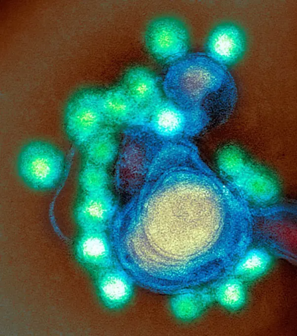
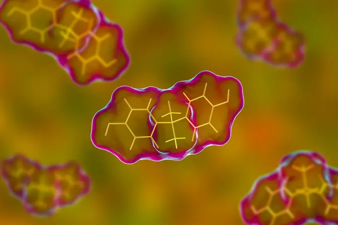
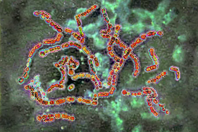
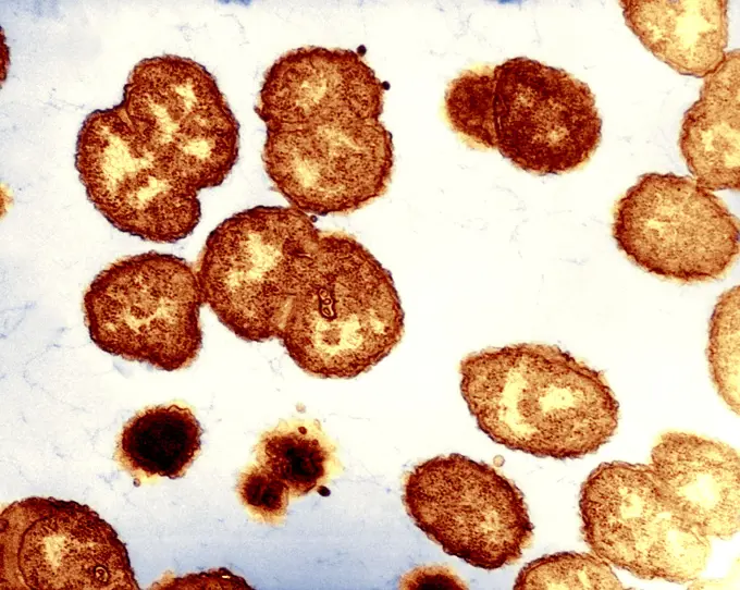
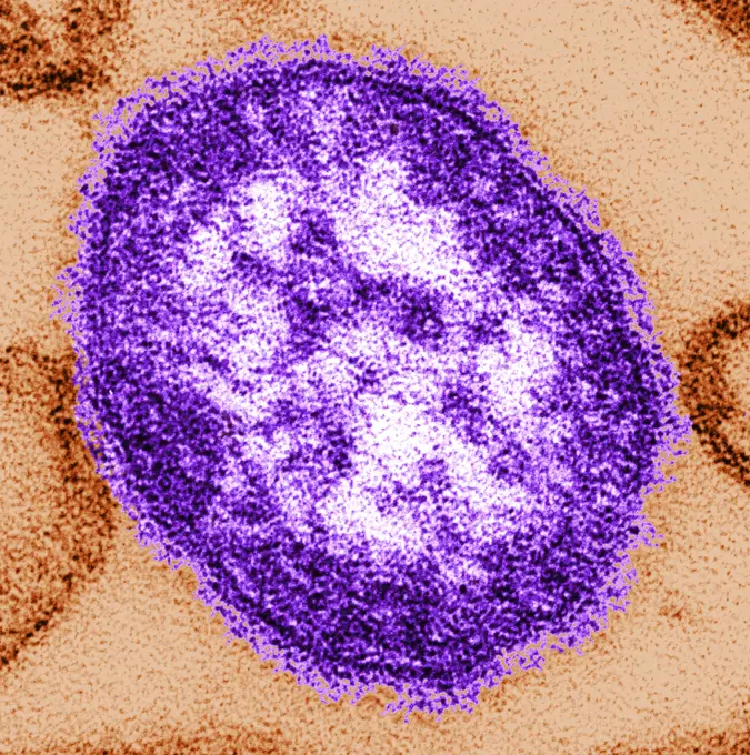
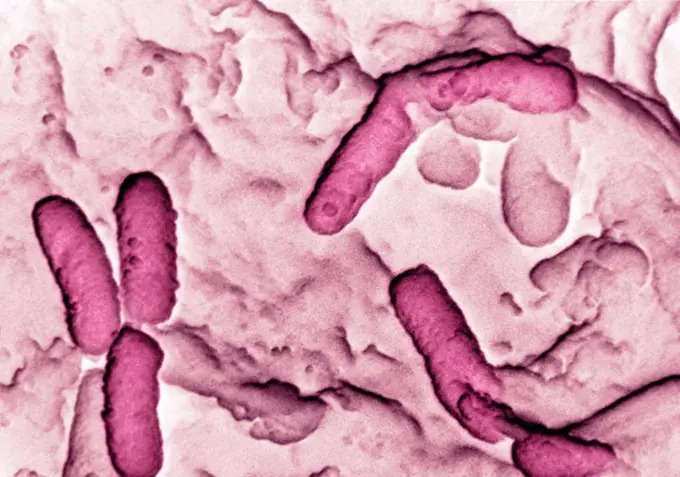
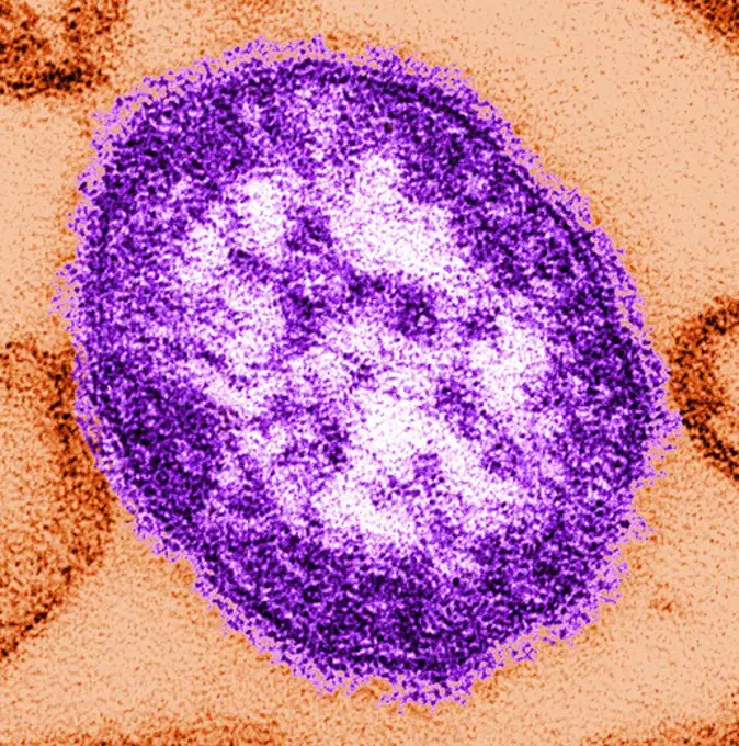
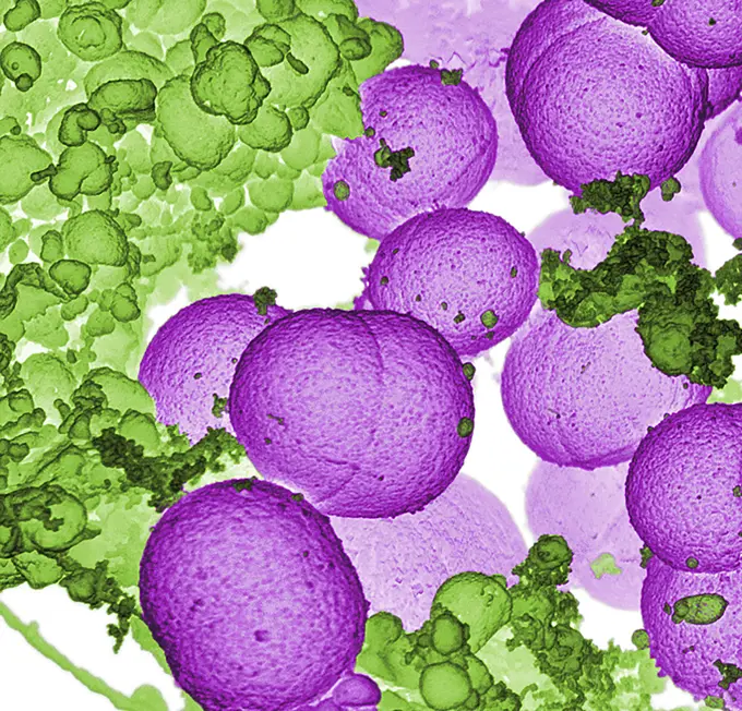
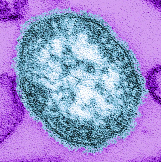
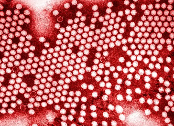
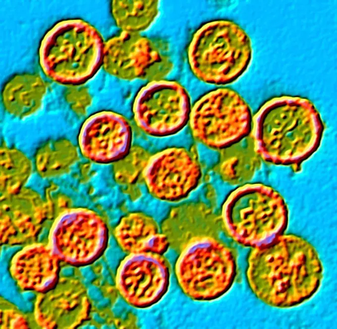
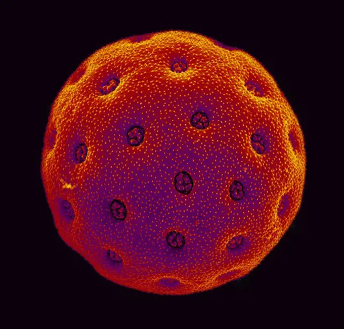
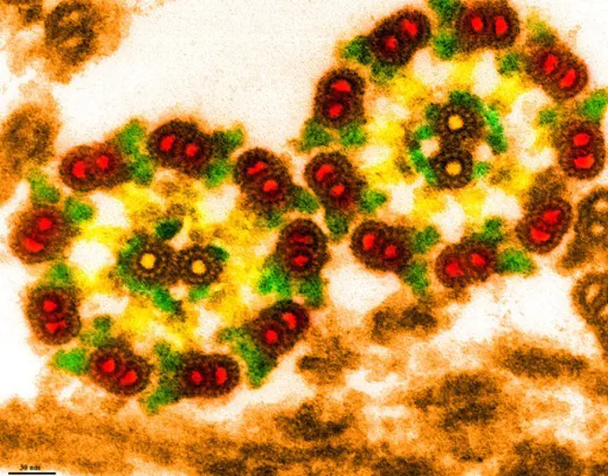
Microscopic images of various viruses, including hepatitis and influenza, highlighting their structure for medical research purposes.

Transmission electron microscopic image of a thin cross section cut through the isolated axoneme of Chlamydomanas reinhardtii, a unicellular flagellate
144 assets in this story
4128R-13409468
Zika virus particles, TEM
824-63208091
Flavivirus seen by transmission electron microscopy. Viral diameter around 40-50 nm.
4128-28767079
Bisphenol A molecule, illustration
1899-61461241
Group A streptococci pyogenes. It is the cause of skin infections (impetigo), abscesses, bronchopulmonary infections, a form of sore throat which can develop into complications such as acute rheumatic fever. Image produced from optical microscopy
4128R-13620405
HIV infection, coloured transmission electron micrograph (TEM). Mature virus and budding release of HIV in human lymph tissue (RNA virus, Retroviridae...
4128-V58615264
824-63123743
Bacteria which is the causative agent of gonorrhea.
1899-53512010
Influenza virus of the Orthomyxoviridae family (respiratory viral infection). Transmission electron microscopy, viral diameter 80 to 120 nanometers.
1525-23769337
stripped paint in the blue wood door and rusty  nail
824-63208093
Hepatitis A virus (HAV). Image produced from an image taken with transmission electron microscopy.
1899-54026853
Meningococcus seen under a scanning electron microscope.
824-63206961
Meningococcus seen under a scanning electron microscope.
824-63195688
This thin-section transmission electron micrograph (TEM) revealed the ultrastructural appearance of a single virus particle, or virion, of measles virus. The measles virus is a paramyxovirus, of the genus Morbillivirus. It is 100-200 nm in diameter, with a core of single-stranded RNA, and is closely related to the Rinderpest and canine distemper viruses. Two membrane envelope proteins are important in pathogenesis. They are the F (fusion) protein, which is responsible for fusion of virus and host cell membranes, viral penetration, and hemolysis, and the H (hemagglutinin) protein, which is responsible for adsorption of virus to cells. There is only one antigenic type of measles virus. Although studies have documented changes in the H glycoprotein, these changes do not appear to be epidemiologically important (ie no change in vaccine efficacy has been observed).
4197-V65369299
1773-97859
Electron Micrograph of Pseudomonas aeruginosa
4297-1826
Thin-section transmission electron micrograph of a single measles virus particle or virion
4128R-11291492
Artwork of the AIDS (acquired immunodeficiency syndrome) virus.
824-63194638
Scanning electron micrograph of S. aureus bacteria escaping destruction by human white blood cells.
4297-1779
Negatively-stained transmission electron micrograph of numerous herpes simplex virions, members of the Herpesviridae virus family
4269-21091508
Zika virus. Coloured transmission electron micrograph (TEM).
824-63179045
Yersinia pestis (Pasteurella pestis) is the bacterium responsible for bubonic plague. Optical microscopy X 2000
4128-V58615260
824-63195690
This thin-section transmission electron micrograph (TEM) revealed the ultrastructural appearance of a single virus particle, or virion, of measles virus. The measles virus is a paramyxovirus, of the genus Morbillivirus. It is 100-200 nm in diameter, with a core of single-stranded RNA, and is closely related to the Rinderpest and canine distemper viruses. Two membrane envelope proteins are important in pathogenesis. They are the F (fusion) protein, which is responsible for fusion of virus and host cell membranes, viral penetration, and hemolysis, and the H (hemagglutinin) protein, which is responsible for adsorption of virus to cells. There is only one antigenic type of measles virus. Although studies have documented changes in the H glycoprotein, these changes do not appear to be epidemiologically important (ie no change in vaccine efficacy has been observed).
4269-21091507
Zika virus. Coloured transmission electron micrograph (TEM).
4128-18801148
Coronavirus particle, illustration
4384-121
This stained transmission electron micrograph (TEM), captured by F.A. Murphy in 1968, depicts a number of Marburg virus virions, which had been grown in an environment of tissue culture cells. Marburg hemorrhagic fever is a rare, severe type of hemorrhagic fever which affects both humans and non-human primates. Caused by a genetically unique zoonotic (that is, animal-borne) RNA virus of the filovirus family, its recognition led to the creation of this virus family. The four species of Ebola viru
4128-V58615209
824-63211594
Meningococci (Neisseria Meningitidis), the bacteria in meningitis. Seen under optical microscopy X 1000.
824-65830682
Transmission electron micrograph showing vesicular stomatitis virus (VSV) particles (yellow) budding from infected cells (burgundy).
824-63194528
This colorized negatively-stained transmission electron micrograph (TEM) captured some of the ultrastructural details exhibited by the new influenza A (H7N9) virus. On April 1, 2013, the World Health Organization (WHO) first reported 3 human infections with a new influenza A (H7N9) virus in China. Since then, additional cases have been reported. Most reported cases have severe respiratory illness and, in some cases, have died. At this time, no cases of H7N9 outside of China have been reported. This new H7N9 virus is an avian (bird) influenza (flu) virus. Human infections with avian influenza (AI, or bird flu) are rare but have occurred in the past, most commonly after exposure to infected poultry. However, this is the first time that this bird flu subtype (H7N9) has been found in people. This virus is very different from other H7N9 viruses previously found in birds.
824-63127917
Agent of feline leucosis, a fatal viral disease specific to cats. This electron micrograph depicts the retrovirus, Feline Leukemia Virus (FeLV). FeLV is a very common, often fatal feline disease passed from one cat to another via adversarial bites, or when one cat sneezes on another, or shares a food bowl. A cat may be vaccinated in order to protect it from acquiring this virus. See image n1302105 on the site.
824-63227274
Colorized transmission electron micrograph MERS virus particles (blue) found near the periphery of an infected VERO E6 cell (yellow).
4269-5507
Transmission Electron Micrograph (TEM) of poliovirus, an RNA enterovirus responsable for poliomyelitis.
4269-27707
Influenza virus. Influenza A virus causes flu in humans, swine, birds, horses and dogs. Which species the virus will infect is determined by the glycoprotein spikes on its outer envelope. They are many strains of influenza A virus, among which the avian flu virus (H5N1), swine flu (H1N1) or human. Transmission Electron Micrograph (TEM).
824-63222517
This transmission electron microscope image shows SARS-CoV-2, the virus that causes COVID-19, isolated from a patient in the United States. Virus particles (golden round objects) emerge from the surface of cells cultured in the laboratory. The spikes on the outer edge of the virus particles give coronaviruses their crown-shaped name. Image captured and colorized at NIAID's Rocky Mountain Laboratories (RML) in Hamilton, Montana. Credits: NIAID
824-63223153
Colorized scanning electron micrograph of Ebola virus particles (yellow) both budding and attached to the surface of infected VERO E6 cells (green). Image captured and color-enhanced at the NIAID Integrated Research Facility in Fort Detrick, Maryland. Credit: NIAID.
824-65830839
Scanning electron micrograph of a human H9 T cell (blue/green) infected with HIV virus particles (yellow).
824-63223141
Colorized scanning electron micrograph of filamentous. Ebola virus particles (red) attached and budding from. a chronically infected VERO E6 cell (blue) (25,000x magnification). Image captured and color-enhanced at the NIAID Integrated Research Facility in Ft. Detrick, Maryland. Credit: NIAID.
4269-24806
Muscle. Colored transmission electron micrograph (TEM) of a longitudinal section through striated skeletal muscle. Skeletal muscle is under voluntary control and moves long skeletal bones.
4128R-14057081
Hantavirus particles, TEM
824-63223181
MERS-CoV particles on camel epithelial cells. Credit: NIAID in collaboration with Colorado State University.
4297-1010
Colorized scanning electron microscope image of amaranthus pollen. The pollen has been acetolyzed to remove cytoplasm and pollenkit in order to reveal the intricate wall structure. Family: Amaranthaceae: Amaranthus opopeo
824-63223138
Colorized scanning electron micrograph of filamentous. Ebola virus particles (green) attached and budding from. a chronically infected VERO E6 cell (orange) (25,000x magnification). Image captured and color-enhanced at the NIAID Integrated Research Facility in Ft. Detrick, Maryland. Credit: NIAID.
4128-V58576918
PREVIOUS
of 2
NEXT
2401 S. Ervay, Suite 206
Dallas, Texas 75215
United States
Get Started
Free ResearchMy BoardsMy Cart
For Creators
How To License Your ContentContributor PortalFrame of Mind
Resources
API accessPricing
Contact
+1 866 236 0087help@viewpictures.co.uk Contact form
©2026 View Pictures. All Rights Reserved. -B
This site is protected by reCAPTCHA and Google Privacy Policy and Terms of Service apply.