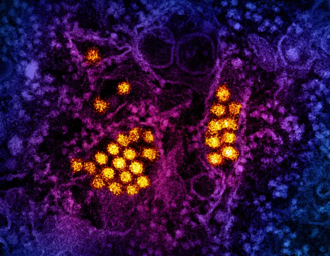
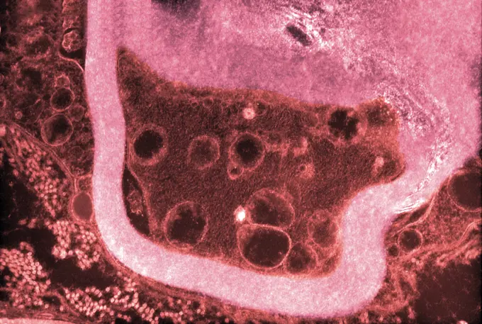
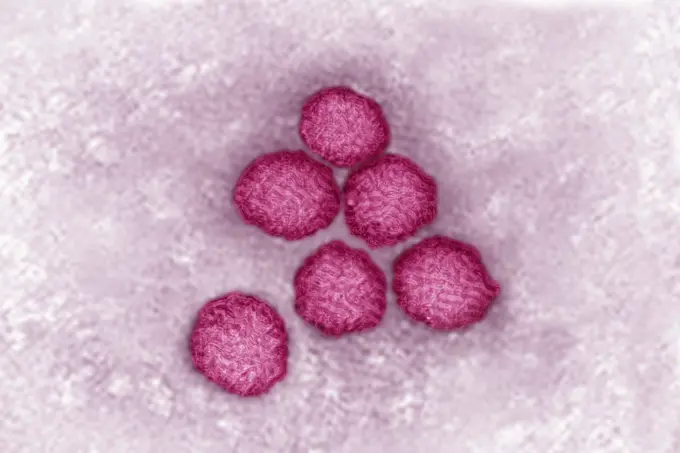
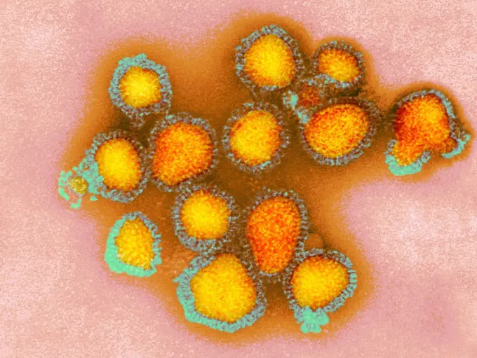
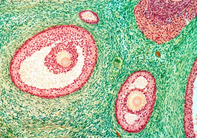
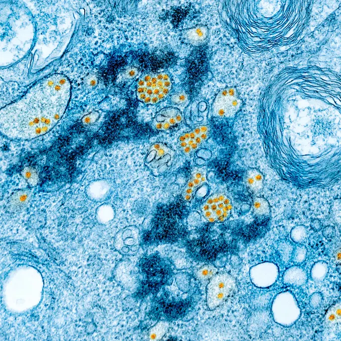
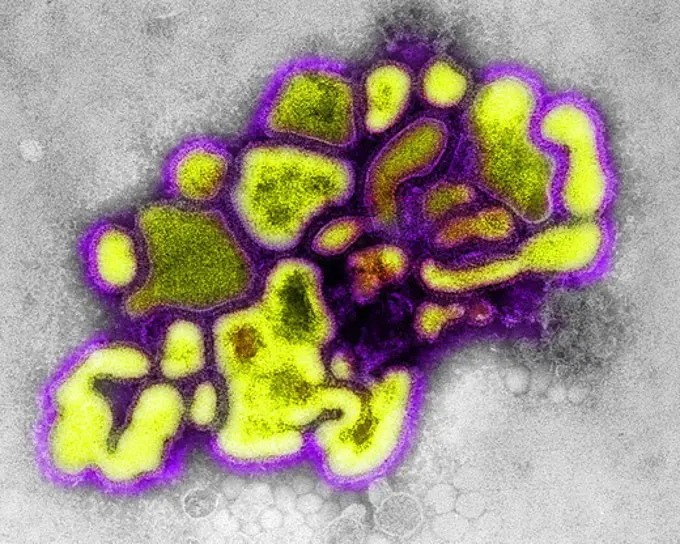
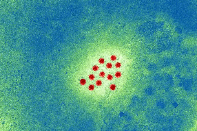
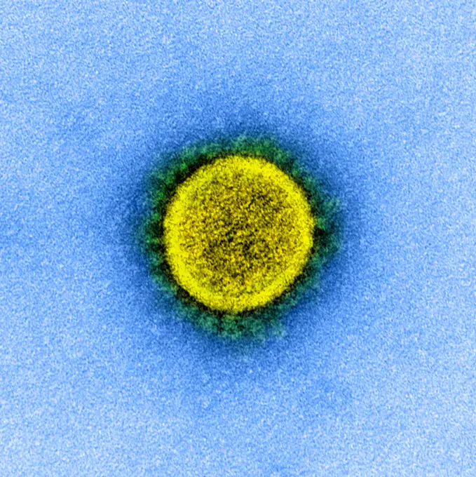
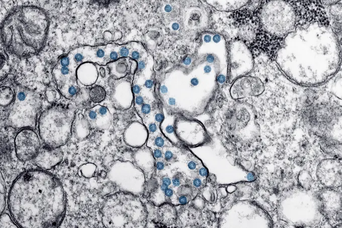
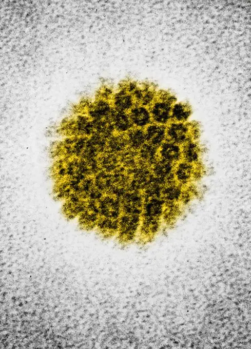
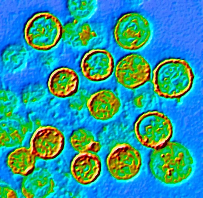
My Boards
View Pictures
  • My Boards

Viral Particles in Microscopy

Colorized transmission electron micrographs showcasing various viruses, including Hepatitis B, HIV-1, and SARS-CoV-2, against vibrant, abstract backgrounds.

Transmission electron micrograph of dengue virus particles (gold).
210 assets in this story
4269-20408835
Coloured transmission electron micrograph (TEM) of a MERS coronavirus particle , this virus (originally novel coronavirus 2012) has been named Middle East respiratory syndrome (MERS) coronavirus.
824-63194906
This colorized transmission electron micrograph (TEM) revealed some of the ultrastructural morphology displayed by norovirus virions, or virus particles.
824-63194535
This colorized negatively-stained transmission electron micrograph (TEM) captured some of the ultrastructural details exhibited by the new influenza A (H7N9) virus. On April 1, 2013, the World Health Organization (WHO) first reported 3 human infections with a new influenza A (H7N9) virus in China. Since then, additional cases have been reported. Most reported cases have severe respiratory illness and, in some cases, have died. At this time, no cases of H7N9 outside of China have been reported. This new H7N9 virus is an avian (bird) influenza (flu) virus. Human infections with avian influenza (AI, or bird flu) are rare but have occurred in the past, most commonly after exposure to infected poultry. However, this is the first time that this bird flu subtype (H7N9) has been found in people. This virus is very different from other H7N9 viruses previously found in birds.
1899-54027727
The mumps virus (paramyovirus). Image produced with transmission electron microscope (viral diameter around 200 nm).
824-63212851
Influenza virus (Myxovirus influenzae) from the Orthomyxoviridae family. Viral diameter from 80 to 120 nanometers, taken using transmission electron microscopy.
4269-6790
Transmission electron micrograph (TEM) of a section of axone. It is protected by a layer of myelin around.
824-63224472
Flavivirus (family Flaviviridae), these viruses are responsible for yellow fever, dengue fever, Japanese encephalitis, Zika virus, West Nile encephalitis. They are transmitted by mosquitoes or ticks. image produced from transmission electron microscopy (viral diameter approximately 40 to 60 nm).
824-29057237
Influenza virus of the Orthomyxoviridae family (respiratory viral infection). Transmission electron microscopy, viral diameter 80 to 120 nanometers.
824-63224471
Coronavirus (CoV). viruses of the Coronaviridae family and the Orthocoronavirinae subfamily. It is a pathogen of respiratory syndromes. Viewed from a transmission electron microscopy (TEM) image. Viral diameter 80nm to 100nm.
4128R-14768294
H3N2 influenza virus particles, TEM
824-63179001
Gonococcus (neisseria gonorrhoeae) is the bacterium responsable for gonorrhea. In men the symptoms are acute burning when urinating and some discharge (clap). In women the symptoms are burning when urinating, bartholin's cyst, cervisitis, salpingitis. Optical microscope x 1850.
6145-29256956
Lung: pulmonary tuberculosis
1990-29051591
Bubbles trapped in frozen surface of pond, Thornton, Ontario, Canada
4128R-13620398
Influenza A virus (RNA virus, Orthomyxoviridae Family), coloured transmission electron micrograph (TEM). 'Influenza Virus', often know as 'The Flu Vir...
824-63212848
Influenza virus (Myxovirus influenzae) from the Orthomyxoviridae family. Viral diameter from 80 to 120 nanometers, taken using transmission electron microscopy.
824-63190248
Pneumococcus (streptococcus pneumoniae). Pneumococcus is the bacterium that causes pneumonia, bronchial pneumonia, purulent pleurisy, bacterial meningitis, ear infections, sinusitis and conjunctivitis. Optical microscopy x 1000.
4128-V58565944
824-63198881
Human coronavirus. Human coronavirus causes respiratory infections and gastroenteritis. Image produced using high-dynamic-range imaging (HDRI) from an image taken with transmission electron microscopy. Viral diameter around 80-160 nm.
824-63227272
The round, pointed objects in the center of the image are MERS coronavirus particles.
4297-1767
Transmission electron micrograph of a group of granules in a human eosinophil
4128R-13409472
Zika virus particles, TEM
824-57657579
Colorized transmission electron micrograph of monkeypox virus particles (orange) cultivated and purified from cell culture.
4128-111581080
Toxic mould spores (Stachybotrys chartarum), computer illustration. Several strains of this fungus (S. atra, S. chartarum and S. alternans) are synony...
824-63184381
Hepatitis B virus (HBV). Image produced using high-dynamic-range imaging (HDRI) from an image taken with transmission electron microscopy. Viral diameter ranges from around 50 to 60 nm.
1899-65662380
Transmission electron micrograph of HIV-1 virus particles (yellow) from infected H9 cells, produced in cell culture. The particles exhibit two stages of replication: the two arcs are immature particles budding from the plasma membrane of the cell, and the center spherical particle is a mature form in extracellular space.
1899-65662345
Colorized transmission electron micrograph of mpox virus particles (pink) found within an infected cell (yellow), cultured in the laboratory.
824-63217788
The human coronavirus is the cause of respiratory infections (colds), and gastroenteritis. View made from a transmission electron microscopy view. Viral diameter approximately: 80 to 160 nm.
4269-5511
Transmission Electron Micrograph (TEM) of poliovirus, an RNA enterovirus responsable for poliomyelitis.
824-63194543
Under a high magnification, this colorized negatively-stained transmission electron micrograph (TEM) captured some of the ultrastructural details exhibited by the new influenza A (H7N9) virus. On April 1, 2013, the World Health Organization (WHO) first reported 3 human infections with a new influenza A (H7N9) virus in China. Since then, additional cases have been reported. Most reported cases have severe respiratory illness and, in some cases, have died. At this time, no cases of H7N9 outside of China have been reported. This new H7N9 virus is an avian (bird) influenza (flu) virus. Human infections with avian influenza (AI, or bird flu) are rare but have occurred in the past, most commonly after exposure to infected poultry. However, this is the first time that this bird flu subtype (H7N9) has been found in people. This virus is very different from other H7N9 viruses previously found in birds.
4297-1224
Transmission electron micrograph, human immunodeficiency virus (HIV), co-cultivated with human lymphocytes
1899-65662346
Colorized transmission electron micrograph of mpox virus particles (red) found within an infected cell (green), cultured in the laboratory.
4269-27244
Syphilis. Light micrograph of a Treponema pallidum. This spirochaete bacterium is the agent of syphilis.
824-63194896
This transmission electron micrograph (TEM) revealed some of the ultrastructural morphology displayed by norovirus virions, or virus particles. Noroviruses belong to the genus Norovirus, and the family Caliciviridae. They are a group of related, single-stranded RNA, nonenveloped viruses that cause acute gastroenteritis in humans. Norovirus was recently approved as the official genus name for the group of viruses provisionally described as Norwalk-like viruses (NLV).
4269-27634
Parainfluenza virus. Transmission electron micrograph (TEM) of parainfluenza virus. This RNA virus of the paramyxovirus class causes minor respiratory infections.
1899-65662377
Transmission electron micrograph of HIV-1 virus particles (teal) from infected H9 cells, produced in cell culture. The particles exhibit two stages of replication: the two Arcs are immature particles budding from the plasma membrane of the cell, and the center spherical particle is a mature form in extracellular space.
824-63194519
This colorized negative-stained transmission electron micrograph (TEM) depicts the ultrastructural details of a number of influenza virus particles, or virions. A member of the taxonomic family Orthomyxoviridae, the influenza virus is a single-stranded RNA organism. The flu is a contagious respiratory illness caused by influenza viruses. It can cause mild to severe illness, and at times can lead to death. The best way to prevent this illness is by getting a flu vaccination each fall.
4269-3233
Transmission electron micrograph (TEM) of the mumps viruses. This RNA virus is a rubulavirus, member of the paramyxovirus.
824-63179018
Cervical cancer. Cellular anomalies seen by the microscopy of a histological section of the endocervical epithelium.
4128R-15405897
Chlamydia trachomatis bacteria, illustration
4269-20408834
Coloured transmission electron micrograph (TEM) of a MERS coronavirus particle , this virus (originally novel coronavirus 2012) has been named Middle East respiratory syndrome (MERS) coronavirus.
824-63208134
Streptococci (Streptococcus pyogenes). Image made from a microscope view.
4128R-13620401
Influenza A virus (RNA virus, Orthomyxoviridae Family), coloured transmission electron micrograph (TEM). 'Influenza Virus', often know as 'The Flu Vir...
824-63123727
Agent of Bolivian hemorrhagic fever. Electron photomicrograph of the Machupo Virus. Machupo Virus is a member of the Arenavirus family, isolated in the Beni province of Bolivia in 1963, It is the agent of Bolivian hemorrhagic fever.
4269-5530
Transmission Electron Micrograph (TEM) of influenza virus particles. Influenza virus are RNA virus (Orthomyxovirus) including Influenza A and B, the two types of influenza viruses.
824-57657678
Colorized transmission electron micrograph of monkeypox virus particles (yellow and green) cultivated and purified from cell culture.
6145-44620201
Springtime for Dunes
824-63223160
Colorized transmission electron micrograph showing H1N1 influenza virus particles. Surface proteins on the virus particles are shown in black. Credit: NIAID.
4269-20408831
Transmission Electron Micrograph (TEM) depicts numbers of H1N1 influenza virus particles.
824-63194523
Colorized transmission electron micrograph of Avian influenza A H5N1 viruses grown in MDCK cells. Avian influenza A viruses do not usually infect humans, however, several instances of human infections and outbreaks have been reported since 1997. When such infections occur, public health authorities monitor these situations closely.
824-63179007
Haemophilus ducreyi, or Ducrey's bacillus, is transmitted by sexual contact (STD), it is an extremely contagious bacterium that does not provide immunity. It causes chancroid (or chancre or Ducrey's chancre), characterized by an ulcerated chancre associated with lymphadenopathy. Visualization by Optical Microscopy
4128R-22523
Ovarian follicles. Light micrograph of a section through secondary follicles in an ovary. Developing eggs oocytes are dark orange. They are surrounded...
1899-53511309
Hepatitis C virus (close to the Flavivirid family). HCV causes blood-borne hepatitis, cirrhosis occurs in 25% of cases, 10 to 20 years after the onset of infection, with a risk of hepatic carcinoma. View produced from a transmission electron microscopy image. Viral diameter approximately: 50 to 60 nm.
824-63227065
Colorized transmission electron micrograph of cultured Vero E6 cells infected with yellow fever virus particles found in distended regions of the endoplasmic reticulum. Image purchased and color-enhanced from the NIAID Integrated Research Facility (IRF) in Fort Detrick, Maryland. Credit: NIAID
824-65830977
Colorized transmission electron micrograph of two mature mpox virus particles (pink) attached to the surface of an infected VERO E6 cell (blue/teal). 2024 : Lower Dose of Mpox Vaccine Is Safe and Generates Six-Week Antibody Response Equivalent to Standard Regimen.
4297-1833
Transmission electron micrograph of the A/New Jersey/76, HSW1N1, virus, a type of swine flu virus, magnification of 37,800X
4269-20408829
Transmission Electron Micrograph (TEM) depicts numbers of H1N1 influenza virus particles.
824-63194572
This digitally-colorized negative-stained transmission electron micograph (TEM) depicted a number of influenza A virions.
824-63211048
The H5N1 avian flu virus seen by transmission electron microscopy (TEM). Viral diameter +/- 100 nm.
1899-65662369
Transmission electron micrograph showing vesicular stomatitis virus (VSV) particles (yellow) budding from infected cells (burgundy).
824-63123744
An electron micrograph of the Hepatitis A virus (HAV), an RNA virus that can survive up to a month at room temperature. This virus enters an organism by ingestion of water and food contaminated by human faeces, and reaches the liver through the bloodstream. HAV infection is endemic in third world countries, and is prevalent in the Far East.
4269-20408833
Coloured transmission electron micrograph (TEM) of a MERS coronavirus particle , this virus (originally novel coronavirus 2012) has been named Middle East respiratory syndrome (MERS) coronavirus.
1899-61460433
Variola virus (poxviruses are large viruses of 300/200/100 nm. They are pathogenic for humans such as smallpox and vaccinia viruses as well as animal viruses that infect humans such as viruses monkeypox, cowpox, milker's nodule, pustular dermatitis in sheep Transmission electron microscopy
4128-V58561253
824-65830792
An electron micrograph showing three Epstein-Barr virus (EBV) particles colorized red-orange. 2024 : scientists Find Weak Points on Epstein-Barr Virus.
4269-5528
Transmission Electron Micrograph (TEM) of the mumps virus.
824-63223218
Transmission electron micrograph of a SARS-CoV-2 virus particle, isolated from a patient. Image captured and color-enhanced at the NIAID Integrated Research Facility (IRF) in Fort Detrick, Maryland. Credit: NIAID.
824-57656524
Transmission electron microscopic image of an isolate from the first U.S. case of COVID-19, formerly known as 2019-nCoV. The spherical viral particles, colorized blue, contain cross-sections through the viral genome, seen as black dots.
824-63194533
This colorized negatively-stained transmission electron micrograph (TEM) captured some of the ultrastructural details exhibited by the new influenza A (H7N9) virus. On April 1, 2013, the World Health Organization (WHO) first reported 3 human infections with a new influenza A (H7N9) virus in China. Since then, additional cases have been reported. Most reported cases have severe respiratory illness and, in some cases, have died. At this time, no cases of H7N9 outside of China have been reported. This new H7N9 virus is an avian (bird) influenza (flu) virus. Human infections with avian influenza (AI, or bird flu) are rare but have occurred in the past, most commonly after exposure to infected poultry. However, this is the first time that this bird flu subtype (H7N9) has been found in people. This virus is very different from other H7N9 viruses previously found in birds.
4297-1780
Transmission electron microscopy image (TEM) showing both HTLV I (Human T-Cell Leukemia Virus) and HIV (Human Immunodeficiency Virus) in a tissue sample
1899-65662350
Transmission electron micrograph of West Nile virus particles (orange/gold) replicating within the cytoplasm of an infected VERO E6 cell (blue).
824-63194530
This colorized negatively-stained transmission electron micrograph (TEM) captured some of the ultrastructural details exhibited by the new influenza A (H7N9) virus. On April 1, 2013, the World Health Organization (WHO) first reported 3 human infections with a new influenza A (H7N9) virus in China. Since then, additional cases have been reported. Most reported cases have severe respiratory illness and, in some cases, have died. At this time, no cases of H7N9 outside of China have been reported. This new H7N9 virus is an avian (bird) influenza (flu) virus. Human infections with avian influenza (AI, or bird flu) are rare but have occurred in the past, most commonly after exposure to infected poultry. However, this is the first time that this bird flu subtype (H7N9) has been found in people. This virus is very different from other H7N9 viruses previously found in birds.
824-63223263
Colorized transmission electron micrograph of negatively stained SW31 (swine strain) influenza virus particles. Credit: NIAID.
4297-1426
Electron micrograph of a negatively stained human papilloma virus (HPV) which occurs in human warts. Warts on the hands and feet have never been known to progress to cancer. However, after many years cervical warts can become cancerous
824-57657670
Colorized transmission electron micrograph of monkeypox virus particles (green) cultivated and purified from cell culture.
5507-47572641
Vector microscopic image of coronavirus
1899-65662322
Colorized transmission electron micrograph of immature mpox virus particles (red with tan viral envelope) found within an infected VERO E6 cell (teal), cultured in the laboratory.
1848-69290407
Structure, pattern and crystals of newly formed ice called the nylas (black ice, skin)
824-29057236
Influenza virus of the Orthomyxoviridae family (respiratory viral infection). Transmission electron microscopy, viral diameter 80 to 120 nanometers.
4269-27431
Hepatitis A virus. Color enhanced Transmission Electron Micrograph (TEM) of the Hepatitis A virus (HAV, Picornaviridae).
824-65831012
Colorized transmission electron micrograph of mature extracellular Nipah virus particles (blue) near the periphery of an infected VERO cell (green).
824-63217810
Influenza virus (from the Orthomyxoviridae family and the Influenzavirus genus). Image made from a transmission electron microscopy view.
824-63223217
Transmission electron micrograph of SARS-CoV-2 virus particles, isolated from a patient. Image captured and color-enhanced at the NIAID Integrated Research Facility (IRF) in Fort Detrick, Maryland. Credit: NIAID.
4269-27408
Marburg virus. Transmission electron micrograph (TEM) of filamentous Marburg virus.This RNA virus of the Filovirus (like Ebola) is known to cause Marburg hemorrhagic fever, which can be fatal.
4128R-14057089
Hantavirus particles, TEM
4297-1881
Scanning electron microscopic image of a Vaccinia virus
4269-24545
Influenza virus. Influenza A virus causes flu in humans, swine, birds, horses and dogs. Which species the virus will infect is determined by the glycoprotein spikes on its outer envelope. They are many strains of influenza A virus, among which the avian flu virus (H5N1), swine flu (H1N1) or human. Transmission Electron Micrograph (TEM).
824-63222221
Coronavirus (CoV). viruses of the Coronaviridae family and the Orthocoronavirinae subfamily. It is a pathogen of respiratory syndromes. Viewed from a transmission electron microscopy (TEM) image. Viral diameter 80nm to 100nm.
4384-128
This negative stained transmission electron micrograph (TEM) shows recreated 1918 influenza virions that were collected from supernatants of 1918-infected Madin-Darby Canine Kidney (MDCK) cells cultures 18 hours after infection. To separate these virions, the MDCK cells are spun down (centrifugation), and the 1918 virus in the fluid is immediately fixed for negative staining. The solid mass in lower center contains MDCK cell debris that did not spin down during the procedure. Dr. Terrence Tumpey
824-63225830
A single elongated CCL-81 cell heavily infected with SARS-CoV-2 virus particles. The small spherical structures in the image are SARS-CoV-2 virus particles. The rod-shaped protrusions from the cells are cell projections or pseudopodium. Image captured at the NIAID Integrated Research Facility (IRF) in Fort Detrick, Maryland.
1899-61460984
This photomicrograph revealed the presence of an Entamoeba histolytica trophozoite, which was photographed here while alive. This organism displayed a limax-like movement, meaning it moved much like a garden slug, crawling or advancing its anterior body, extending a single, large pseudopodia, while pulling itself forward. CDC/Armed Forces Pathology Institute (AFIP) 1964
824-65830714
Colorized transmission electron micrograph of Ebola virus particles (gold) found both extracellularly and budding from chronically infected African green monkey kidney cells (green)
4269-7076
Herpes simplex virus (HSV) is a viral infection characterized by blisters and sores that usually occur around the mouth and nose or on the genitals and buttocks. There are two types, Type 1 (cold sores) an Type 2 (genital sores), that are transmitted by contact with an infected person.
1899-54027974
Adenovirus (highly contagious virus), responsible for: Colds, pharyngitis, bronchitis, pneumonia, gastroenteritis, conjunctivitis and keratitis. Image made from a transmission electron microscopy view.
824-63217784
Adenovirus (highly contagious virus), responsible for: Colds, pharyngitis, bronchitis, pneumonia, gastroenteritis, conjunctivitis and keratitis. Image made from a transmission electron microscopy view.
4269-7075
Herpes simplex virus (HSV) is a viral infection characterized by blisters and sores that usually occur around the mouth and nose or on the genitals and buttocks. There are two types, Type 1 (cold sores) an Type 2 (genital sores), that are transmitted by contact with an infected person.
824-63208095
Hepatitis B virus. Image produced from an image taken with transmission electron microscopy.
4269-27432
Hepatitis A virus. Color enhanced Transmission Electron Micrograph (TEM) of the Hepatitis A virus (HAV, Picornaviridae).
1899-61460429
Variola virus (poxviruses are large viruses of 300/200/100 nm. They are pathogenic for humans such as smallpox and vaccinia viruses as well as animal viruses that infect humans such as viruses monkeypox, cowpox, milker's nodule, pustular dermatitis in sheep Transmission electron microscopy
824-57657589
Highly magnified negative-stain transmission electron micrograph (TEM) image revealing a type M monkeypox virus, or mulberry, particle that has been found in human vesicular fluid. The surface of M-type virions is covered with short, whorled filaments, while C-type virions, or capsular-type virions, are penetrated by the stain and, therefore, will appear as a dense, well-defined nucleus, surrounded of several stratified zones of different densities.
4297-1822
Scanning electron micrograph of Leptospira interrogans strain RGA showing two spirochetes bound to a 0.2 µm filter
PREVIOUS
of 3
NEXT
2401 S. Ervay, Suite 206
Dallas, Texas 75215
United States
Get Started
Free ResearchMy BoardsMy Cart
For Creators
How To License Your ContentContributor PortalFrame of Mind
Resources
API accessPricing
Contact
+1 866 236 0087help@viewpictures.co.uk Contact form
©2026 View Pictures. All Rights Reserved. -A
This site is protected by reCAPTCHA and Google Privacy Policy and Terms of Service apply.