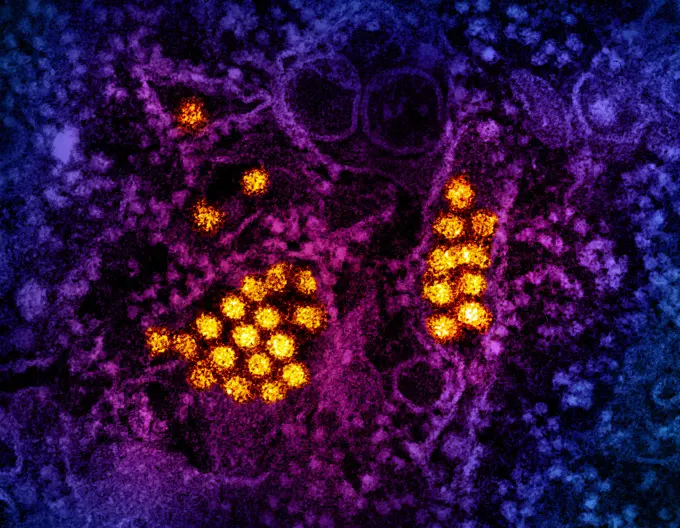
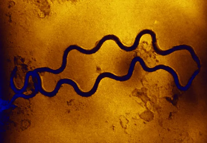
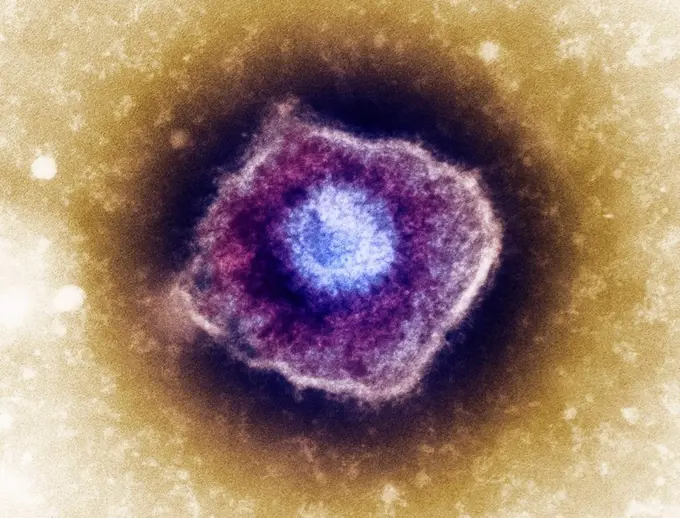
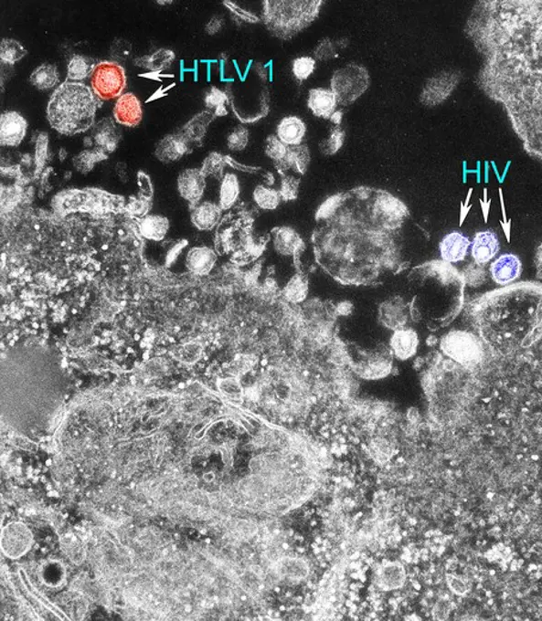
My Boards
View Pictures
  • My Boards

Viral Particles in Microscopy

Colorized transmission electron micrographs showcasing various viruses, including Hepatitis B, HIV-1, and SARS-CoV-2, against vibrant, abstract backgrounds.

Transmission electron micrograph of dengue virus particles (gold).
210 assets in this story
824-63127939
Bacteria responsible for syphilis. An electron photomicrograph of two spiral-shaped Treponema pallidum bacteria. Here we see two Treponema pallidum bacteria scanned by an electron microscope, magnified 36,000XT pallidum is the causative agent of syphilis. It contains one of the smallest prokaryotic genomes consisting of about 1000 kilobase pairs. See image n0831005 on the site.
824-65830914
Three influenza A (H5N1/bird flu) virus particles (rod-shaped purple).
4128R-3138
Varicella zoster virus particle, TEM
4297-1417
Transmission electron micrograph showing the presence of both the human T-cell leukemia type-1 virus (HTLV-1) and the human immunodeficiency virus (HIV)
824-63194385
This digitally-colorized scanning electron micrograph (SEM) depicts a single filamentous Ebola virus particle that had been bended from the surface of a VERO cell of the African green monkey kidney epithelial cell line. Ebola hemorrhagic fever (Ebola HF) is one of numerous Viral Hemorrhagic Fevers. It is a severe, often fatal disease in humans and nonhuman primates (such as monkeys, gorillas, and chimpanzees). Ebola HF is caused by infection with a virus of the family Filoviridae, genus Ebolavirus. When infection occurs, symptoms usually begin abruptly. The first Ebolavirus species was discovered in 1976 in what is now the Democratic Republic of the Congo near the Ebola River.
824-63123709
Thic electron micrograph reveals the morphologic traits of the Respiratory Syncytial Virus (RSV). The virion is variable in shape, and size (average diameter of between 120-300nm). RSV is the most common cause of bronchiolitis and pneumonia among infants
4297-1882
Scanning electron microscopic image of a Vaccinia virus
824-63194492
This colorized negative stained transmission electron micrograph (TEM) shows recreated 1918 influenza virions that were collected from supernatants of 1918-infected Madin-Darby Canine Kidney (MDCK) cells cultures 18 hours after infection. To separate these virions, the MDCK cells are spun down (centrifugation), and the 1918 virus in the fluid is immediately fixed for negative staining. The solid mass in lower center contains MDCK cell debris that did not spin down during the procedure.
1848-55194877
Wheals and nodules on an arm after an allergic reaction to harvest mites (Neotrombicula autumnalis)
1848-55194878
Wheals and nodules on a forearm after an allergic reaction to harvest mites (Neotrombicula autumnalis)
PREVIOUS
of 3
NEXT
2401 S. Ervay, Suite 206
Dallas, Texas 75215
United States
Get Started
Free ResearchMy BoardsMy Cart
For Creators
How To License Your ContentContributor PortalFrame of Mind
Resources
API accessPricing
Contact
+1 866 236 0087help@viewpictures.co.uk Contact form
©2026 View Pictures. All Rights Reserved. -A
This site is protected by reCAPTCHA and Google Privacy Policy and Terms of Service apply.