My Boards
View Pictures
  • My Boards

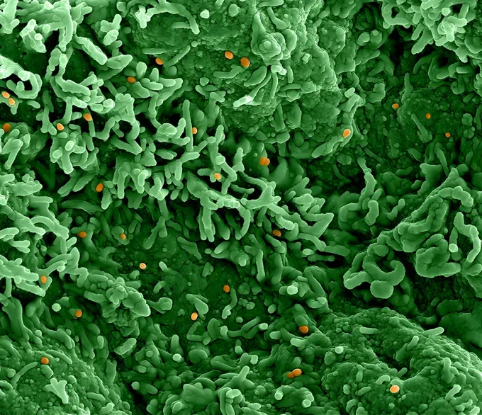
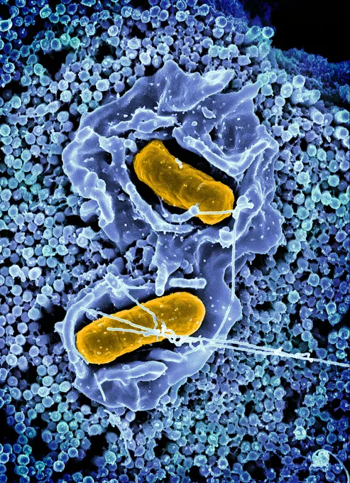
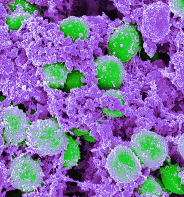
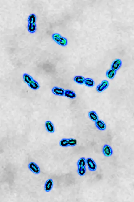
Virus Particles Under Electron Microscopy

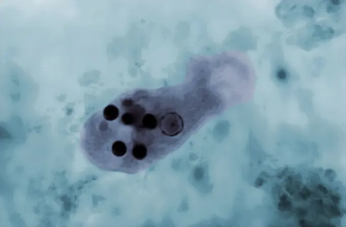
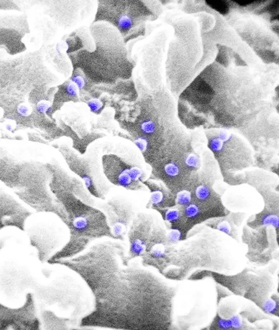
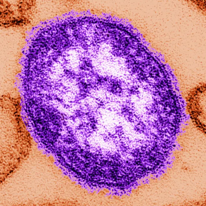
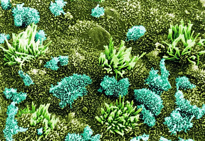
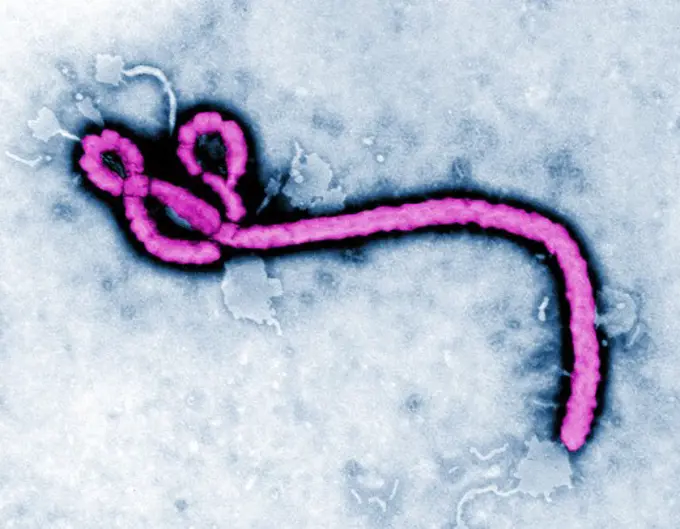
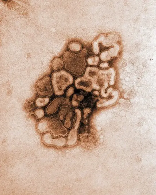
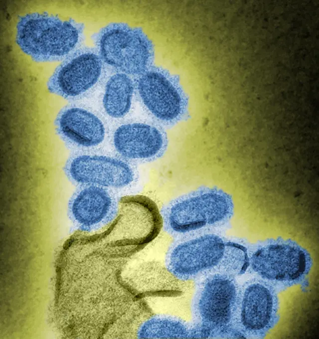
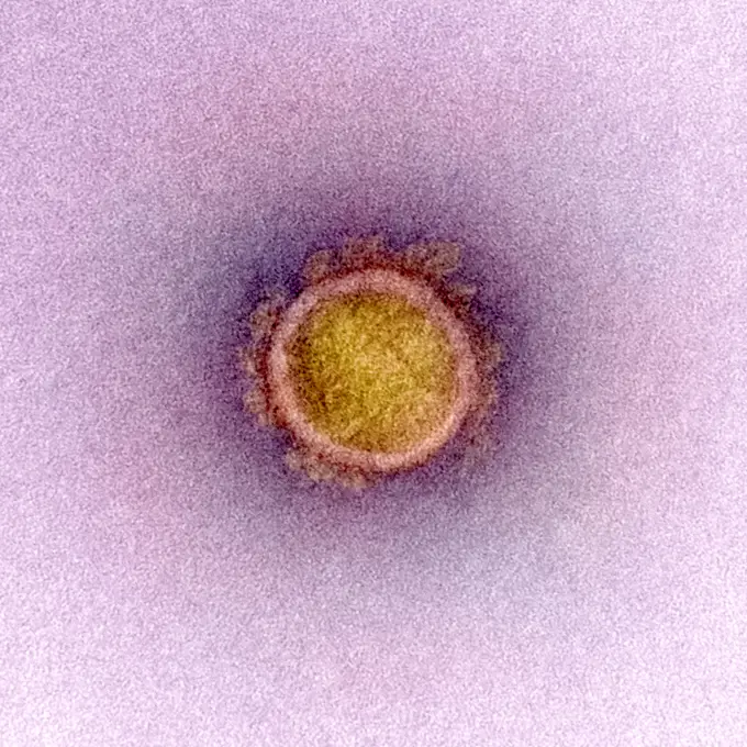
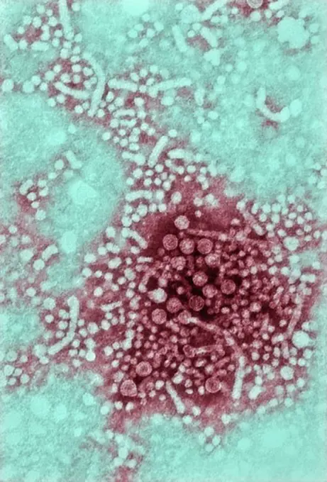
Colorized images of various virus particles including hepatitis B, herpes simplex, bunyavirus, smallpox, and influenza, showcasing detailed structures captured through transmission electron microscopy

Colorized negatively-stained transmission electron micrograph of hepatitis B virus, HBV, virions, also know as Dane particles
254 assets in this story
824-57657549
Colorized scanning electron micrograph of monkeypox virus (orange) on the surface of infected VERO E6 cells (green).
824-65831054
Scanning electron micrograph of Salmonella Typhimurium invading a human epithelial cell.
824-63194631
Scanning electromicrograph of Staphylococcus aureus bacteria.
824-63225823
Colorized scanning electron micrograph of a cell (red) heavily infected with SARS-CoV-2 virus particles (green), isolated from a patient sample. Image captured at the NIAID Integrated Research Facility (IRF) in Fort Detrick, Maryland.
824-63222973
Pneumococcal bacteria (streptococcus pneumoniae). Pneumococcus is an important pathogen in humans. It is responsible for many infections (pneumonia). He was responsible for pneumonia during the Spanish flu pandemic). Visualized by optical microscopy.
824-63194481
Under a plate magnification of 37,800X, this colorized transmission electron micrograph (TEM) depicted a strain of swine flu, the A/New Jersey/76 (Hsw1N1) virus, while in the virus' first developmental passage through a chicken egg. Swine Influenza (swine flu) is a respiratory disease of pigs caused by type A influenza that regularly cause outbreaks of influenza among pigs. Swine flu viruses cause high levels of illness and low death rates among pigs. Swine influenza viruses may circulate in swine throughout the year, but most outbreaks among swine herds occur during the late fall and winter months similar to humans. The classical swine flu virus (an influenza type A H1N1 virus) was first isolated from a pig in 1930.
824-63197031
Human papilloma virus (HPV). Image produced using high-dynamic-range imaging (HDRI) from an image taken with transmission electron microscopy. Viral diameter around 55 nm.
1899-65662326
Colorized transmission electron micrograph of an mpox virus particle (yellow and red) found within an infected VERO E6 cell (blue), cultured in the laboratory.
1899-61460356
Colorized scanning electron micrograph of monkeypox virus (orange) on the surface of infected VERO E6 cells (green)
1899-61460869
This photomicrograph of a trichrome-stained specimen revealed the presence of an Entamoeba histolytica trophozoite, in which a number of phagocytosed erythrocytes could be seen as round, dark inclusions. CDC/ Dr Mae Melvin Dr Greene 1966
4128-111584780
Convolvulus pollen grains. Coloured scanning electron micrograph (SEM) of pollen grains from a convolvulus flower.Convolvulus is a genus of about 200 ...
4128-V58562081
6145-46770845
Micrograph of milk. Milk stored 1 week in the refrigerator. Sample no. 34.
4297-1221
Scanning electron micrograph of human immunodeficiency virus (HIV), grown in cultured lymphocytes. Virions are seen as small spheres on the surface of the cells
4384-115
This thin-section transmission electron micrograph (TEM) revealed the ultrastructural appearance of a single virus particle, or virion, of measles virus. The measles virus is a  paramyxovirus, genus  Morbillivirus. It is 100-200 nm in diameter, with a core of single-stranded RNA, and is closely related to the rinderpest and  canine distemper  viruses. Two membrane envelope proteins are important in pathogenesis. They are the F (fusion) protein, which is responsible for fusion of virus and host
4128R-2143
Coloured SEM of uterus wall early proliferation
1899-65662463
Colorized scanning electron micrograph of mpox virus (purple) on the surface of infected VERO E6 cells (tan).
4239-18641827
25,000x magnified colorized scanning electron micrograph of filamentous Ebola virus particles (red) budding from a chronically infected VERO E6 cell (blue).
824-63191208
Scanning electron micrograph of HIV particles infecting a human T cell.
824-57659471
This photomicrograph revealed the presence of an Entamoeba histolytica trophozoite, which was photographed here while alive. This organism displayed a limax-like movement, meaning it moved much like a garden slug, crawling or advancing its anterior body, extending a single, large pseudopodia, while pulling itself forward. CDC/Armed Forces Pathology Institute (AFIP) 1964
4201-66340
Sole (Solea sp) SEM close-up view of scales at 42x magnification
1899-65662397
Colorized transmission electron micrograph of Ebola virus particles (gold) found both extracellularly and budding from chronically infected African green monkey kidney cells (green).
4128-111612486
Insulin bound to insulin receptors and glucose transport into the cell, illustration. Insulin receptors (blue) are transmembrane proteins, that are ac...
824-63179048
Pasteurella multocida or Pasteurella septica is the bacillus involved in the infection of wounds by animal bites or scratches. After an incubation of a few hours appears a Pasteurellian phlegmon characterized by lymphangitis. Optical microscopy.
4128-20043925
Nuclear pore complex, molecular model
824-63227448
Zika virus particles (red) shown in African green monkey kidney cells.
4269-27737
Leptospira. Scanning electron micrograph (SEM) of the Gram-negative bacteria Leptospira sp. Leptospira is a type of spirochete. Many species are pathogenic, causing leptospirosis (Weil's disease) or other diseases with similar symptoms, attacking the liver (jaundice) or the meninges of the brain (meningitis).
824-63194483
Under a plate magnification of 37,800X, this colorized transmission electron micrograph (TEM) depicted a strain of swine flu, the A/New Jersey/76 (Hsw1N1) virus, while in the virus' first developmental passage through a chicken egg. Swine Influenza (swine flu) is a respiratory disease of pigs caused by type A influenza that regularly cause outbreaks of influenza among pigs. Swine flu viruses cause high levels of illness and low death rates among pigs. Swine influenza viruses may circulate in swine throughout the year, but most outbreaks among swine herds occur during the late fall and winter months similar to humans. The classical swine flu virus (an influenza type A H1N1 virus) was first isolated from a pig in 1930.
824-63223257
Colorized scanning electron micrograph of a cell heavily infected with SARS-CoV-2 virus particles (yellow), isolated from a patient sample. The black area in the image is extracellular space between the cells. Image captured at the NIAID Integrated Research Facility (IRF) in Fort Detrick, Maryland. Credit: NIAID.
4297-1009
Colorized scanning electron microscope image of amaranthus pollen. The pollen has been acetolyzed to remove cytoplasm and pollenkit in order to reveal the intricate wall structure. Family: Amaranthaceae: Amaranthus opopeo
824-57657697
Transmission electron micrograph of West Nile virus particles (gold) replicating within the cytoplasm of an infected VERO E6 cell (pink).
824-66066020
A 3D rendering of an antibody (foreground left), and examples of high-priority &quotprototype&quot pathogens that have the potential to threaten human health/are the focus of pandemic preparedness research efforts (left to right): hantavirus, yellow fever virus, Nipah virus, picornavirus, and Chikungunya. As of September 2024, The National Institutes of Health (NIH) has established a pandemic preparedness research network known as ReVAMPP to conduct research on high-priority pathogens with the goal of developing effective vaccines and monoclonal antibodies. Credit: NIAID See related September 13, 2024 NIAID news release, &quotNIH Awards Establish Pandemic Preparedness Research Network,&quot at.
824-65830824
Colorized transmission electron micrograph of an extracellular HIV-1 virus particle (blue) found outside of an H9 T cell (light green).
4128-17857706
Helianthus stem. Light micrograph (LM) of the stem of a perennial sunflower (Helianthus sp.) in cross-section showing the vascular bundles (green, yel...
824-65830825
Colorized transmission electron micrograph of an extracellular HIV-1 virus particle (red) found outside of an H9 T cell (light blue). 2024 : A Change in Drug Regimen is Associated with Temporary Increases in Dormant HIV.
4179-42166
Isopod (Idotea sp) Tatoosh Island, Washington
4197-V65369290
1788-111174239
Green alga, Chlorella vulgaris, seen under a microscope, at x670 magnification.
4269-5518
Color enhanced Transmission Electron Micrograph (TEM) of the ebola virus, an RNA virus (filovirus) causing hemorrhagic fever.
824-63179035
Leptospira interrogans, bacterium responsible for icterohemorrhagic leptospirosis or Weil's disease which is the severe form of leptospirosis, it associates: hemorrhage, acute renal failure, and neurological impairment. Optical microscopy X 1000
6177-V53548826
4269-7092
Transmission electron micrograph (TEM) of two avian influenza A H5N1 viruses. Magnification x 108,000.
4297-1454
Colorized transmission electron micrograph revealed some of the ultrastructural morphology displayed by an Ebola virus virion
4384-226
Swine Flu Virus
4128R-16119
Mitosis nuclear division, computer artwork.
4269-5520
Color enhanced Transmission Electron Micrograph (TEM) of the ebola virus, an RNA virus (filovirus) causing hemorrhagic fever.
824-63123773
Scanning electron micrograph of Leptospira interrogans strain RGA. Two spirochetes bound to a 0.2 µm filter. Strain RGA was isolated in 1915 by Uhlenhuth and Fromme from the blood of a soldier in Belgium. Leptospira is known to cause the infectious disea
824-63123784
Ebola virus seen at the MET. The Ebola virus is an RNA virus. It is responsible for severe hemorrhagic fevers. This microorganism is on the CDC's list of infectious agents most likely to be used as biological weapons, due to its high virulence, its ease of dissemination and the significant mortality it induces.
4384-295
Scanning Electron Micrograph of Borrelia burgdorferi Bacteria
824-63194420
Under a very high magnification of 50,000x, this scanning electron micrograph (SEM) shows a strain of Staphylococcus aureus bacteria taken from a vancomycin intermediate resistant culture (VISA). Under SEM, one can not tell the difference between bacteria that are susceptible, or multidrug resistant, but with transmission electron microscopy (TEM), VISA isolates exhibit a thickening in the cell wall that may attribute to their reduced susceptibility to vancomycin . VISA and VRSA are specific types of antimicrobial-resistant staph bacteria. While most staph bacteria are susceptible to the antimicrobial agent vancomycin some have developed resistance. VISA and VRSA cannot be successfully treated with vancomycin because these organisms are no longer susceptibile to vancomycin. However, to date, all VISA and VRSA isolates have been susceptible to other Food and Drug Administration (FDA) approved drugs.
824-63194495
This colorized negative stained transmission electron micrograph (TEM) shows recreated 1918 influenza virions that were collected from supernatants of 1918-infected Madin-Darby Canine Kidney (MDCK) cells cultures 18 hours after infection. To separate these virions, the MDCK cells are spun down (centrifugation), and the 1918 virus in the fluid is immediately fixed for negative staining. The solid mass in lower center contains MDCK cell debris that did not spin down during the procedure.
824-63179039
Meningococcus or Neisseria meningitidis, is a bacterium that can cause meningitis. The bacteria are germs that can be found in the rhinopharynx, where it can cause benign rhinopharyngitis or an asymptomatic state. Only one in 400 carriers become victim to a serious meningococcal disease, mostly commonly acute purulent meningitis. Seen through an optical microscope x 1850.
824-63223214
Transmission electron micrograph of a SARS-CoV-2 virus particle, isolated from a patient. Image captured and color-enhanced at the NIAID Integrated Research Facility (IRF) in Fort Detrick, Maryland. Credit: NIAID.
1746-111525222
Colorized transmission electron micrograph (TEM); Created by CDC microbiologist Cynthia Goldsmith; ultra structural morphology displayed by an Ebola virus virion
PREVIOUS
of 3
NEXT
2401 S. Ervay, Suite 206
Dallas, Texas 75215
United States
Get Started
Free ResearchMy BoardsMy Cart
For Creators
How To License Your ContentContributor PortalFrame of Mind
Resources
API accessPricing
Contact
+1 866 236 0087help@viewpictures.co.uk Contact form
©2026 View Pictures. All Rights Reserved. -B
This site is protected by reCAPTCHA and Google Privacy Policy and Terms of Service apply.