My Boards
View Pictures
  • My Boards

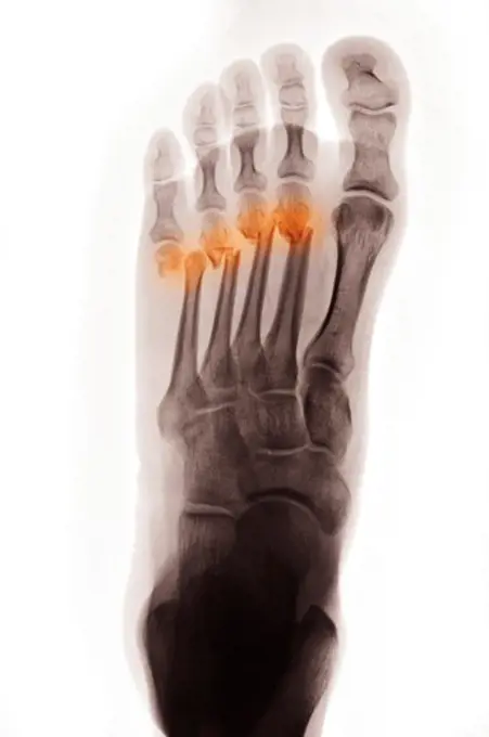
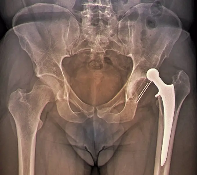
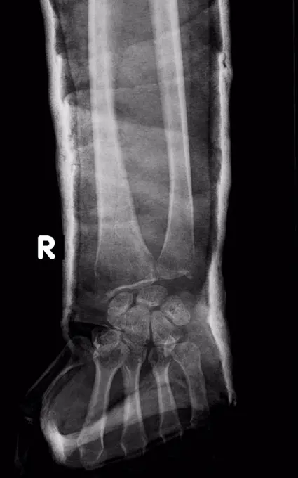
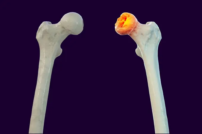
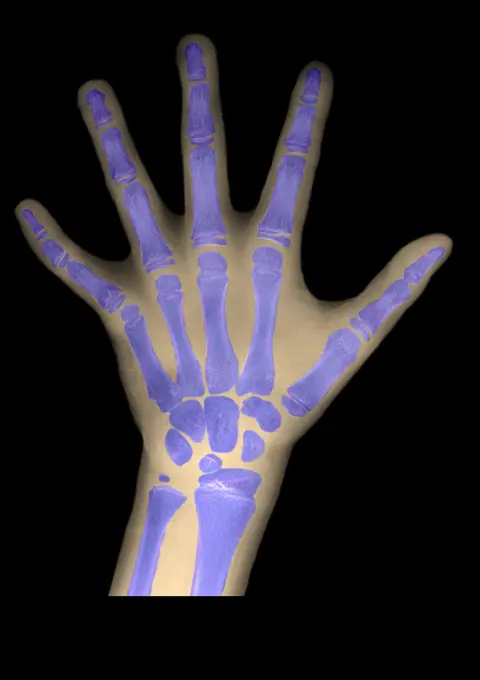
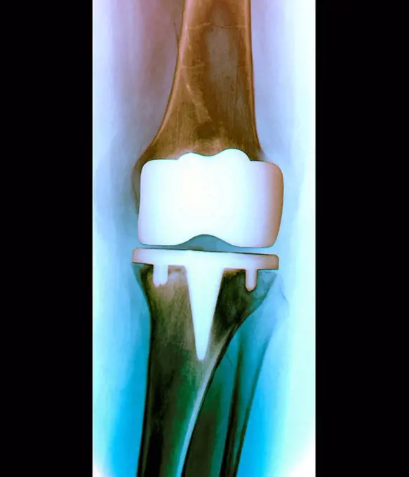
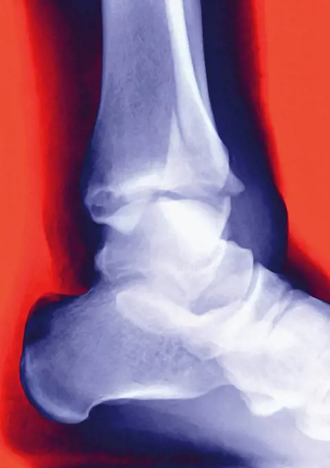
X-ray and Medical Imaging

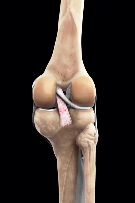
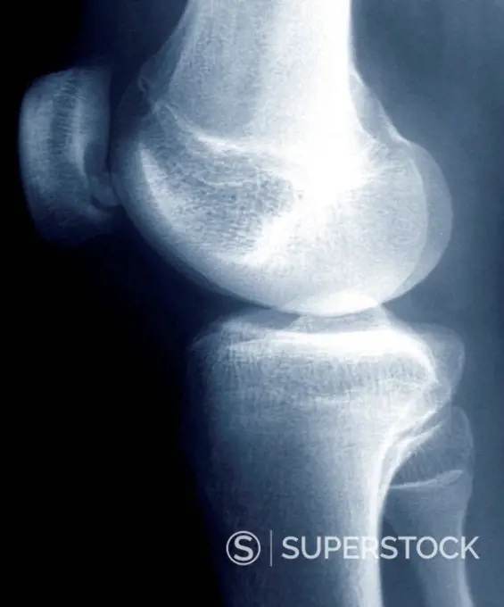
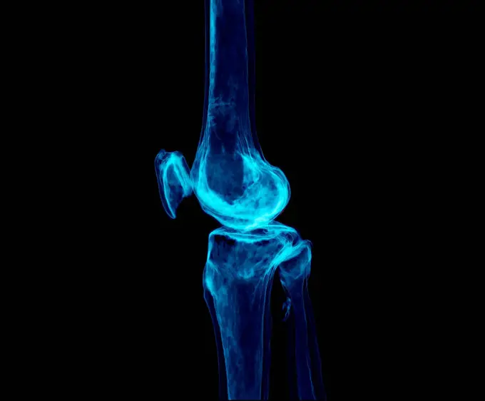
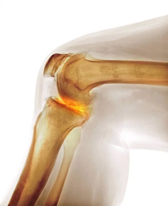
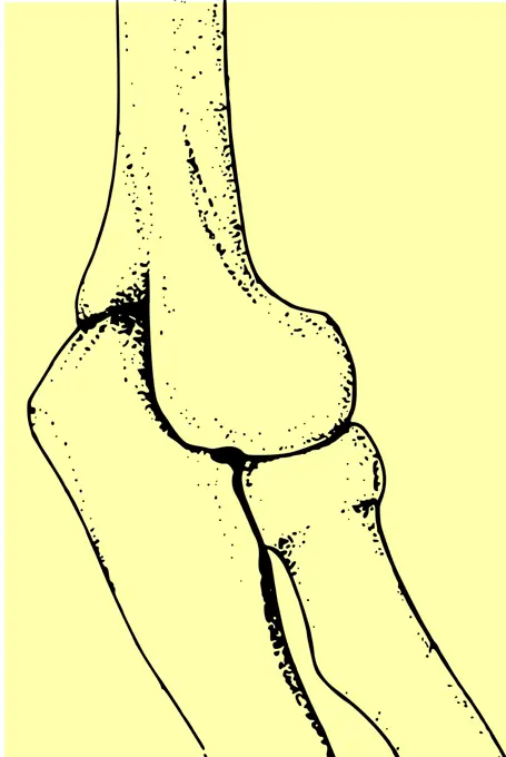
Detailed images of knee joints showcased through X-rays and MRIs, highlighting both healthy and injured conditions to represent medical assessments.

308 assets in this story
6188-55490768
X-ray picture of human knee joint X-ray picture of human knee joint isolated on white background ,property released Copyright: xZoonar.com/ValeryxVoennyyx 7103478 ,model released, Symbolfoto ,property released
1525R-178783
1556R-0432
X-ray of broken leg
6188-66528731
Genu valgum knock knees. Genu valgum is a deformity of the lower limb corresponding to an outward deviation of the leg the knees protrude inwards, the ankles do not touch. We speak of genu valgum when the angle between the femur and the tibia is less than 170. Genu valgum drawing 016238_053
824-63212803
Arthrodesis of the ankle involving the tibia, the talus and the heel bone. Saggital plane x-ray of the right ankle.
1525-24588656
Human bones joint vector. Isolated knee or elbow bones, orthopedics and arthritis. Knee or elbow bones joint isolated
1574R-018854
X-ray of a foot
824-63178679
X ray of the left foot of a 29 years old male patient. Normal
4128-16072254
Arm and hand in profile, X-ray
4128R-11287832
Osteoarthritis of the ankle. Coloured X-ray of the ankle of an 80 year old female patient with severe osteoarthritis.
4128R-15515047
Illustration of the posterior cruciate ligament
4128R-15465
Normal shoulder. X_ray of the healthy left shoulder of a 28 year old patient.
1899-53511737
Osteoarthritis of the left hip (coxarthrosis) visualized by a pelvic x-ray of the front. (Photo by: CAVALLINI JAMES/BSIP/UIG)
1525-28446598
x-ray of the leg. A steel plate holding together the tibia of the leg.. steel plate holding together the tibia of the leg.
4128R-30951
Healthy ankle joint. Coloured frontal X-ray of the left ankle of a 21 year old patient. Strapping around the ankle is visible on this X-ray.
4128R-3331
Prosthetic knee joint, X_ray
4128R-15516615
Illustration of an elbow implant
1525-22128975
X-ray of a human leg with metal screws and rods
4378-20013580
Medial Tibial Stress Syndrome
4297-1058
Normal hand x-ray of a 19 year old boy
4297-1205
X-ray showing metatarsal fractures in a 37 year old woman
1848-53980274
Screwed
824-63190339
Healthy knee seen from the side.
4128-V58576822
4128-16071979
Hand with thumb up, X-ray
4128R-30955
Dislocated hip replacement. Coloured frontal X-ray of the pelvis of a 35 year old patient with an artificial left hip (right) that has become dislocat...
1899-53508087
X-ray of the hip before fitting a hip prosthesis.
824-63205978
Nephritic colic seen on a frontal x-ray of the abdomen.
4378-20013602
Right Knee Bones
6188-55566899
Xray Medical X-Ray imaging of a shoulder, used in diagnostic radiology of skeleton bones Copyright: xZoonar.com/ClaudioxDiviziax 823019
4128-19248933
Painful knee joints, illustration.
4378-5459
Posterior Cruciate Ligament Injury
4128-18929627
Fractured hand in cast, X-ray
4128R-12658
Arthritis of the knee. Frontal coloured X_ray showing the knee of a 59 year old patient with arthritis.
1899-86171
Knee X Ray
4128R-13412585
Bones of Foot
1525-22722422
Human joints vector icons for orthopedics and surgery medical design. Vector isolated icons of leg knee or arm and hand joints with cartilage synovial fluid for orthopedics treatment medicine pills. Vector icons of joints and bones
1838-76240503
X-ray of adult man's left knee after knee replacement surgery
4128-18929608
Supracondylar humerus fracture, X-ray
1899-53509069
Bone system, the bony joint of the hip.
4297-1154
Colorized x-ray of the hand of a 40 year old man showing the surgical repair of a boxer's fracture with a plate and screws
4128-20043164
Broken hand, X-ray
1848-55284665
X-ray after osterosynthesis, metal implant for correction of joint position
4128R-12769
Normal knee. Coloured X_ray of the knee of a 44 year old woman.
4128-V58575029
4239-18641990
Hand showing osteoarthritic joints.
1899-85990
Foot X Ray
824-63206084
Illustration of osteoarthritis in the shoulder. Bone and cartilage damage most often due to age, which deforms the joint and inflames the synovial membrane. The inter-joint area diminishes, pain due to inflammation if felt.
4378-20013943
Right Knee Ligaments
824-63224568
Fracture of the foot on the wedge bone. Foot scanner
1525R-178839
4128-16224241
Hand with jewellery, X-ray
4128-28970849
Healthy elbow joint, X-ray
4128R-11544872
Human knee joint, computer illustration.
1838-76240502
X-ray of broken toe on left foot
4128-19489678
Humerus fracture, X-ray
4128R-15516396
Illustration of a bunion
4239-18641984
Front view of knee showing lateral collateral ligament tear.
4378-20014114
Posterior Cruciate Ligament Injury
824-63163786
Bone system: silhouette of a 70-year-old woman with severe osteoporosis.
4128-19357528
Contrast barium enema X-ray of a patient with chronic diarrhoea and anaemia showing a filling defect with an irregular margin called the 'apple core s...
4297-1517
Colorized x-ray showing the surgical repair of an ankle fracture in a 61 year old man
4128R-13374634
Foot bones
4128R-29893
Ankle pain, conceptual computer artwork.
824-63178629
Calculation of the bone age with a radiography. 12 years old hand x ray. Normal size
1848-55989191
Doctor analyzing an X-ray radiograph
4128-20243407
Hip osteoarthritis, CT scan
824-63217799
Normal knee visualized by MRI in sagittal section.
1848-56124319
X-ray of a leg fracture and external fixation
4128-30420597
Fractured leg, X-ray
6177-V66000595
4128-30420317
Arthritis of the foot, conceptual illustration
4128R-30963
Knee disease. Coloured X-ray of the right knee of an 18 year old patient with osteochondritis dissecans (OCD) of the knee. A small piece of bone and c...
4128-28514565
Knee, CT scan
4128R-12781
Arthritis of the knee. Coloured X_ray the arthritic knee of a 37 year old man. The knee had previously been damaged in a motorbike accident.
6188-55478601
X-ray image of human shoulder joint film with X-ray image of front view of human shoulder joint isolated on white background Copyright: xZoonar.com/ValeryxVoennyyx 13808382
824-63224569
Fracture of the foot on the wedge bone. Foot scanner
4128R-7476
Normal elbow joint, X_ray
1525R-103025
Mid-adult Caucasian male holding a x-ray.
1525-23789132
The fibula helps stabilize and support your leg, body, ankle, and leg muscles 3d illustration. The fibula helps stabilize and support your leg, body, ankle, and leg muscles
4128R-12964474
Human knee joint, Illustration
4128-19357537
Bone tumour of the elbow. X-ray showing the humerus osteosarcoma with an ill-divined lytic lesion with wide zone transition and a Codman triangle, a t...
4128-28968679
Legg-Calve-Perthes disease, illustration
824-63178641
6 years old child radiography. Normal bone age
4378-20393201
Knee Replacement
4128R-5201
Prosthetic knee joint, X_ray
4128R-2213
Fractured ankle, X_ray
4269-27494
Shoulder chondrosarcoma. X-ray of the shoulder showing a chondrosarcoma of the shoulder blade. The tumor is evidenced by the presence of a vascularized heterogenous lytic area with bone calcifications.
4128-28769199
Healthy foot, X-ray
1525-22197504
painful ankle
4239-18641997
Front view and cross section of knee showing moderate osteoarthritis.
4128-19358205
Left oblique hysterosalpingography (X-ray) of a 35 year old women with infertility for 3 years showing bilateral patent tuba fallopian and bilateral c...
824-63163800
Bone system: silhouette of a 60-year-old woman with moderate osteoporosis.
4128R-11298421
Knock knee, computer artwork.
4128R-13412586
Bones of Foot
824-72488521
Genu valgum (knock knees). Genu valgum is a deformity of the lower limb corresponding to an outward deviation of the leg (the knees protrude inwards, the ankles do not touch). We speak of genu valgum when the angle between the femur and the tibia is less than 170.
4128R-12577968
Artificial heart valves. Coloured X-ray of the chest of a 59 year old patient with heart failure who has had both the mitral and aortic valves of thei...
824-63203271
Osseous edema on the knee. Sub-cortical lesion of the condyle, seen on a frontal section MRI.
6188-55565476
Der Ellenbogen Der Ellenbogen Copyright: xZoonar.com/curryx 2482837
1525-26590585
Knee pain 2D vector isolated illustration. Ruptured ligament risk flat sticker on cartoon background. Arthritis conditions. Overuse injury colourful scene for mobile, website, presentation. Knee pain 2D vector isolated illustration
PREVIOUS
of 4
NEXT
2401 S. Ervay, Suite 206
Dallas, Texas 75215
United States
Get Started
Free ResearchMy BoardsMy Cart
For Creators
How To License Your ContentContributor PortalFrame of Mind
Resources
API accessPricing
Contact
+1 866 236 0087help@viewpictures.co.uk Contact form
©2026 View Pictures. All Rights Reserved. -A
This site is protected by reCAPTCHA and Google Privacy Policy and Terms of Service apply.