My Boards
View Pictures
  • My Boards
← Medical & Healthcare

X-ray and Medical Imaging

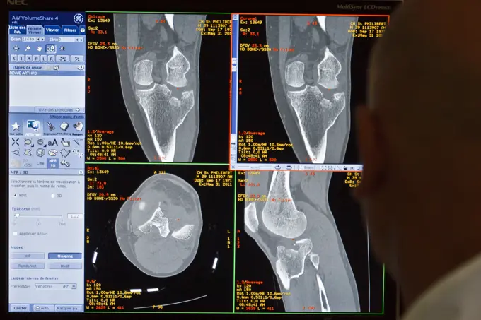
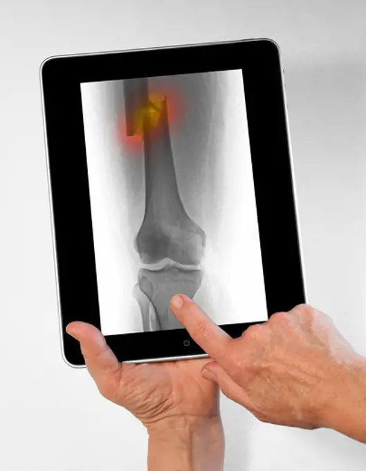
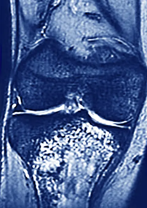
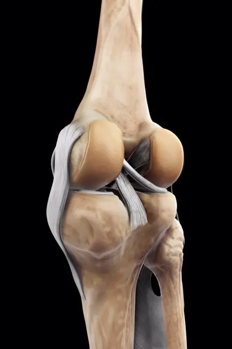
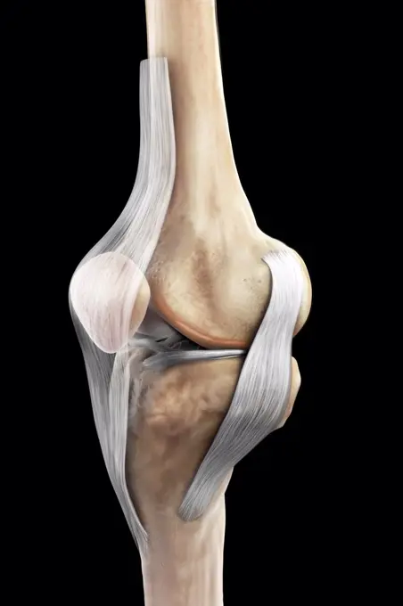
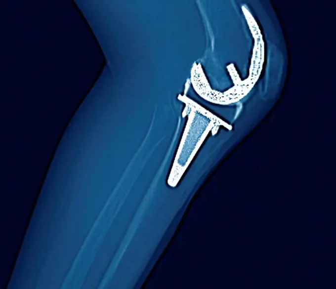
Detailed images of knee joints showcased through X-rays and MRIs, highlighting both healthy and injured conditions to represent medical assessments.

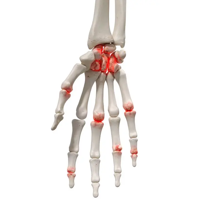
308 assets in this story
4128-V58575031
4128-V58575817
4128R-13374722
Foot bones
824-63198848
Degenerative disk disease. Severe vertebral osteoarthritis with osteoporsis and osteophytes, seen on a frontal lumbar x-ray.
4128-28968666
Legg-Calve-Perthes disease, illustration
4378-20393496
Tennis Elbow
4378-20013598
Right Knee Ligaments
824-63190359
Illustration of torn ligaments on the internal side of the ankle, as a result of an evarsion of the foot.
4269-24800
Rheumatoid polyarthritis. X-ray of a shoulder affected by a rheumatoid polyarthritis on the shoulder, evidenced by the narrowing of the joint space and the erosion of the cartilage and bones.
4239-18641934
Side and back view of ruptured achilles tendon.
824-63211027
Prostatic adenoma seen on a frontal pelvic x-ray.
4128R-6325
Arthritic hand, X_ray
4128R-11287594
Dislocated shoulder. Coloured X-ray of a 77 year old female patient with a dislocated shoulder.
4128R-11287837
Foreign body in finger. Coloured X-ray of the finger of a 60 year old male patient that contains a foreign body that is causing swelling of the soft t...
4128R-7133
Total hip replacement, X_ray
4128-30420307
Arthritis of the foot, conceptual illustration
824-63198626
Representation of a hallux valgus. Also called bunion, hallux valgus corresponds to an abnormal deviation of the big toe, which causes a deformation of the forefoot at the level of the first metatarsal and the big toe.
4128R-14637716
Arthritis in the shoulder, illustration
4128-V58567700
824-63178645
Delay bone age Hand xray 12 years and 10 months old child measuring 1meter 35 which is the height of a 10 years old girl.
4408-1001
Left Hand, Medical - Xray / Radiology
1848-61113911
Cervical spine
824-72488514
Healthy lumbar vertebrae. See images 1530707 for spondylarthrosis with interapophyseal osteoarthritis and spinal cord compression, 1530807 for simple discarthrosis.
1525-56450594
Knee problem icon flat vector. Joint pain. Doctor bone. Knee problem icon flat vector. Joint pain
824-63173351
Ct scanner examination
4128-16073322
Scans of feet and ankles side, MRI
824-63194015
Pelvic x-ray on a 93-year old woman, showing widespread bone demineralisation.
4297R-2004
Digital tablet showing an x-ray of a femur fracture
1525-24357295
Human knee joint
824-63170764
Osteosarcoma of the upper part of the tibia. MRI of the knee in front view.
824-63198858
Degenerative disk disease. Severe vertebral osteoarthritis with osteoporsis and osteophytes, seen on a side lumbar x-ray.
4128-18929585
Rectal diverticulum and proctitis, X-ray
4128-28575124
Mammogram
824-57655647
X-ray of rheumatism in the hands.
824-63214260
Diverticulum in the bladder. Frontal pelvic x-ray.
1899-54027989
Normal knee seen on a sagittal section MRI scan.
4128-28968674
Legg-Calve-Perthes disease, illustration
4269-25673
Knee arthrosis. X-ray of knees affected by arthosis (green), also called osteoarthritis or gonarthrosis. It is a joint disease that can cause cartilage destruction (seen as a reduction in the joint space), bone erosions, and tendon inflammation.
4128-20043275
Rectovesical fistula, X-ray
4128R-11287871
Fractured collarbone. Coloured X-ray of a 16 year old male with a fractured clavicle (collar bone).
4128-18929621
Healthy bladder and urethra, X-ray
4128-19358342
Achilles tendon injury, illustration.
4128-V58573742
4128-24795475
Sinusitis, X-ray
4128-18929503
Colles fracture, X-ray
1899-54027131
Arteritis seen in an angiogram of the left hand.
6188-55562556
Computer Key Springing Up holding Human Hand X-ray Computer Key Springing Up holding Human Hand X-ray Copyright: xZoonar.com/yogeshxmorex 5529176
1899-54026848
Congential dislocation of the right hip in a 2 and a half-year old child, seen on a frontal pelvic x-ray.
4128R-13326081
Human elbow ligament, illustration.
4297-1077
Lumbar spine x-ray of a 41 year old man showing minor degenerative changes
824-63178583
Nose-forehead x-ray of the sinuses of a 42-year-old woman. Normal transparency of all the sinuses of the face. Bilateral hypertrophy of the inferior turbinates.
4443-21167902
roux's amputation, medical equipment, surgical instrument, history of medicine
4128R-13410633
Leg foot
4378-5559
Right Knee Ligaments
1525-56234371
A tear in the anterior cruciate ligament is one of the most common knee injuries. 3D rendering. A tear in the anterior cruciate ligament is one of the most common knee injuries.
4128R-14637724
Arthritis in the hand, illustration
4378-20013559
Right Knee Ligaments
1439-57940803
Xray of a shoulder with orthopedic hardware
4443-21168015
eicision of first metatarsal bone, flap, medical equipment, surgical instrument, history of medicine
824-63178438
MRI of the right knee. Adult male No pathology
824-63194026
Pelvic x-ray on a 93-year old woman, showing widespread bone demineralisation.
1525-28452033
Dental x ray film for dental care concept
1841R-111799
824-63178642
6 years old child radiography. Normal bone age
4378-20393302
Knee Replacement
6188-55584280
Xray Medical X-Ray imaging of hand fingers used in diagnostic radiology of skeleton bones Copyright: xZoonar.com/ClaudioxDiviziax 7589775
4378-5222
Right Knee Ligaments
1525-26700106
Side view of the spine on a white background. Vector illustration. Side view of the spine on a white background
1838-14333
X-Ray of Foot on Computer Screen
4128-V58575824
824-63210142
Reportage on a mountain rescue doctor working in both the Carroz and de Flaine ski resorts in Haute-Savoie, France. He treats skiers and snowboarders who have been injured on the slopes. X-ray of a patient following a skiing accident. He has a displaced fracture of the tibia and fibula.
4269-19001
This 2D and 3D digital X-ray imaging equipment is dedicated to the orthopedic practice and permits a static study of the musculoskeletal system. It takes by low dose X-ray scanning two simultaneous, perpendicular planar views in the standing position. A reconstruction provides a 2 and 3D image of the osteoarticular structures of the patient. The university hospital of Bordeaux is the first hospital to use this diagnostic technique in France.
4128R-4939
Temporary ankle immobilisation, X_ray
824-63178615
Renal lithiasis, x-ray
1525-26855065
RX diagnostic of neck. Element of design.
1525-56599220
Sick red place. Crack in White bone. Magnifying glass. Logo in circle. Vertebral column. X-ray of internal organs. Medical care. Fracture of intervertebral discs. Problems with spine. Posture issues
4128-18929565
Gun shot in leg, X-ray
4408-1003
Skull and Neck, Medical - Xray / Radiology
6188-62271154
Magnetic resonance imaging of the cervical spine. Magnetic resonance imaging of the cervical spine. MRI vertebral discs in different views Copyright: xZoonar.com/MaximxxPavlovx 9378556
824-63208947
Reportage in a radiology centre in Haute-Savoie, France. X-ray of a patient with a knee prosthesis.
6019-29996487
Neck and skull base x-ray film
4443-21167896
disarticulation of the great toe, medical equipment, surgical instrument, history of medicine
4128R-14637719
Arthritis in the cervical spine, illustration
4128R-6280
Knee replacement, CT scan
1525-24760350
Orthopedic and spine icons set. Cartoon illustration of 16 orthopedic and spine vector icons for web. Orthopedic and spine icons set, cartoon style
4409-17348996
Knee joint-cartilage.
824-63190062
Reportage at the Maussins-Nollet Clinic in Paris. X-ray of a hip prosthesis.
824-63224951
Dental panoramic of a 70 year old man with an implant and many crowns, the crosses visualizing the teeth that will need care. Paris, Ile-de-France, France.
824-63178596
Sinus X-ray of a 35-year-old woman. No anomaly noted
1525-26870450
Foot Bone Anatomy Vector Illustration
824-63191356
Illustration of the various types of fracture in the proximal part of the femur. From top to bottom : -subcapital fracture -transcervical fracture -petrochanteric fracture -subtrochanteric fracture
1439-57940801
Medical Xray showing spinal fixation devices
824-29055559
X-ray of the knees of a patient wearing a prosthesis.
4128-20043126
Kidney stones, X-ray
1525-76145721
Problems with spine. Posture issues. Sick red place. Crack in White bone. Magnifying glass. Logo in circle. Vertebral column. X-ray of internal organs. Medical care. Fracture of intervertebral discs. Problems with spine. Posture issues
4129-1469
X-ray of wounded spine
1899-54027171
Rheumatoid arthritis seen on an x-ray of the hand.
4297-1075
Lumbar spine of an 80 year old man with spinal fusion hardware
4128-19358071
Lateral left decubitus (LLD) abdominal X-ray of a patient with abdominal pain showing a crescent shape radiolucent lesion above the liver suggestive o...
6188-65130956
Kniebish leg with kneeclover
PREVIOUS
of 4
NEXT
2401 S. Ervay, Suite 206
Dallas, Texas 75215
United States
Get Started
Free ResearchMy BoardsMy Cart
For Creators
How To License Your ContentContributor PortalFrame of Mind
Resources
API accessPricing
Contact
+1 866 236 0087help@viewpictures.co.uk Contact form
©2026 View Pictures. All Rights Reserved. -A
This site is protected by reCAPTCHA and Google Privacy Policy and Terms of Service apply.