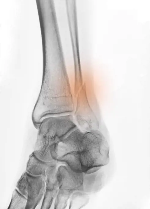
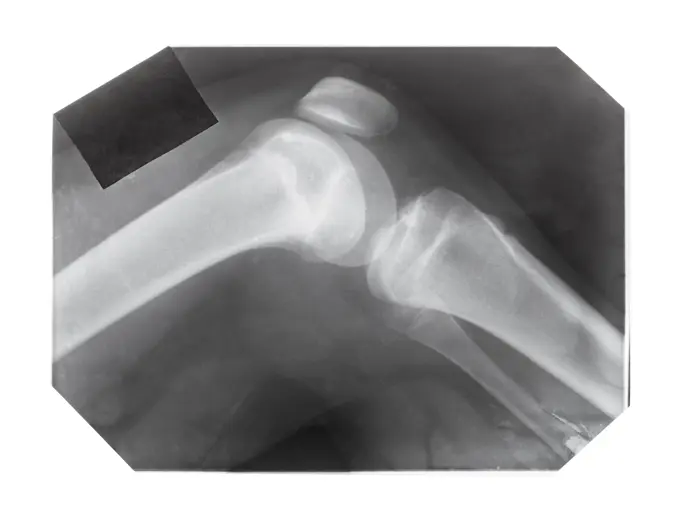
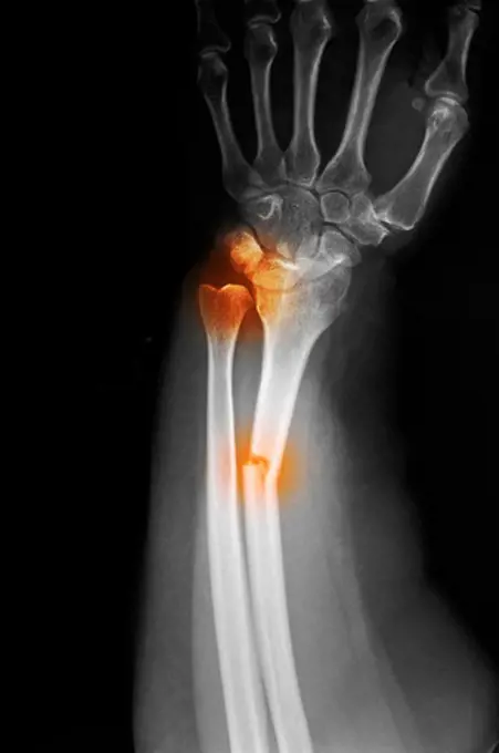
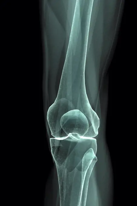
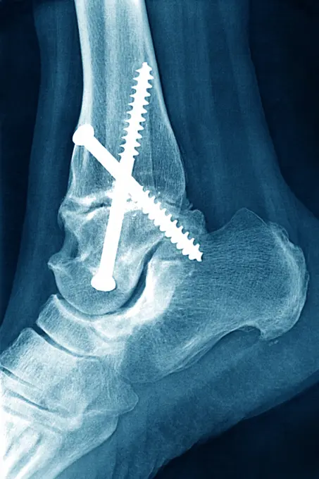
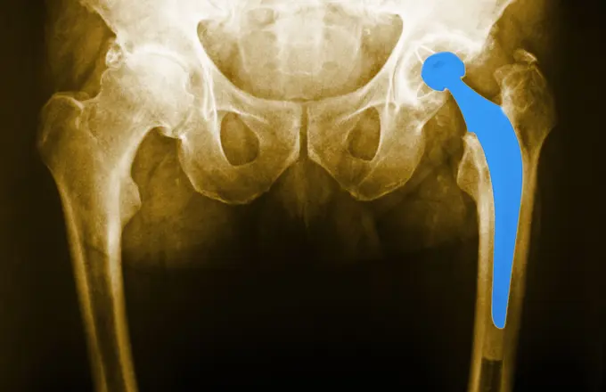
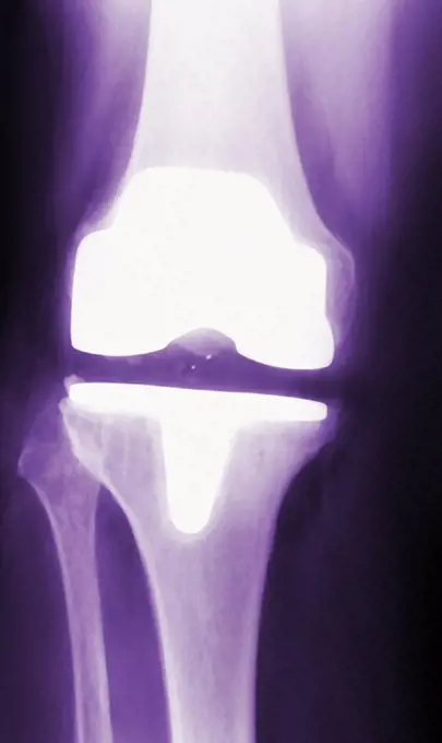
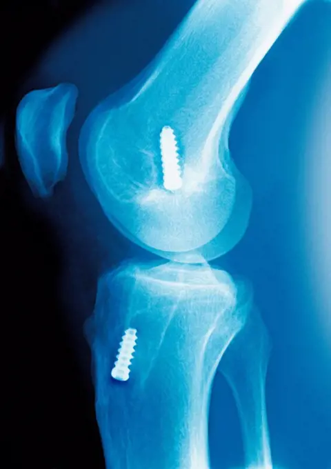
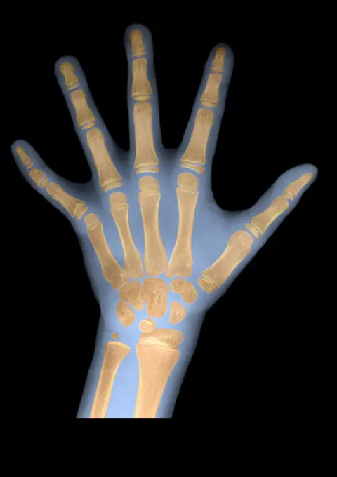
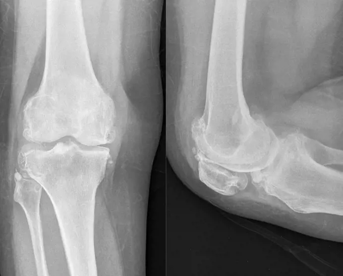
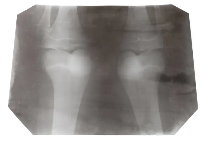
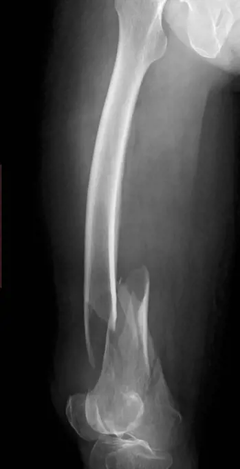
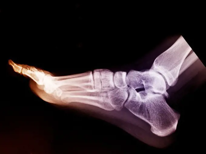
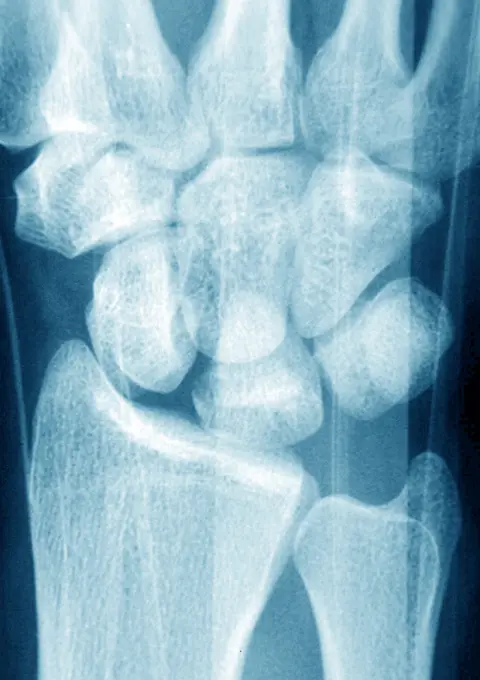
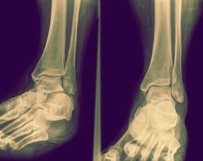
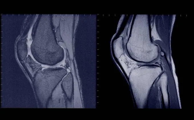
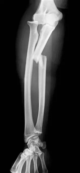
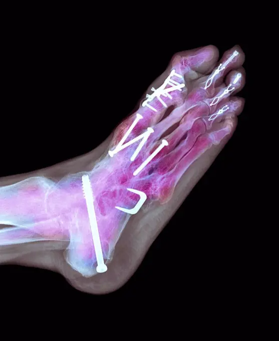
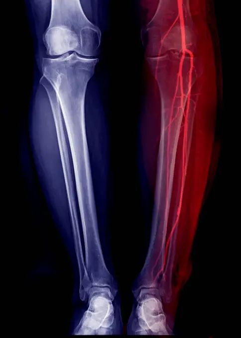
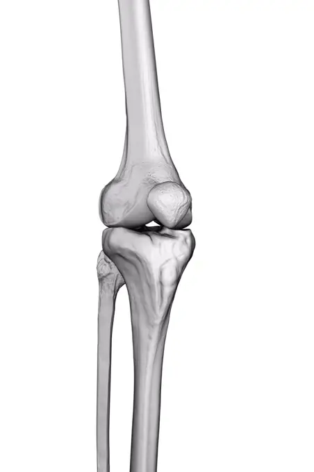
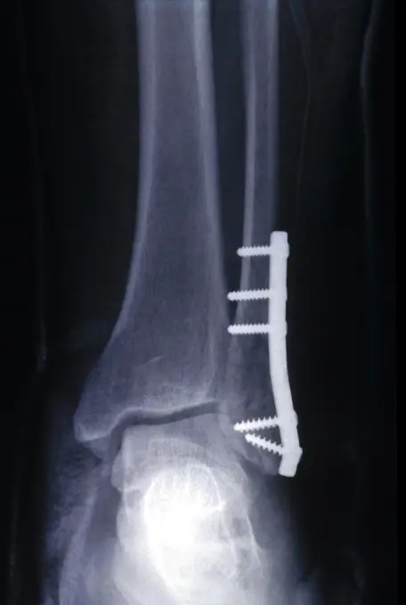
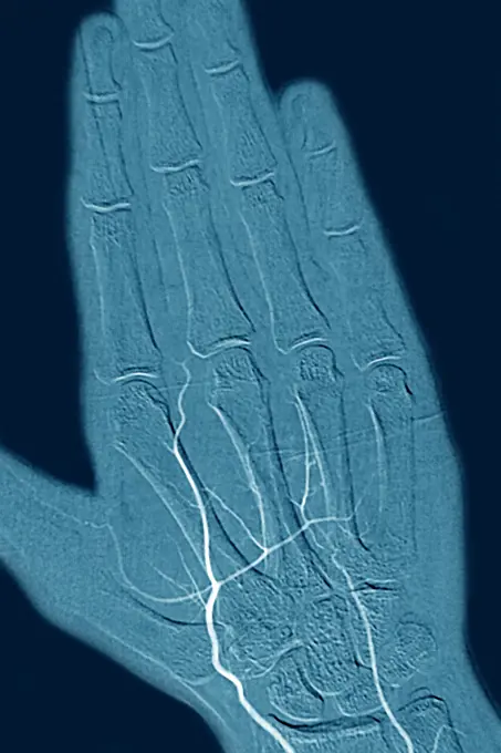
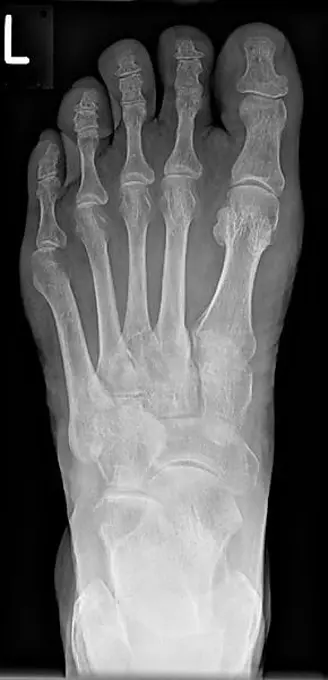
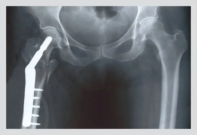
My Boards
View Pictures
  • My Boards

X-Ray Imaging of Bone Injuries

A collection of x-ray images showcasing various bone injuries including fractures in the thumb, ankle, and knee, as well as a normal elbow x-ray.

An x-ray of a forefoot mid-tarsal dislocation.
215 assets in this story
824-63211054
Fractured thumb at the metacarpal, seen on a hand x-ray.
6114-50976649
An x-ray of a forefoot mid-tarsal dislocation.
6114-50976688
An x-ray of an ankle fracture.
824-63178630
12 year old child. Normal radio. Elbow
4128R-15730
Broken knee. X_ray of the knee of a 38 year old patient with a fractured patella kneecap.
4129-126
knee x-ray
824-63178452
X-ray of the left and right knee of a 40-year-old woman. No anomaly
1525-24966668
x-ray of ankle
4128R-4342
Pinned wrist fracture, X_ray
4128-19357559
X-ray of a knee, taken from a patient with knee pain, showing a calcified mass within Hoffa's fat pad. This is suggestive Hoffa's fat pad syndrome.
4128-28766897
Healthy elbow joint, X-ray
824-63178448
X-ray of the left and right knee of a 40-year-old woman. No anomaly
1899-85887
Foot X Ray
6188-65147228
x-ray of knee with screws lateral seitliche Röntgenaufnahme eines männlichen Knies mit verschraubter Kniescheibe ,model released, Symbolfoto Copyright: xZoonar.com/AndyxNowackx 4662076 ,model released, Symbolfoto ,property released
4128R-3302
Normal shoulder joint, X_ray
4297-1350
Colorized x-ray of an ankle fracture of the fibula in a 50 year old man
4128-28766917
Healthy wrists, X-ray
4129-132
Hand x-ray
6188-55478597
film with X-ray image of knee joint with patella film with X-ray image of human knee joint with patella isolated on white background Copyright: xZoonar.com/ValeryxVoennyyx 13808341
4269-27501
Chondroma. Sagittal MRI of the knee showing a chondroma of the femur (thigh bone). This benign bone tumor is formed by a calcified cartilage matrix giving the bone marrow its white spotted aspect.
4128-24795320
Fabella, X-ray
4128R-1432
Normal ankle joint, X_ray
4128-19489815
Tibial plateau fracture, X-ray
4297-1095
X-ray of a radius forearm fracture of a 52 year old woman with an associated wrist dislocation
4378-1946
X-Ray image showing the knee joint.
4128R-12577981
Profile X-ray showing the right knee of a 46 year old obese patient with arthritis.
4128R-13374584
Foot bones
824-63198910
Hallux valgus on the big toe, seen on an x-ray of the right foot.
4297-1096
X-ray of a radius forearm fracture of a 52 year old woman with an associated wrist dislocation
4128-19489576
Broken knee cap, X-ray
4128R-26163
Healthy knee, computer artwork.
1899-61460128
Knee fracture (caused by a violent shock, causing trauma with serious lesions to the tibial plateau and to the lower end of the femur. X-ray of the left knee from the front
824-63178474
Severe osteonecrosis of the knee of a 78 years old patient. X Ray
1841R-111842
824-63178621
Child of 15 years measuring 1.48 m which corresponds to 13 years and 4 months. Bone age is calculated on both the hand and the elbow.
4297-1042
Normal foot x-ray of a 17 year old girl
824-63178424
Normal X-ray of the knee of a young child
1525-28448896
X-ray film with doctor’s hand
824-63212802
Arthrodesis of the ankle involving the tibia, the talus and the heel bone. Saggital plane x-ray of the right ankle.
824-63128448
Here left total prosthesis. Frontal x-ray of the hips. See image n0623005 on the site.
4128R-5615
Prosthetic knee, X_ray
4128R-2731
Anterior cruciate ligament repair, X_ray
824-63209001
Reportage in a radiology centre in Haute-Savoie, France. X-ray result of a hip with osteoarthritis.
824-63178640
6 years old child radiography. Normal bone age
4128-20043169
Knee osteoarthritis, X-ray
6188-55490839
X-ray picture of two human knee-joints isolated X-ray picture of two human knee-joints isolated on white background ,property released Copyright: xZoonar.com/ValeryxVoennyyx 7103559 ,model released, Symbolfoto ,property released
4128-19499654
Comminuted femur fracture, X-ray
4128-19499651
Comminuted femur fracture, X-ray
824-63211055
Fractured thumb at the metacarpal, seen on a hand x-ray.
824-63178620
Child of 15 years measuring 1.48 m which corresponds to 13 years and 4 months. Bone age is calculated on both the hand and the elbow.
4128-20239838
Segond fracture, X-ray
6188-55645104
Hand, man X -ray, man, hand copyright: xzoonar.com/dr.xnorbertxlangeex 13793128
4128R-5927
Arthritic hand, X_ray
4128-19357536
X-ray showing the dislocated ankle of a patient in a traffic accident.
1525-28446597
Fused bones of the lower leg after removing the steel bonding plate, x-ray of the leg. Fused bones of the lower leg after removing the steel bonding plate
824-63194042
Knee x-ray on a 73-year old woman. The exam shows widespread bone demineralisation, a discrete pinching in the internal femorotibial joint space and an osteosynthesis of the left tibial plateau (screw).
824-57655648
X-ray of rheumatism in the hands.
6114-50976800
X-ray of fracture distal tibia and fibula.
824-63217828
Screw and plate osteosynthesis on a fracture of the fibula. Frontal x-ray: tibia, fibula, malleolus and astragalus.
4128-28514384
Foot, 3D CT scan
824-63207452
Rheumatoid arthritis seen on an x-ray of the hand.
4128-28514553
Foot, 3D CT scan
4297-1041
Normal foot x-ray of a 17 year old girl
824-63194049
Knee x-ray on a 73-year old woman. The exam shows widespread bone demineralisation, a discrete pinching in the internal femorotibial joint space and an osteosynthesis of the left tibial plateau (screw).
824-63170785
Fracture of the carpal scaphoid. X-ray of a left wrist.
1439-57940613
Sidebyside Xray images showing the bones of a foot and ankle.
4128-16073312
Knee joints side view, MRI
1525-22424681
X-ray of mature woman feet.
1899-53511598
Osteosynthesis of the hip by a nail-plate prosthesis, following a fracture of the neck of the femoral head. X-ray of the left hip.
1841R-111773
4128-19489665
Humerus fracture, X-ray
824-63208953
Reportage in a radiology centre in Haute-Savoie, France. A knee x-ray on a patient with the beginnings of osteoarthritis.
4297-1397
Colorized x-ray of an ankle fracture in a 19 year old woman surgically repaired with two large screws
4269-27199
Knee. Sagittal frontal Magnetic Resonance Image (MRI) of the knee showing the tibial collateral ligament (blue), between the femur and the fibula.
4128R-12964774
Broken upper leg bone, Illustration
824-63178617
Delay bone age 15 years old child mesearing 1meter 48 high which correspond to the size of a 13 years and 4 months old child. The bone age is calculated on the hand or the elbow radiography
4128-19358066
X-ray of a proximal ulnar fracture with radial head dislocation, known as a Monteggia fracture.
4128-16072198
Arm and hand, X-ray
4128-18929519
Fractured leg, X-ray
824-63178490
Knee, x-ray
6114-50976851
X-ray of the HAND AND DIGITS of a child whose HANDS HAVE BEEN  COCOONED IN SKIN FORMED DUE TO THE CONDITION EPIDERMOLYSIS BULLOSA DYSTROPHICA.
1525-28446599
x-ray of the leg. A steel plate holding together the tibia of the leg.. steel plate holding together the tibia of the leg.
1841R-111807
4128-19499645
Comminuted femur fracture, X-ray
824-63123153
Hysterosalpingography.
824-29055572
In a radiology center, Telos dynamic radiography of a knee, following a ruptured ligament.
4128-28514396
Foot, 3D CT scan
4128-19358212
X-ray of a proximal ulnar fracture with radial head dislocation, known as a Monteggia fracture.
824-63194019
Pelvic x-ray on a 93-year old woman, showing widespread bone demineralisation.
4128-15750367
Surgical fixation for rheumatoid arthritis of feet, X-ray
4128-19489718
Humerus fracture, X-ray
4128-28767004
Lower legs, X-rays
4378-20393119
Knee Joint
6114-50976772
X-ray of a plate fitted for ankle fracture.
824-63207413
Arteritis seen in an angiogram of the left hand.
1848-50928896
X-ray, left foot with advanced arthrosis, Germany, Europe
1525R-76796
X-ray of pins in a wrist
4408-1012
Femur Repair, Medical - Xray / Radiology
824-63204757
Complete hip prostheses (treatment for the destruction of hip cartilage through arthritis). Frontal pelvic CT scan.
4297-1361
Normal hand x-ray of a 21 year old man
PREVIOUS
of 3
NEXT
2401 S. Ervay, Suite 206
Dallas, Texas 75215
United States
Get Started
Free ResearchMy BoardsMy Cart
For Creators
How To License Your ContentContributor PortalFrame of Mind
Resources
API accessPricing
Contact
+1 866 236 0087help@viewpictures.co.uk Contact form
©2026 View Pictures. All Rights Reserved. -A
This site is protected by reCAPTCHA and Google Privacy Policy and Terms of Service apply.