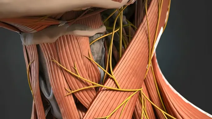
My Boards
View Pictures
  • My Boards

Human Neck And Shoulder Anatomy

Detailed anatomical illustrations showing the muscles, nerves, and structures of the human neck and shoulder. Bright colors highlight anatomical features.

Shoulder anatomy
Shoulder anatomy
139 assets in this story
4378-2554
Model showing the human knee joint and its connecting bones.
4128R-13723619
Illustration of a cross-section through a human spinal cord.
4128-19247913
Foot bones, illustration.
4128R-33400
Human leg musculature, computer artwork.
1525-56184116
The fibula or calf bone is the bone located on the lateral side of the shinbone and attached to the tibia both from above and below. 3D illustration. Fibula bone that carries the body
824-63164352
Neurogenesis: brain of an adult rodent with migration of neuroblasts.
1525-26244497
Human Neck Muscles And Nerves 3d illustration. Human Neck Muscles And Nerves
1525-56198066
When your head moves around, the liquid inside the semicircular canals sloshes around and moves the tiny hairs that line each canal. 3d illustration. Your semicircular canals are three tiny, fluid-filled tubes in your inner ear that help you keep your balance.
4269-24553
Streptococcus. Artwork
1848-56638818
Anatomy of the eye
4128R-13586453
Illustration of spinal cord anatomy.
824-63208848
Schematic illustration of the anatomy of a tendon from its insertion in the bone to the beginning of muscle structure. From left to right: -bone structure (beige) -tendon (white) -synovial sheath (green, cut on the right) -fibrous white sheath which surrounds the synovial sheath (cut here as it usually maintains the tendon in contact with the bone structure) -muscle (pink)
4128R-11324971
Human leg muscles, computer artwork.
1815-18355919
3D illustration of machine downloading human consciousness into cyborg body
1525-56198071
When your head moves around, the liquid inside the semicircular canals sloshes around and moves the tiny hairs that line each canal. 3d illustration. Your semicircular canals are three tiny, fluid-filled tubes in your inner ear that help you keep your balance.
1558-14088997
England, London, Southwark, The Old Operating Theatre and Herb Garrett Museum, Display of Historical Anatomical Model used for Teaching
1525-56198335
When light hits the retina , special cells called photoreceptors turn the light into electrical signals. These electrical signals travel from the retina through the optic nerve to the brain. Then the brain turns the signals into the images you see. 3d illustration. The mechanism of vision in the human eye
4378-3969
Male figure swinging a baseball bat. The internal anatomy of the heart is visible within the body.
824-72488654
The vocal cords. The vocal cords correspond to folds of the mucosa of the larynx. They are stretched between the arytenoid cartilage (above) and the thyroid cartilage (below). The vocal cords are supported by another pair of folds, the ventricular bands. The vocal cords are in an open position during breathing, and closed during phonation, vibrating as air passes through and thus allowing sounds to be produced. The transition between these two states is made possible by several muscles: the lateral cricoarytenoid muscles (on the sides of the cricoid cartilage) and posterior cricoarytenoid muscles (above the cricoid cartilage), the arytenoid muscle (below the cricoid cartilage). Contraction of the posterior cricoarytenoid muscles moves the thyroid cartilage, separating the vocal cords. Conversely, contraction of the lateral cricoarytenoid muscles or the arytenoid muscle brings the vocal cords together. The vocal muscles (or thyroarytenoids), running along the vocal cords, ensure their t
1525-23828412
3D Medical illustration Female Reproductive System, Menstrual Cycle 3D illustration. 3D Medical illustration Female Reproductive System, Menstrual Cycle
4128R-13723727
Illustration of human brain structures.
1525-26275240
The lumbar spine refers to the lower back, where the spine curves inward toward the abdomen. 3D illustration. 3D Medical 3D illustration of transparent human Lumbar Spine Anatomy
1525-56183990
long bone microscopic view anatomy 3d illustration. long bone microscopic view anatomy
4128R-11312903
Human spinal chord, computer artwork.
1525-56854530
Stage I breast tumors are invasive and have started to spread to the fatty breast tissue or to nearby lymph nodes. 3D rendering. Stage I breast tumors are invasive and have started to spread to the fatty breast tissue or to nearby lymph nodes.
1788-20762639
Circulatory system, surgical removal of lipid plaque in an artery through a tiny drill inserted through a catheter, drawing.
4128R-11478212
Human hand ligaments, computer artwork.
1815-16187704
Studio shot of breathing device
1525-20582529
Osteoporosis medical illustration concept as a close up diagram of the inside of a human bone from a skeletal hip joint as a normal healthy condition gradually degrades to abnormal unhealthy bone mass on a white background.
1525-56149788
A slipped disc may also be called a prolapsed or herniated disc. 3d illustration. A slipped disc may also be called a prolapsed or herniated disc.
1525-56190033
The pivot joint, also called the rotary joint or trochoid joint, is a freely movable joint (diarthrosis) in vertebrate anatomy that allows rotational motion around only one axis. 3d illustration. The pivot joint is also called the rotary joint or the trochoid joint.
4128R-11312472
Human spinal chord, computer artwork.
4239-19301026
Kentrosaurus dinosaur, side view on white background with copy space.
1525-56198125
The proximal tubule is the segment of the nephron in kidneys which begins from the renal pole of the Bowman’s capsule to the beginning of loop of Henle. 3D illustration. The proximal convoluted tubule is responsible for the most amount reabsorption of almost all filtered substances
4128R-13409964
Human brain anatomy, illustration
4128R-33586
Human lymphatic system, computer artwork.
4128-30420584
Tetanus, illustration
1525-25154124
Image of the sun on your body girl
4128R-13244815
Human brain, illustration.
PREVIOUS
of 2
NEXT
2401 S. Ervay, Suite 206
Dallas, Texas 75215
United States
Get Started
Free ResearchMy BoardsMy Cart
For Creators
How To License Your ContentContributor PortalFrame of Mind
Resources
API accessPricing
Contact
+1 866 236 0087help@viewpictures.co.uk Contact form
©2026 View Pictures. All Rights Reserved. -A
This site is protected by reCAPTCHA and Google Privacy Policy and Terms of Service apply.