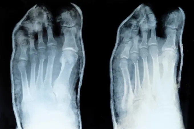
My Boards
View Pictures
  • My Boards
← Medical & Healthcare

Knee X-ray Images

Various X-ray images showing knee joints in detail. Images feature bones and joints from different angles, highlighting medical analysis.

184 assets in this story
1841R-111819
824-63178623
Child of 15 years measuring 1.48 m which corresponds to 13 years and 4 months. Bone age is calculated on both the hand and the elbow.
1899-66333
X-ray image of female knee joint, with knee cap
1841R-111777
1841R-111798
4128R-12800
Normal knee. Coloured X_ray of the knee of a 44 year old woman.
1841R-111791
4297-1068
Normal knee x-ray of a 49 year old woman
4297-1097
X-ray of a radius forearm fracture of a 52 year old woman with an associated wrist dislocation
4128R-15732
Broken knee. Coloured X_ray of the knee of a 38 year old patient with a fractured patella kneecap.
1899-61460133
Knee fracture (caused by a violent shock, causing trauma with serious lesions to the tibial plateau and to the lower end of the femur. X-ray of the left knee from the front
4128-19489625
Broken knee cap, X-ray
4128-28766919
Fractured ulna, X-ray
4128-28766898
Fractured ulna, X-ray
6114-50976695
A lateral view  x-ray of an ankle fracture.
1525-28468530
X ray film for human hands and legs
4297-1349
Normal x-ray of the ankle of a 12 year old girl
1848-51215048
X-ray of a patient after tibial plateau fracture with two screws
4128R-12577978
Profile X-ray showing the left knee of a 46 year old obese patient with arthritis.
4128-20043620
Knee osteoarthritis, X-ray scans
824-63178556
4 month old child. Search for morphological abnormality between the two legs. normal radio
824-29055702
Osteosynthesis: septic surgery for the removal of an internal ankle fixator following an infection.
1841R-111784
4128-18929498
Colles fracture, X-ray
4128-18929612
Tibial plateau fracture, X-ray
4128-19489704
Humerus fracture, X-ray
824-63188610
Hip osteoarthritis seen on a frontal x-ray.
1899-53508085
Osteosynthesis, septic surgery for the removal of an internal ankle fixator following an infection.
824-63204756
Complete hip prostheses (treatment for the destruction of hip cartilage through arthritis). Frontal pelvic CT scan.
1525-26837125
X ray film with doctor’s hand to examine
1525-27166709
Image Of Knee X-Ray. Detecting Radiographic Knee Osteoarthritis.
4408-1002
Shattered Elbow Repair, Medical - Xray / Radiology
1439-57940359
Radiographic revelation a glimpse into human bones
4128-19489562
Broken knee cap, X-ray
4128-19489783
Tibial plateau fracture, X-ray
4128-19489806
Tibial plateau fracture, X-ray
824-63194034
Knee x-ray on a 73-year old woman. The exam shows widespread bone demineralisation, a discrete pinching in the internal femorotibial joint space and an osteosynthesis of the left tibial plateau (screw).
4128R-15727
Rickets. X_ray of the legs of a young child with rickets. This condition, which causes soft bones, is caused by a lack of vitamin D.
4128-28769182
Hip replacement, MRI scan and X-ray
824-63176453
Reportage in the orthopaedic surgery unit in the Diaconesses Croix Saint Simon hospital in Paris. Placing a second hip prosthesis. X-ray of the patient before the operation.
4128-19489601
Broken knee cap, X-ray
824-63178434
X-ray after arthroscopy on the left knee. You can see the slightly pinched joint.
4128-28769208
Healthy wrist, X-rays
4128-28769181
Hip replacement, X-ray
824-63178653
Trauma-contusion of the right thumb after a shock. No fracture. Bone tearing
4128R-15197
Normal wrist, coloured X_ray.
4128-19489765
Supracondylar fracture, X-ray
824-63225293
Osteosynthesis of the hip by a nail-plate prosthesis, following a fracture of the neck of the femoral head. x-ray of the left hip.
4128R-12577976
X-rays of the right shoulder of a 36 year old patient that has repeatedly dislocated the joint. At left the shoulder is in neutral rotation, at centre...
824-63128352
Ankle, x-ray
824-63194047
Knee x-ray on a 73-year old woman. The exam shows widespread bone demineralisation, a discrete pinching in the internal femorotibial joint space and an osteosynthesis of the left tibial plateau (screw).
6188-69050244
Röntgen Aufnahme Becken mit Hüfte Röntgen Aufnahme Becken mit Hüfte LicenseRF Copyright: xZoonar.com/HGVorndranx 23243026
4128-19489685
Humerus fracture, X-ray
4128-19357550
X-ray of a tibial fracture with internal and external fixation.
1525-21261267
X-Ray leg shot isolated on white
4128R-7848
Prosthetic knee joint, X_ray
824-63178491
Knee, x-ray
824-63211557
Osteoarthritis of the hip seen on a frontal x-ray.
4297-1528
Colorized x-ray of a hand showing a middle metacarpal fracture
4128-19489643
Obstructed fallopian tube, X-ray
824-63215601
Foot x-ray.
4128R-12922491
Total hip replacement, X-ray
4297-1598
Hand x-ray of an adult man showing a dislocation of the fifth finger at the proximal interphalangeal joint
4128-16073317
Foot and ankle, MRI
1848-49597915
X-ray of a patient with a distal radial extension fracture on the right with PSU avulsion, stabilised by plate osteosynthesis with angular stable plate
4128-28769207
Healthy wrist, X-ray
4128-24795650
Dislocated shoulder, X-ray
4408-1011
Two Ankles, Medical - Xray / Radiology
4297-1044
Normal foot x-ray of a 59 year old woman
824-63170758
Osteosarcoma of the upper part of the tibia. MRI of a lateral view of a knee.
4128-30419974
Fractured knee, CT scan
4297-1040
Foot x-ray of a 52 year old man with a fracture of his great toe, highlighted in color
1899-66325
X-ray image image of female foot
4128-28766921
Healthy lower body, X-rays
4297-1054
Normal hand x-ray of a 17 year old girl
4297-1358
X-ray of a 46 year old woman who dislocated her elbow after a fall
1899-86254
Sciatalgia
6188-55645105
Hand, Mann Röntgenbild, Mann, Hand Copyright: xZoonar.com/Dr.xNorbertxLangex 13793143
1525-56312857
x-ray of pelvic with external fixation device fixed in femur and pelvic bones
1889R-9919
X-ray of a hip replacement
6188-55508653
Roentgenogram X-ray of hand and foot on black background Copyright: xZoonar.com/NatalliaxYaumenenkax 3559074
4128-16073513
Two outstretched hands, X-ray
4128-28970817
Obstructed fallopian tubes, X-ray
4491-111865801
X ray of side view and top view of deformed bones of feet with foreign bodies ;Broken bones of feet after accident on x ray
4128R-30953
Healthy ankle joint. Coloured profile X-ray of the left ankle of a 21 year old patient. Strapping around the ankle is visible on this X-ray.
4128-19499640
Comminuted femur fracture, X-ray
1899-85965
Retrograde Cystography
4269-27149
Capillaro-veinous malformation. Colored arteriogram of the right hand showing a malformation of the capillaro-veinous vessels seen as red spots under the little finger.
1899-86368
Knee scan
1525R-81238
X-ray of a human leg with metal screws and rods
4128-V58576835
1525-22149170
X-Ray Hands
4128-30422359
Damaged knee cartilage, illustration
4128R-12922499
Hip before hip replacement surgery, X-ray
1899-54027403
Diverticulum in the bladder. Frontal pelvic x-ray. (Photo by: BSIP/UIG via Getty Images)
6188-66548179
Röntgenbild der Zehen, mit verschobenem kleinen Finger x-ray of toes, with displaced little finger Copyright: xZoonar.com/KASHAPOVAx 22737986
1848-49588960
X-ray of a patient with a distal radial extension fracture on the right with PSU avulsion
824-63170779
This frontal MRI image of the knee shows a exostosis of the thighbone.
824-63173333
MRI, Magnetic Resonance Imaging. Knee.
4297-1055
Hand x-ray of a 17 year old woman showing a small fracture at the base of the of the thumb
PREVIOUS
of 2
NEXT
2401 S. Ervay, Suite 206
Dallas, Texas 75215
United States
Get Started
Free ResearchMy BoardsMy Cart
For Creators
How To License Your ContentContributor PortalFrame of Mind
Resources
API accessPricing
Contact
+1 866 236 0087help@viewpictures.co.uk Contact form
©2026 View Pictures. All Rights Reserved. -A
This site is protected by reCAPTCHA and Google Privacy Policy and Terms of Service apply.