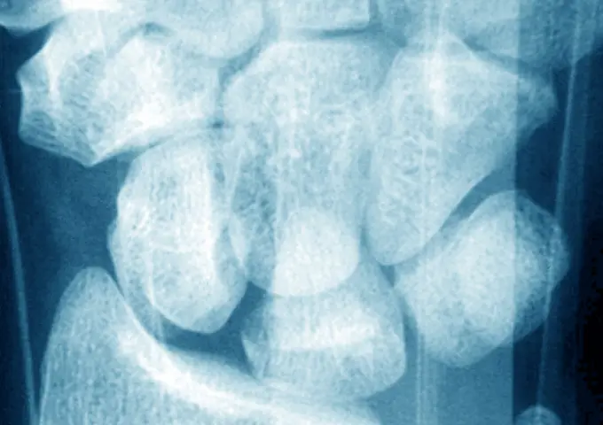
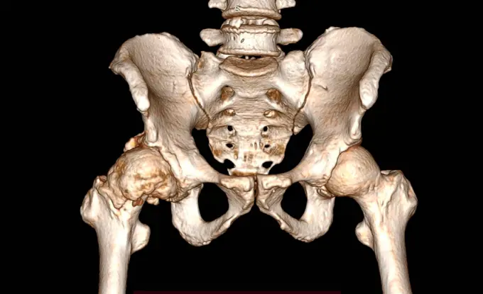
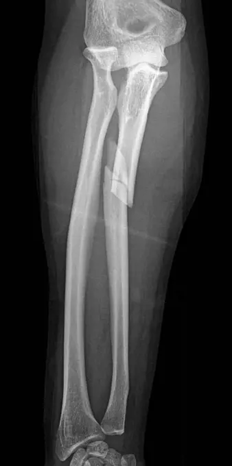
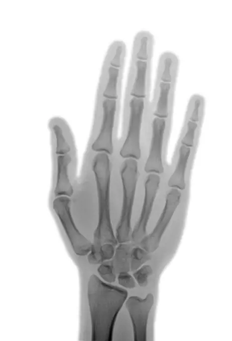
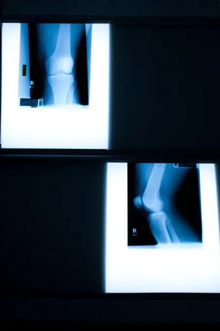
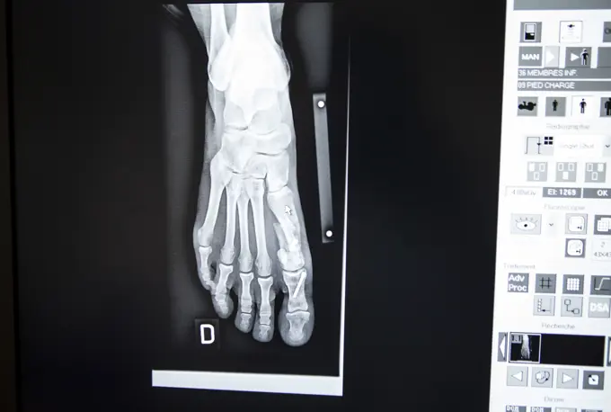
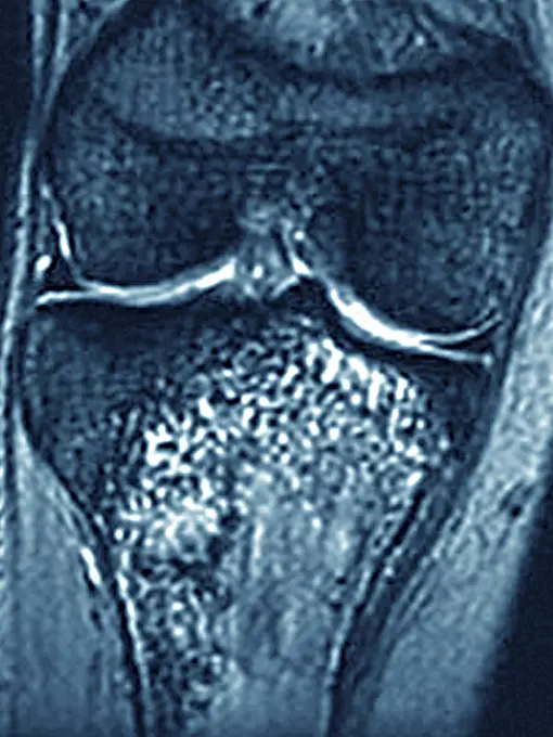
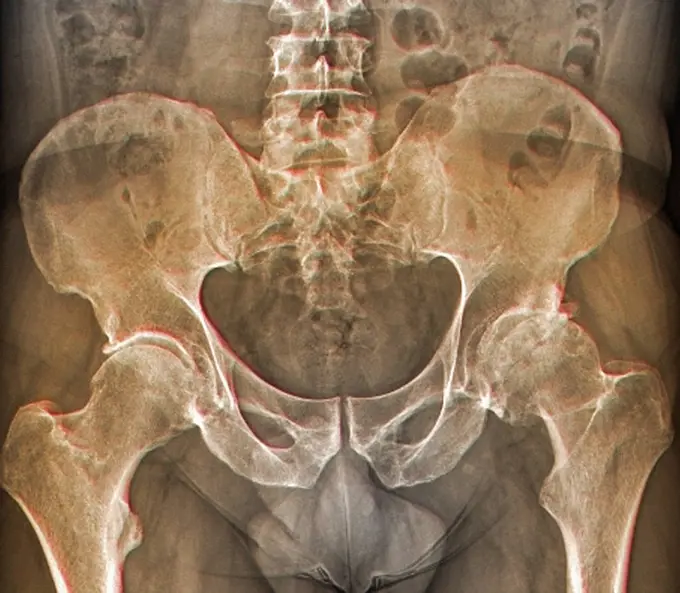
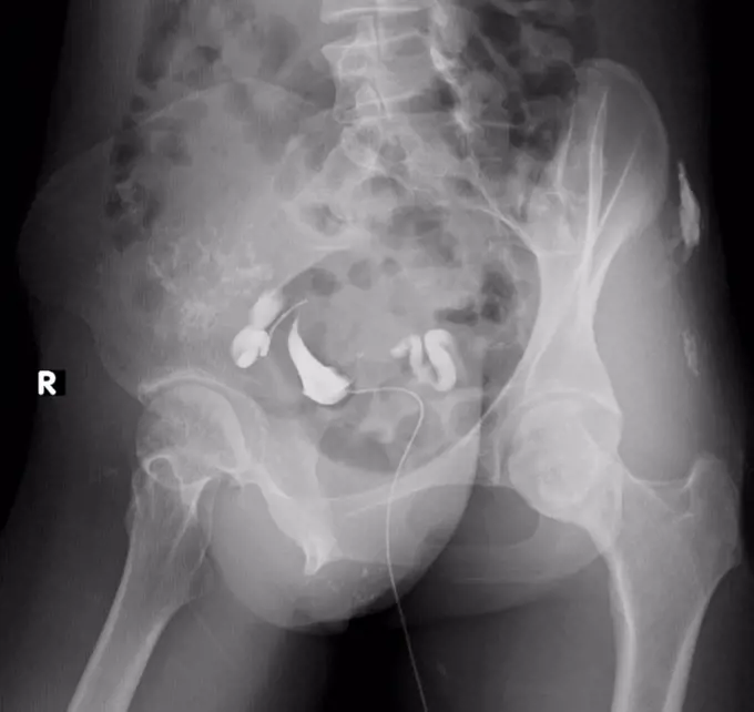
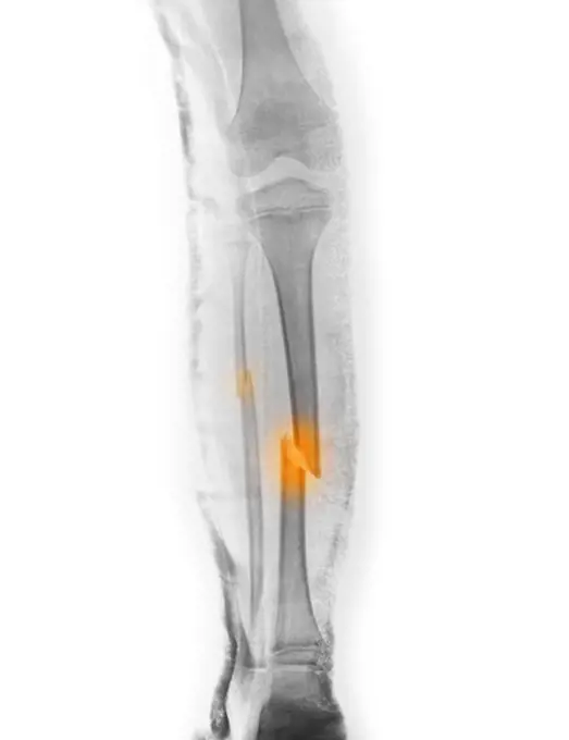
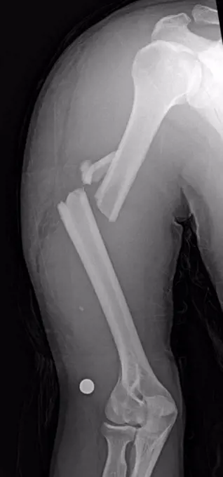
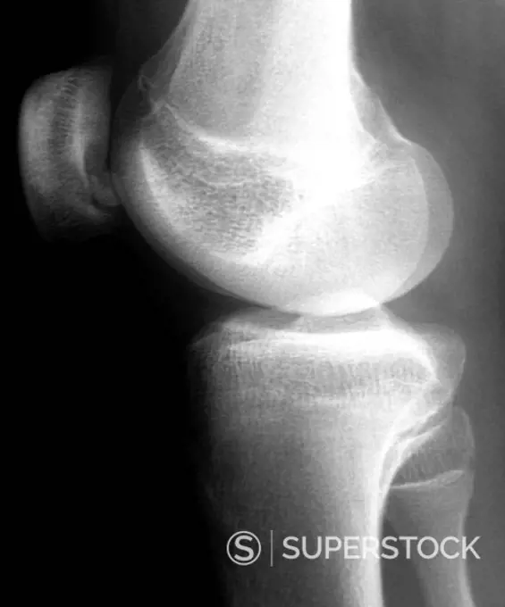
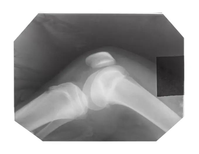
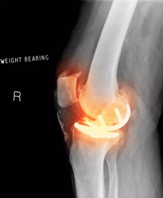
My Boards
View Pictures
  • My Boards
← Medical & Healthcare

Knee X-ray Images

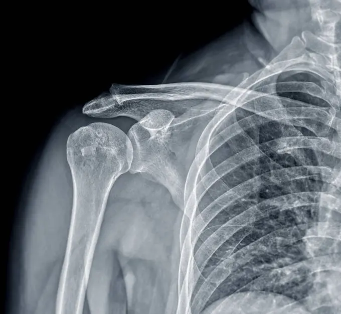
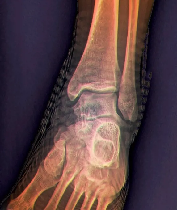
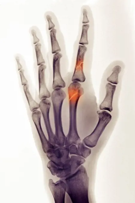
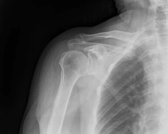
Various X-ray images showing knee joints in detail. Images feature bones and joints from different angles, highlighting medical analysis.

184 assets in this story
4128-28766913
Fractured shoulder, X-ray
4128-19357560
X-ray showing a patient with oblique proximal phalanx fracture.
4128R-30952
Healthy ankle joint. Coloured frontal X-ray of the left ankle of a 21 year old patient. Strapping around the ankle is visible on this X-ray.
4297-1182
Colorized x-ray of the hand of a 30 year old man showing fractures of the index finger proximal phalanx and metacarpal
1439-57940804
X-ray of dislocated shoulder with Calcific Tendonitis, 75 year old male
824-63170796
Fracture of the carpal scaphoid. X-ray of a left wrist.
4128R-15464
Normal shoulder. Coloured X_ray of the healthy right shoulder of a 28 year old patient.
824-63188654
Ankylosing spondylitis in the lumbar vertebrae, seen on a frontal x-ray.
1525-22336811
Close-up of a dental X-Ray
1525-22721012
X-ray image film detail of neck and red zone pain.
4128-24795468
Osteoarthritis of the hip, 3D CT scan
1525-28468531
X ray film for human hands and legs
4128-19358065
X-ray showing a proximal ulnar fracture with radial head dislocation, known as a Monteggia fracture.
1889R-8481
X-ray of foot
824-63194018
Pelvic x-ray on a 93-year old woman, showing widespread bone demineralisation.
4297-1362
Normal hand x-ray of a 21 year old man
4408-1005
Infant Chest, Medical - Xray / Radiology
1848-55301631
X-ray of a broken upper arm after surgery, showing plates and screws, 50 year-old woman
1525-19848394
Panoramic x-ray image scan of humans teeth
824-63225294
Osteosynthesis of the hip by a nail-plate prosthesis, following a fracture of the neck of the femoral head. x-ray of the left hip.
1841R-111812
824-63214256
Nephritic colic caused by a stenosis on the left ureter. Frontal abdominal-pelvic x-ray.
4409-17333417
RADIOGRAFIA DE TIBIA CON ROTURA Y TORNILLOS EN EL TOBILLO.
1525-26414886
different types of wisdom teeth problems concept, problem teeth X-ray image scanned, panoramic image
4128-20043646
Pleural effusion, X-ray
4128R-1175
Pinned broken leg, X_ray
4297-1364
Normal hand x-ray of a 68 year old man who is wearing a wedding ring
1525-27162143
Dental x-ray with braces. Radiography for teeth straightening and dental structures research concept.
1525-56254744
x-ray with external fixation device fixed in femur and pelvic bones
1660R-55737
Male hands holding xrays
1899-54027992
Normal knee seen on a frontal MRI scan.
824-63208920
Reportage in a radiology centre in Haute-Savoie, France. A technician carries out a foot x-ray on a patient who had an operation on a hallux valgus.
824-63188648
Osteosarcoma in the tibia, seen in a frontal MRI scan.
1899-54028018
Screw and plate osteosynthesis on a fracture of the fibula. Frontal x-ray: tibia, fibula, malleolus and astragalus.
6188-55577731
Retro look Xray Vintage looking Medical X-Ray imaging of hand fingers used in diagnostic radiology of skeleton bones Copyright: xZoonar.com/ClaudioxDiviziax 5827458
4128R-26912
Osteoarthritis of the hip. Coloured frontal X-ray (front view) of the pelvis in a 59 year old patient showing osteoarthritis of both hip joints. The b...
4128-18929603
Swollen fallopian tubes, X-ray
4297-1525
Colorized lateral skull x-ray of a child with orthodontic braces
4297-1086
Panorex x-ray of the jaw of a 65 year old man showing dental fillings and missing teeth
824-63194013
Pelvic x-ray on a 93-year old woman, showing widespread bone demineralisation.
824-63217842
Osteosarcoma of the fibula. Knee seen on a frontal MRI scan.
4297-1389
Lower leg x-ray of a 7 year old boy who fractured his tibia and fibula. This is the pre-operative x-ray with the leg in a splint
4128-28968671
Legg-Calve-Perthes disease, illustration
4128-18929452
Fractured arm, X-ray
4128-19357516
Lateral cervical X-ray of patient with neck pain showing apophyseal fusion.
4380-323
X-ray of a Human Hand with Thumb and Pointer Finger Touching
4128R-30962
Knee disease. X-ray of the right knee of an 18 year old patient with osteochondritis dissecans (OCD) of the knee. A small piece of bone and cartilage ...
6188-55478593
film with X-ray image of knee-joint with kneecap film with X-ray image of human knee-joint with kneecap isolated on white background Copyright: xZoonar.com/ValeryxVoennyyx 13808299
6114-50976639
An x-ray showing a swallowed coin in a patients oesophagus, the part of the alimentary canal that connects the throat to the stomach.
1848-49516150
Old mid 20th century X-ray photograph, radiograph showing shoulder tendinitis, tendinopathy, inflammation of the tendons
1525-28468603
Dental x ray film for dental care concept
1525-26510351
Xray scan skull nose medical exam results from hospital clinic.
1525-19826767
Panoramic and detail x-ray image scan of humans teeth
4297-1363
Normal hand x-ray of a 68 year old man who is wearing a wedding ring
4128-V58567377
6145-46419867
Pair of hands. .
4128R-14637717
Arthritis in the shoulder, illustration
4128-19358208
90 minutes intravenous urography (X-ray) of a 30 year old women with right abdominal pain, showing right hydroureteronephrosis. Hydroureteronephrosis ...
4128R-5881
Heart pacemaker, X_ray
1525-27074321
Dental x ray film for dental care concept
5507-47999335
Realistic X-ray Hand Medical Image
1525-27074748
Dental x ray film for dental care concept
1525-27074749
Dental x ray film for dental care concept
824-63208973
Reportage in a radiology centre in Haute-Savoie, France. The results of a teenage girls cranial x-ray.
824-63209339
Reportage on the Rossetti health centre in Nice, France. This rehabilitation centre is a hub of excellence with cutting-edge technology. The EOS low-emission radiology system: it enables simultaneous frontal and profile images to be produced from head to foot on a standing patient, and enables the 3D reconstruction of all the osteo-articular areas, with a considerable reduction in x-ray levels. A sports patient suffering from a knee injury and articular rheumatism. Severe scoliosis is also noted.
6188-67177086
ribs and lungs on x -ray image to see health exam thorax 1077405822
4128-V58573265
1525-22403756
X-Rays on light board.
4128R-12791
Loosened hip replacement. Coloured X_ray of a loosened total hip replacement of the left hip right of a female. The right hip left is showing signs of...
4128R-14566
Degenerative disc disease. Coloured 3D computed tomography CT scan of the lumbosacral spine of a 74 year old patient with degenerative disc disease. T...
824-63178427
MRI of the left knee. The patient will undergo arthroscopy to remove the damaged part of the meniscus.
4128R-11287690
Osteoarthritis of the knee, coloured X-ray.
4128-V58567355
4269-1061
Three-dimensional computed tomographic reconstruction of the neck and the head. The carotids are seen on both side of the cervical vertebraes, above the lower part of the skull with the jaws.
4129-129
feet x-ray
4128-20043143
Swallowed bracelet, X-ray
4128R-12922505
Degenerative foot deformation, X-ray
4128-24796142
Doctor examining hand X-rays
824-63194020
Pelvic x-ray on a 93-year old woman, showing widespread bone demineralisation.
4128-18680263
Spondylolysis and spondylolisthesis, X-rays
6188-54821319
X-ray of toes. Foot on Xray. bone research X-ray of toes. Foot on X-ray. bone research Copyright: xZoonar.com/LeonidxEremeychukx 14189991
6177-V53474219
4128-V58567370
1525-22341744
X-Ray images over a hospital screen
PREVIOUS
of 2
NEXT
2401 S. Ervay, Suite 206
Dallas, Texas 75215
United States
Get Started
Free ResearchMy BoardsMy Cart
For Creators
How To License Your ContentContributor PortalFrame of Mind
Resources
API accessPricing
Contact
+1 866 236 0087help@viewpictures.co.uk Contact form
©2026 View Pictures. All Rights Reserved. -A
This site is protected by reCAPTCHA and Google Privacy Policy and Terms of Service apply.