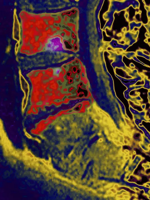
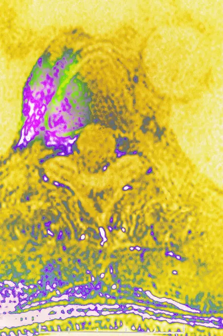
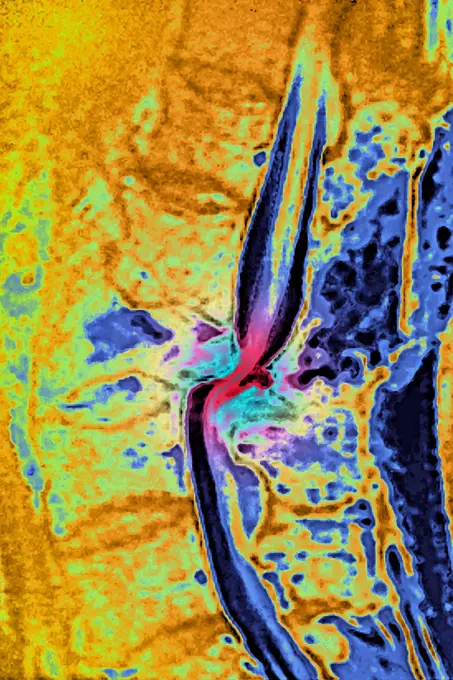
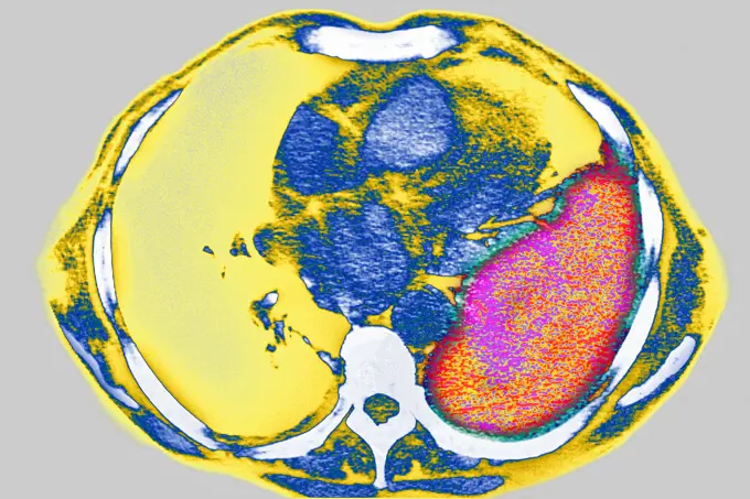
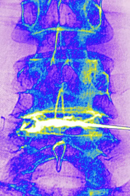
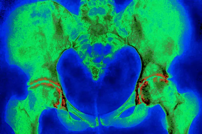
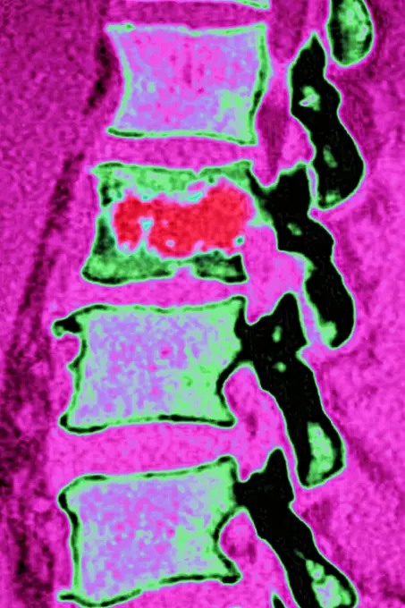
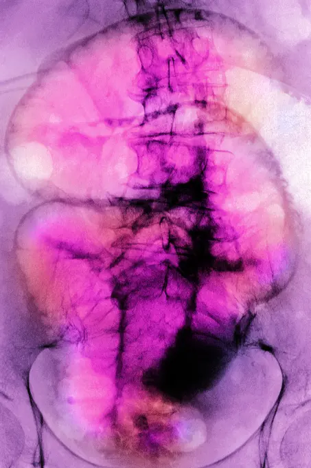
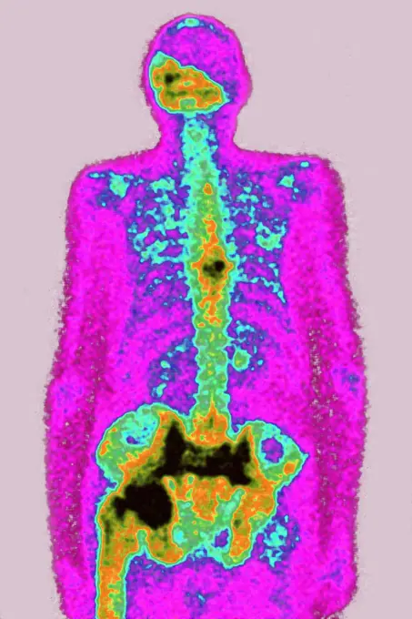
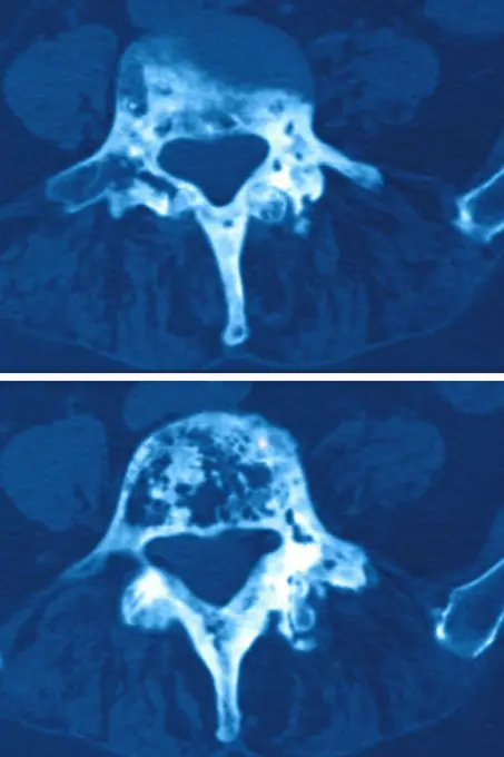
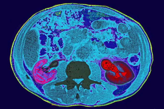
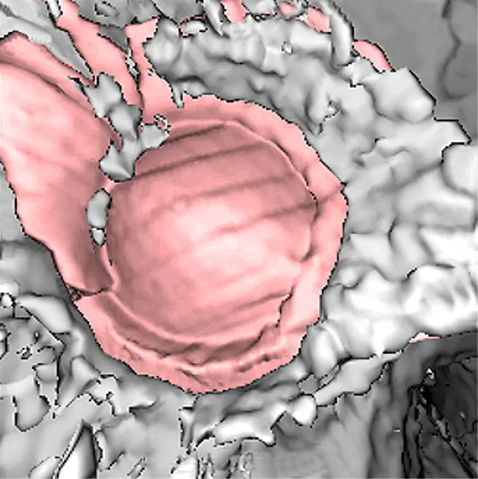
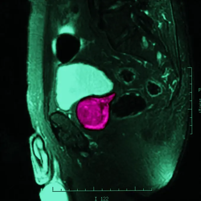
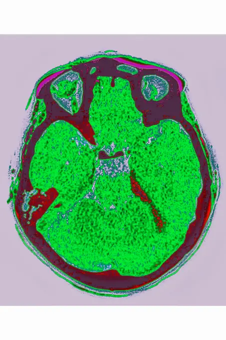
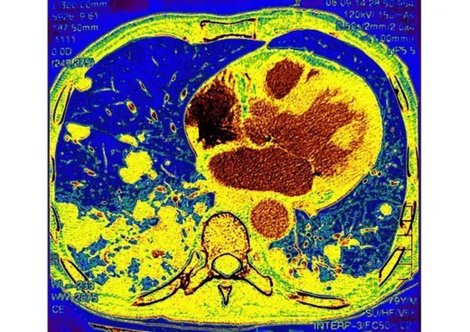
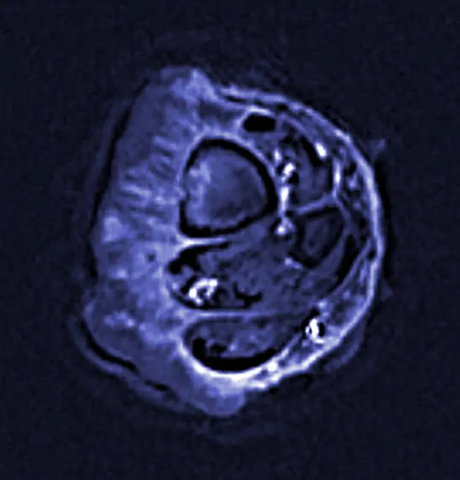
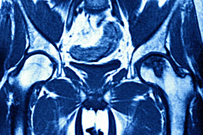
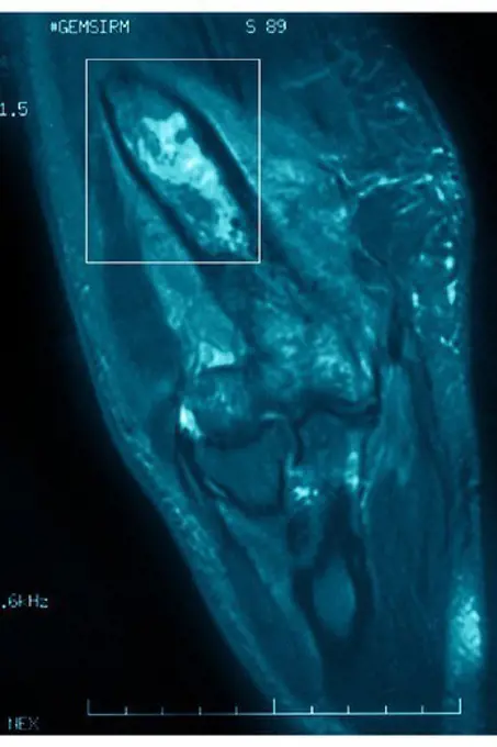
My Boards
View Pictures
  • My Boards

Medical Imaging and Conditions

Colorful MRI and CT scan images visualizing various medical conditions, showcasing detailed anatomical structures and abnormalities.

Osteitis on the L4-L5 lumbar vertebra, seen on a saggital plane MRI scan.
199 assets in this story
824-29057226
Retinal detachment of the left eye, visualized by radial section MRI.
1899-61460152
Vertebral osteitis (inflammation of bone tissue in a dorsal vertebra, caused by a bacterial infection). radial x-ray of a dorsal vertebra
1899-53511902
Dislocation of the lumbar vertebrae by spinal trauma, between the 3rd and 4th lumbar vertebrae, visualized by MRI in sagittal section. (Photo by: CAVALLINI JAMES/BSIP/UIG)
824-63188636
Osteitis on the L4-L5 lumbar vertebra, seen on a saggital plane MRI scan.
1899-53511641
Sinusitis in the right maxillary sinus, visualized by cranial CT scan in frontal section.
1899-53511529
Uterine cancer visualized by frontal MRI.
824-63206257
Aortic dissection in the ascending aorta, seen on a sagittal MRI scan of the thorax.
824-63170760
This sagital MRI image of the thoracic spine shows a malignant tumor of a intervertebral disc with medullar compression.
824-63215974
Pleurisy (left lung), seen on a radial section chest scan.
824-57656898
Vertebral osteitis (inflammation of the bone tissue of two dorsal vertebrae, caused by a bacterial infection). sagittal radiograph of the dorsal vertebrae.
824-65830297
Lumbar osteoporosis (disease causing compression and fractures of the lumbar vertebrae). X-ray of the lumbar vertebrae seen from the front.
824-57656867
Lung cancer (cancerous tumor consisting of a group of cancerous cells that invade nearby tissues and destroy them, they can also spread in metastases). Lungs visualized by thoracic scanner in radial section.
824-63205972
Adenopathy (spleen lymph gland). Cross-section abdominal scan.
1899-54027127
Hip algodystrophy, seen in a frontal section MRI scan.
824-63217805
Normal knee seen on a frontal MRI scan.
4269-24972
Aorta. Normal thoracic aorta MRI (Magnetic Resonance Imaging) and angiography
4269-27157
Osteoid osteoma. Color enhanced axial computed tomography (CT) scan of the thigh showing osteoid osteoma (benign bone tumor) on the femur. The osteoid osteoma is revealed by a thickening of the bone connective tissues (pink) at the surface of the femur (white).
824-63226774
Malignant tumor localized on the cervical vertebrae, in transverse section.
824-63170804
This sagittal MRI image of the lumbar spine reveals a spondylodiscitis at the L3 L4 level.
824-63206252
Cancer of the right kidney, seen on a cross-section abdominal scan.
824-63203255
Ensethopathy of the supeficial plantar fascia. Sagittal section CT scan of the foot.
824-57656871
Knee fracture (caused by a violent shock, causing trauma with serious lesions to the tibial plateau and to the lower end of the femur. X-ray of the left knee from the front.
824-29057242
Normal knee visualized by MRI in sagittal section.
824-63222980
Myocardial infarction with a significant thrombus in the right coronary artery and extends into the left retro ventricular and the posterior interventricular artery. Coronary angiography.
824-63214246
X-ray of urinary schistosomiasis. Frontal pelvic x-ray.
824-63195633
CT scan showing a pancreatic cyst.
824-29057221
Retinal detachment of the left eye, visualized by radial section MRI.
824-29057219
Retinal detachment of the left eye, visualized by radial section MRI.
824-29057222
Retinal detachment of the left eye, visualized by radial section MRI.
1899-54027719
Pulmonary tuberculosis seen on a radial cross-section chest CT scan.
824-63188665
Secondary malignant tumour on a lumbar vertebra, seen on a saggital plane MRI scan.
4269-25370
Myocardial infarctus. 3D CT Scan et 2D
824-63224763
Uterine cancer visualized by frontal MRI.
824-63212857
Osseous edema of the knee. Subcortical lesion of the condyle, radial cross-section MRI scan of the knee.
1899-53511727
Right coronary artery visualized by frontal thoracic angiography. (Photo by: CAVALLINI JAMES/BSIP/UIG)
824-63225960
Right coronary artery visualized by frontal thoracic angiography.
4269-6991
Ultrasound scan of a 3 months old fetus sucking his thumb.
824-63206013
Myocardial infarction caused by a stenosis of the right coronary artery. Seen via coronarography.
4269-27197
Sacroiliitis. Frontal colored Computed Tomography (CT) scan of the pelvis with sacroiliitis : inflammation of the sacro-iliac joint causing bone erosion (blue) and condensation at the surface on the bone (white).
824-63175252
CT scan of internal ear.
1899-53511903
Dislocation of the lumbar vertebrae by spinal trauma, between the 3rd and 4th lumbar vertebrae, visualized by MRI in sagittal section. (Photo by: CAVALLINI JAMES/BSIP/UIG)
1899-53511620
Corticosteroid infiltration between the fourth and fifth lumbar vertebrae, done under radiographic control.
4128R-13587323
Mycobacterium, illustration.
824-63218215
Fracture of the skull, seen on a radial section scanner.
824-63206281
Multiple cysts on the right kidney, seen on a frontal abdominal x-ray.
824-63226771
Malignant tumor localized on the cervical vertebrae, in transverse section.
824-63218248
Spinal neuromas (schwannomas) in cervical vertebrae, seen on a sagittal section MRI scan.
824-63195597
Abdominal CT scan showing a stomach tumour (gastric cancer).
1899-53511743
Osteoarthritis of the hips (coxarthrosis) visualized by a pelvic x-ray of the front. (Photo by: CAVALLINI JAMES/BSIP/UIG)
824-63225976
Osteoarthritis of the hips (coxarthrosis) visualized by a pelvic x-ray of the front.
1899-61460145
Vertebral osteitis (inflammation of the bone tissue of a lumbar vertebra, caused by a bacterial infection). sagittal x-ray of the lumbar vertebrae
1899-53511990
Chronic colitis (inflammatory bowel disease), revealed by a frontal abdominal x-ray.
4128-18929506
Cannonball pulmonary metastases, CT scan
824-63225336
Paget's disease (bone involvement), visualized by scintigraphy.
1899-54027158
Paget"s disease, seen in a sagittal-plane MRI scan of the L5 lumbar vertebra.
824-63207449
Pagets disease, seen in a radial cross-section MRI scan of the L4 and L5 lumbar vertebrae.
824-63207437
Kidney infarction seen in an abdominal angiogram.
824-63211044
Renal colic of the left kidney, blockage in the urethra by a nephrolith with upstream renal dilation. Radial section abdominal scanner.
824-63226754
Pulmonary metastases (secondary to kidney cancer), visualized by chest CT scan in axial section.
824-63211556
Cerebrovascular accident caused by thrombosis of an artery in the left hemisphere. Saggital plane cross-section brain scan.
1899-54027395
X-ray of urinary schistosomiasis. Frontal abdominal-pelvic x-ray. (Photo by: BSIP/UIG via Getty Images)
4269-27666
Cerebral aneurysm. Three-dimensional computed tomographic (CT) scan reconstruction of a skull showing an intracranial aneurysm (yellow ballon shaped swelling) located in the arterial circle of Willis  (where the basilar artery and internal carotid arteries meet). An aneurysm is a bulge of a vessel due to the dilatation of its wall. A ruptured aneurysm can be fatal.
824-63211572
Vertebral osteoarthritis with osteophytes (bone spurs) seen on a saggital plant CT-scan of the lumbar vertebrae.
4269-25421
Cardiac valve. 3D computed tomographic (CT) scan (virtual endoscopy) of a cardiac valve.
4269-39005
Supra-aortic trunks (TSA) showing the collateral branches of the aorta from the aortic arch: the brachiocephalic arterial trunk, the left common carotid artery (centre), the subclavian artery, which supply blood to the upper body. Magnetic resonance angiography.
824-63128444
Pelvic MRI of a 60-year-old man. Moderate prostatic hypertrophy. See image n0839805 on the site.
824-63225315
Herniated disc between L4-L5 lumbar vertebrae (lesion compressing the spinal cord by displacement of part of an intervertebral disc outside the limits of its normal situation). Lumbar spinal MRI in sagittal section.
824-63188609
Diffused joint chondrocalcinosis located between the top of the radius and the lunate and triquetral bones. Frontal wrist x-ray.
824-694
Human Kidney
4128-30415816
Sacrum, illustration
4269-27876
Prostate cancer. Colored MRI (Magnetic Resonance Imaging) showing a prostate cancer seen as a colored sport at center.
4297-1729
CT scan of abdomen showing an aortic aneurysm and extensive arterial atherosclerosis
824-63128433
Regular foot. See image n1081805 on the site.
824-63204734
Multiple hemorrhagic foci, in contact with the meninges. Cross-section brain scan.
1899-53511611
Stress fractures on the shins, visualized by scintigraphy.
824-63225309
Stress fractures on the shins, visualized by scintigraphy.
824-63227449
Zinc is necessary for the functioning of more than 300 enzymes, including those that help regulate gene expression, in various organisms, including humans. Researchers are studying how plants acquire, sequester and distribute zinc to find ways to increase the zinc content of crops to improve human health. Using synchrotron X-ray fluorescence technology, they created this heat map of zinc levels in an Arabidopsis thaliana plant leaf.
4128-20043080
Normal lung, CT scan
4128R-5282
Chromosome, artwork
1899-53511998
Retinal detachment of the left eye, visualized by radial section MRI.
824-63224754
Uterine cancer visualized by MRI in radial section.
824-80841
LUNG METASTASIS, SCAN. Pulmonary metastases secondary cancer, carcinoma in a 79_year_old man. Thoracic scan in axial section.
4269-27497
Epidermoid carcinoma. Colored enhanced Axial Computed Tomography (CT) scan of the calf showing a epidermoid carcinoma, a type of malignant skin  cancer. The tumor appears as the fleshy thickening at left. The section of the tibia is seen at top left, the section of the fibula, smaller, is below at right.
824-29057220
Retinal detachment of the left eye, visualized by radial section MRI.
6145-44565633
This anaglyph view of 'Chimp,' south southwest of the lander, was produced by NASA's Mars Pathfinder's Imager camera. 3D glasses are necessary to identify surface detail.
824-63188658
Spondylodiscitis on the intervertebral lumbar disc, seen on a saggital plane MRI scan.
824-63206940
Osteoarthritis of the left fermoral head, seen on a frontal pelvic MRI scan.
824-63224561
Right coronary artery. Angiography with injection of contrast product.
824-63170791
This frontal MRI image of the left hip reveals an osteonecrosis (bone death) of the femoral head.
4269-24975
Endoprosthesis. 3D computed tomography (CT) scan of a post-traumatic dissecting aneurism of the abdominal aorta treated with an endoprosthesis (yellow).
1899-54027991
Normal knee seen on a sagittal section MRI scan.
824-63175250
CT scan of internal ear.
4128R-16867
Calabi_yau manifolds, computer artwork. These six_dimensional shapes are thought to be the location of the extra six dimensions on top of the four kno...
824-63175257
X-ray of the skull showing sinonasal polyposis.
4269-27189
Arterious system. Colored Computed Tomography (CT) scan of the whole arterious system. Is seen at center the aorta, the main artery of he body. At top, the aortic arch branching in the brachiocephalic artery, the left carotid artery and the left subclavian artery. Going down : the renal arteries, and the division in 2 iliac arteries and their branches. The white spots on the aorta are atheromathous plaques.
824-63224681
Angiography of myocardial infarction caused by atheromatous plaque in the left coronary artery. Angiographic visualization is produced by injection of contrast product.
824-63206002
Fractures of the tibia and fibula seen on a frontal x-ray.
824-63206289
Evolutionary metastases of kidney cancer, seen on a dorsal view scintiscan.
6188-64561333
Cosmic images computer processed images of galaxies Copyright: xAshaxAdatia/DinodiaxPhotox
824-112260
MYELOMA, MRI. MYELOMA, MRI Femur, frontal view.
PREVIOUS
of 2
NEXT
2401 S. Ervay, Suite 206
Dallas, Texas 75215
United States
Get Started
Free ResearchMy BoardsMy Cart
For Creators
How To License Your ContentContributor PortalFrame of Mind
Resources
API accessPricing
Contact
+1 866 236 0087help@viewpictures.co.uk Contact form
©2026 View Pictures. All Rights Reserved. -A
This site is protected by reCAPTCHA and Google Privacy Policy and Terms of Service apply.