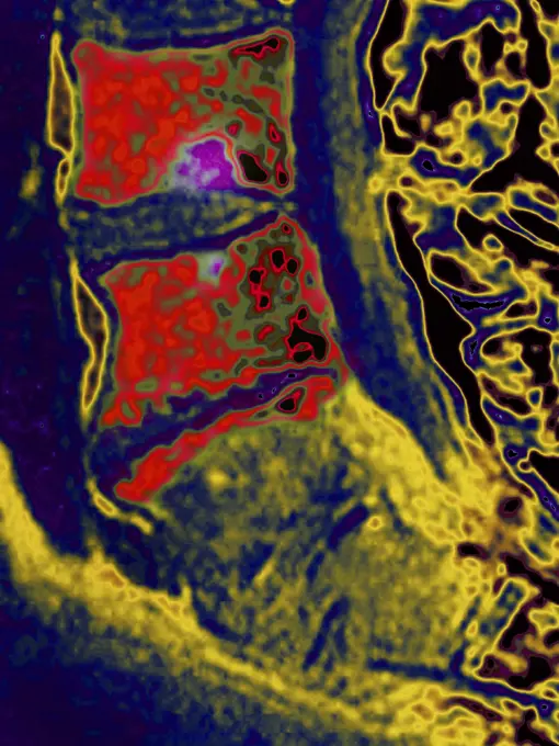
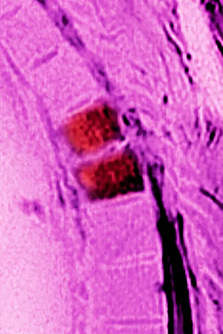
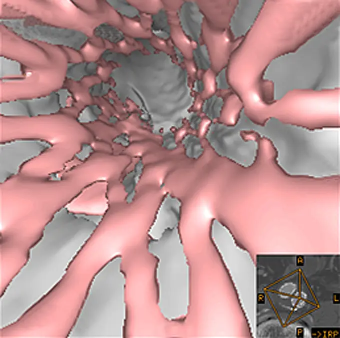
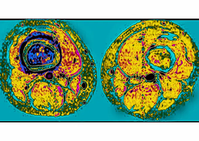
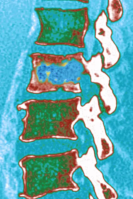
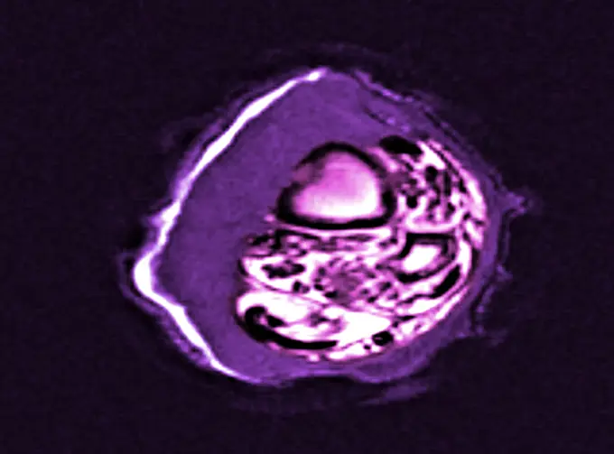
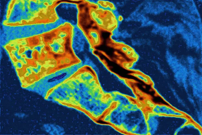
My Boards
View Pictures
  • My Boards
← Science

Medical Imaging and Conditions

Colorful MRI and CT scan images visualizing various medical conditions, showcasing detailed anatomical structures and abnormalities.

Osteitis on the L4-L5 lumbar vertebra, seen on a saggital plane MRI scan.
199 assets in this story
4128-30419383
Prostate, illustration
1525-62544451
Green slime animated sprite, animation effect showing stages of dripping and splashing toxic liquid phlegm for video game. Cartoon vector sequence frame of gooey, slimy poison drip, gui storyboard. Green slime animated sprite, animation effect
824-63226773
Malignant tumor localized on the cervical vertebrae, in transverse section.
824-63178734
(Angiography exam of the eyes. Small irritation of the left eye (here clear area) situated just in the middle of the macula (Here dark area) creating for the patient a discomfort in reading. This irritation can be due to big stress and should disappear in some weeks
1899-53513307
Myocardial infarction with two stenosis in the right coronary artery. Coronary angiography.
824-29057262
Prostate (healthy prostate gland), visualized in ultrasound in frontal view.
1899-53511907
Pulmonary metastases (secondary to kidney cancer), visualized by chest CT scan in axial section. (Photo by: CAVALLINI JAMES/BSIP/UIG)
824-57656897
Vertebral osteitis (inflammation of the bone tissue of two dorsal vertebrae, caused by a bacterial infection). sagittal radiograph of the dorsal vertebrae.
4269-25393
Stent prosthesis. Virtual endoscopic view of a stent prosthesis. Stent are mesh-wire like endoprosthesis (pink) inserted into a vessel during angioplasty to support tissue. It is used to treat stenosis or aneurysm of blood vessels.
824-63211585
Prostate cancer (adenocarcinoma) with metastases, seen on a frontal scintiscan.
1899-53512028
Prostate (healthy prostate gland), visualized in ultrasound in frontal view.
6145-44518529
Tracking a Feature on Eros
824-63205984
Nephritic colic seen on a frontal x-ray of the abdomen.
824-63215988
Thymus cancer seen on a cross-section chest CT scan.
824-63170786
This axial MRI image reveals an ostéosarcoma of the right thighbone.
824-63222982
Myocardial infarction with two stenosis in the right coronary artery. Coronary angiography.
824-63217821
Slipped disc compression of the medulla lumbar vertebra.
6145-44513247
Mars Orbiter Laser Altimiter (MOLA) Globe
824-63206288
Evolutionary metastases of kidney cancer, seen on a dorsal view scintiscan.
824-57656896
Vertebral osteitis (inflammation of the bone tissue of a lumbar vertebra, caused by a bacterial infection). sagittal x-ray of the lumbar vertebrae.
4413-97268
Wood melick ears
6188-63949563
Spices, saffron thread in water on lavender background Copyright: xHarixMahidhar/DinodiaxPhotox
6145-44566166
Moons Around Saturn
824-63217272
Pulmonary tuberculosis seen on a radial cross-section chest CT scan.
824-63226775
Malignant tumor localized on the cervical vertebrae, in transverse section.
4269-27499
Epidermoid carcinoma. Colored enhanced Axial Computed Tomography (CT) scan of the calf showing a epidermoid carcinoma, a type of malignant skin  cancer. The tumor appears as the fleshy thickening at left. The section of the tibia is seen at top left, the section of the fibula, smaller, is below at right.
4128-19249581
Psilocybin mushrooms, computer illustration. Commonly known as magic mushrooms, these are a group of fungi that contain psilocybin which turns into ps...
824-63198853
Degenerative disk disease. Dorsal vertebrae osteoarthritis seen on a side x-ray of the thorax.
4269-27276
Cerebral aneurysm. 3D reconstruction from CT scanner and angiography of the skull and its vascularization system showing a brain aneurysm (square). An aneurysm is a bulge of a vessel due to the dilatation of its wall. A ruptured aneurysm can be fatal.
1899-53511494
Lumpy tumor of the thyroid gland (benign tumor), thyroid ultrasound.
6145-44530226
S73-32883 (20 Aug. 1973) --- This false color isophote, processed from an Aug. 20, 1973 television transmission of Apollo Telescope Mount (ATM) experiments from Skylab 3, dramatically reveals a significant change in the coronal hole as compared to the previous day. Solar rotation accounts for the new location of the coronal hole.
6145-45260709
The complexity and richness of Jupiter's southern lights (also known as auroras) are on display in this animation of false-color maps from NASA's Juno spacecraft. Auroras result when energetic electrons from the magnetosphere crash into the molecular hydrogen in the Jovian upper atmosphere. The data for this animation were obtained by Juno's Ultraviolet Spectrograph. The images are centered on the south pole and extend to latitudes of 50 degrees south. Each frame of the animation includes data from 30 consecutive Juno spins (about 15 minutes), just after the spacecraft's fifth close approach to Jupiter on February 2, 2017. The eight frames of the animation cover the period from 13 40 to 15 40 UTC at Juno. During that time, the spacecraft was receding from 35,000 miles to 153,900 miles (56,300 kilometers to 247,600 kilometers) above the aurora; this large change in distance accounts for the increasing fuzziness of the features. Jupiter's prime meridian is toward the bottom, and longitud
4269-25940
Pulmonary embolism. Gamma scans of lungs showing pulmonary embolism. This occurs when one of the pulmonary arteries is blocked, usually by an abnormal blood clot (thrombus).
1525-26226400
Iridescent film of oil spill isolated on black background
824-63211611
Pyogenic streptococci A. Bacteria causing skin infections (impetigo), abscesses, bronchopulmonary infections. Optical microscopy X 1000.
824-66065375
Brain scans of healthy volunteers have shown that human brains may drain waste through lymphatic vessels, the bodys sewer system.
1899-54027160
Paget"s disease, seen in a sagittal-plane MRI scan of the L5 lumbar vertebra.
824-63207443
Pagets disease, seen in a sagittal-plane MRI scan of the L5 lumbar vertebra.
824-63212836
Colon cancer (tumors on the caecum). Visualisation using 3D reconstruction from a CT scan (virtual coloscopy).
6145-44494877
Mars Gravity Map (RS)
1899-54028019
Screw and plate osteosynthesis on a fracture of the fibula. Frontal x-ray: tibia, fibula, malleolus and astragalus.
6188-66537417
Menschliches Atmungssystem Zwerchfell Anatomie 3D Illustration Concept of Human Respiratory System Diaphragm Anatomy Copyright: xZoonar.com/magicminex 22683539
824-63170772
This sagittal MRI image of the lumbar spine reveals a spondylodiscitis at the L3 L4 level.
824-63211033
Cancer of the right kidney, seen on an abdominal ultrasound scan.
6188-54824490
Snake Charmer in scientific high-tech thermal imager Snake Charmer in scientific high-tech thermal imager on black background isolated Copyright: xZoonar.com/Yrax 16556346
4128R-15729
Brain abscess. Coloured computed tomography CT scan of an axial section through the brain of a 41 year old patient with a cerebral abscess. An abscess...
824-63211573
Vertebral osteoarthritis with osteophytes (bone spurs) seen on a saggital plant CT-scan of the lumbar vertebrae.
824-72488772
Finger osteoarthritis. Left: healthy joint. Middle: moderate osteoarthritis. The cartilage is worn, the cartilaginous debris causes inflammation of the joint. Right: erosive osteoarthritis. The cartilage has completely disappeared, there is disc pinching with bone erosion. The joint is very inflamed and swollen. Bone remodeling occurs: condensation of the bone (osteocondensation) in places and geodes (cavities) in the contiguous regions. Osteophytes (bony growths) appear causing a Heberden's node (cyst).
1525-26227048
Natural iridescense of oil film isolated on black background
824-63211059
Fracture of a dorsal vertebra, with damage to the spinal cord with a bony fragment in the neuroforamen. Cross-section scanner.
4128-19358534
Fibroadenoma, illustration.
4128-16224292
Shawl, X-ray
824-29057209
Lung cancer (right), visualized by an AP chest X-ray.
4269-24994
Shoulder blade. 3D computed tomographic (CT) scan reconstruction of the shoulder showing a displaced transverse scapula fracture (yellow). In white, the humerus.
1773-100563
Thermal image of plate of food
1899-53511325
Arteritis in a lower limb (progressive occlusion of the arteries supplying the limbs and organs), angiography of a lower limb.
824-63224525
Arteritis in a lower limb (progressive occlusion of the arteries supplying the limbs and organs), angiography of a lower limb.
6176-67063913
Andromeda Galaxy. Artist: Unknown
6145-46708993
Skiopticon image from the Department of Photography at the Royal Institute of Technology. Use by Professor Helmer Bäckström as lecture material. Bäckström was Sweden's first professor in photography at the Royal Institute of Technology in Stockholm 1948-1958.The developmental temperature scale on crakeled photography.
4128R-7857
Normal stomach, X_ray
824-63224725
Lumpy tumor of the thyroid gland (benign tumor), thyroid ultrasound.
4128-30419280
Female bladder, illustration
1899-53513308
Myocardial infarction with two stenosis in the right coronary artery. Coronary angiography.
824-63224551
Seminoma-type testicular cancer visualized by ultrasound.
1899-54027123
Prostatic adenocarcinoma seen in a transverse section ultrasound scan.
6145-44738611
C-130 aircraft Shoreline Amphitheater Thermal IR Imagery
6214-V61192471
6145-45002287
As NASA's Mars rover Curiosity is progressing toward Mount Sharp, researchers are using the rover's RIM and ChemCam instruments to examine soils and rocks in Gale Crater.
6145-44627997
Opportunity's View on Sol 354 (Polar)
1525-22988759
Panoramic radiograph is a scanning dental X-ray of the upper jaw maxilla and lower jawbone mandible. The photo shows a child aged 7 seven years. Colored images panoramic dental x-ray of child
824-63179521
Reportage at the Neuroimaging research centre in Pitié Salpêtrière hospital in Paris, France. Magnetoencephalography (MEG) platform. MEG detects variations in the brain's magnetic field during various types of cerebral activity. It studies normal and abnormal brain function. Recording the magnetic field produced by neuronal currents requires ultra sensitive sensors called SQUIDs (Superconducting Quantum Interference Device). The 306 sensors spread over 102 areas allow both near and far magnetic fields to be measured, and the brain's deep structures to be 'seen'. Results of a MEG carried out on a healthy patient receiving visual stimulation. The curves at bottom left show magnetic fields both before and during visual stimulation. The right-hand image combines an MRI scan with the MEG. The active areas of the brain during stimulation can be seen (red line). The top left-hand image shows the brain's magnetic flux.
1525-26226408
Vivid multicolored shapes created by iridescent oil spill film isolated on black background
4269-39211
Ultrasound scan of a cancer of the prostate, seen in red.
4269-38845
Colored X-ray showing the head of a man using a smartphone. The use of mobile phone generates microwaves that penetrate in the brain.
4269-38844
Colored X-ray showing the head of a man using a smartphone. The use of mobile phone generates microwaves that penetrate in the brain.
6145-44562048
This series of six images shows the evolution of atmospheric water vapor over the Pacific Ocean during the 1998 El Nio condition.
4239-18641870
Microscopic view of the cryptosporidium parasite, on black background.
6145-44737258
Saturn's rings, as photographed by Voyager 2.
824-47218
ADENOCARCINOMA OF COLON. ADENOCARCINOMA OF COLON Institute of Nuclear Medicine, University Hospital of Lille, France.   PET Scan (Positron emission tomography).  Adenocarcinoma right side of colon, due to Metastasis of lung tumor (right middle lobe).
6145-44546733
This image is a false color version of a near infrared map of lower level clouds on the night side of Venus, obtained by the Near Infrared Mapping Spectrometer aboard the Galileo spacecraft as it approached the planet Feb. 10, 1990.
4115-5359
Giant ostracod Gigantocypris mulleri} from between 867 and 1805m, Mid-Atlantic Ridge, North Atlantic Ocean
824-30915
LUNG TUMOR. LUNG TUMOR Institute of Nuclear Medicine, University Hospital of Lille, France.   PET Scan (Positron emission tomography). Tumor in left, lower lobe.
6145-44631934
Map of Titan in Infrared
4269-2888
3D endoscopic view of the atrium of the heart from Computed Tomography (CT) scans. Image obtained with a 64-Slice CT Scanner. Producing 64 slices per rotation, this advanced equipment is the first in France used only for heart diagnostic. The very high definition 3D images produced in less than 10 secondes are a usefull non invasive technique to observe a mobile organ like the heart. Heart and Lung Center, department of Pr Allal, University Hospital of Poitiers, France.
4128-38524542
Quantum fluctuations, conceptual illustration
824-63214271
Vesico-sigmoid fistulae. Frontal pelvic x-ray.
824-98507
LYMPHOMA IN THE LUNG. LYMPHOMA IN THE LUNG Institute of Nuclear Medicine, University Hospital of Lille, France.   PET Scan (Positron emission tomography).  Recurring pulmonary MALT lymphoma. Several metastatic tumors in soft tissue (left arm, side, right inguinal region, thigh).
6145-44564598
This perspective view of Venus, generated by computer from NASA's Magellan data and color-coded with emissivity, shows the boundary between the lowland plains and characteristic Venusian highland terrain in Ovda Regio.
6188-54824741
willow warbler in scientific high-tech thermal imager willow warbler in scientific high-tech thermal imager on black background isolated Copyright: xZoonar.com/MaximilianxBuzunx 16869051
6188-55650233
Blue Bike infrared Blue Bike out of whack . Thermal inpressionism. Image from thermal imager device. Copyright: xZoonar.com/MaximilianxBuzunx 20661288
4269-25941
Pulmonary embolism. Gamma scans of lungs showing pulmonary embolism. This occurs when one of the pulmonary arteries is blocked, usually by an abnormal blood clot (thrombus).
4269-12920
Centre de lutte contre le cancer Paul Strauss Chirurgie oncologique Chirurgie du cancer du sein
6145-44591830
Manicouagan Crater is one of the world's largest and oldest known impact craters and perhaps the one most readily apparent to astronauts in orbit. The age of the impact is estimated at 214 million years before present.
6145-45079338
NASA's Cassini's radar instrument images show that a bright feature appeared in Kraken Mare, Titan's largest sea.
1848-69294537
Snowboarding. Man in ski suit and helmet goes down ski slope. Scanning people's body temperature with thermal imager
1525-26654979
Thermal image of steam train
1525-26687026
Thermal image of apartment building
1525-26687015
Thermal image of woman eating ice cream
1525-27453841
MRI research of organs medicine. Vector magnetic resonance imaging medical diagnostic. Healthcare clinic radiology, MRI analysis, diagnostics of digestive, respiratory and urogenital system posters. MRI research of organs medicine vector posters
PREVIOUS
of 2
NEXT
2401 S. Ervay, Suite 206
Dallas, Texas 75215
United States
Get Started
Free ResearchMy BoardsMy Cart
For Creators
How To License Your ContentContributor PortalFrame of Mind
Resources
API accessPricing
Contact
+1 866 236 0087help@viewpictures.co.uk Contact form
©2026 View Pictures. All Rights Reserved. -A
This site is protected by reCAPTCHA and Google Privacy Policy and Terms of Service apply.