My Boards
View Pictures
  • My Boards

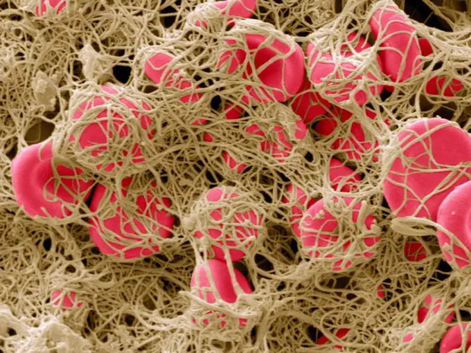
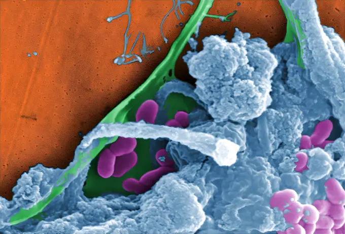
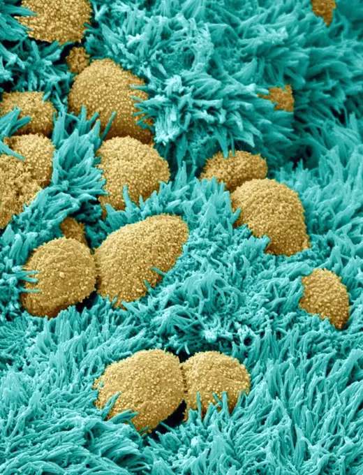
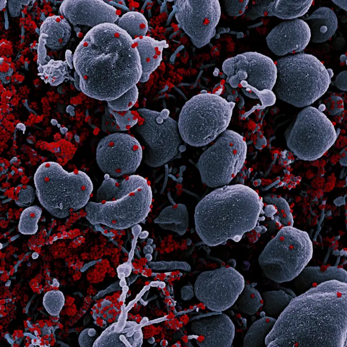
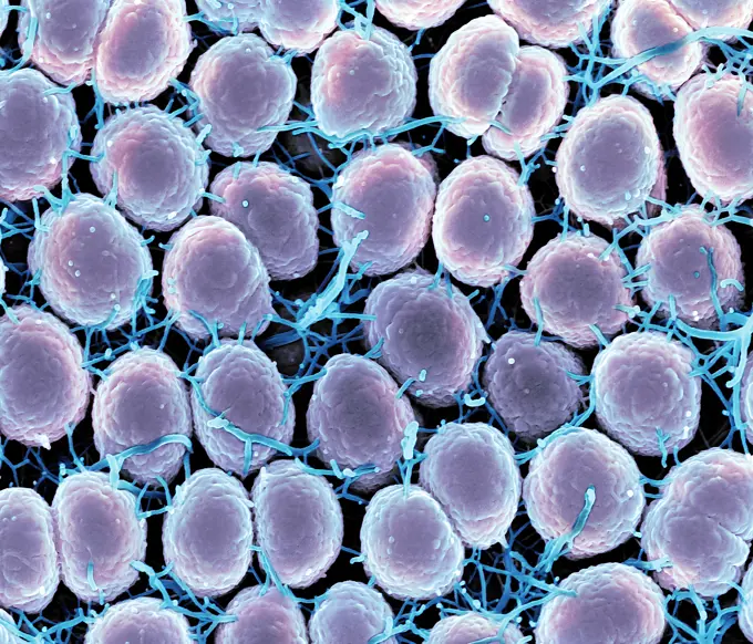
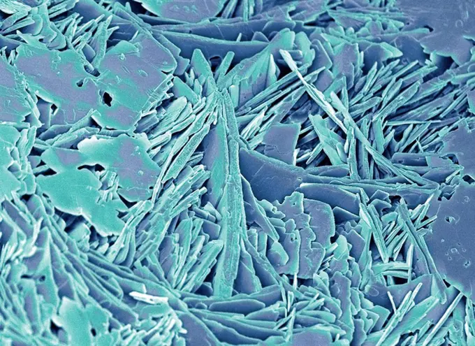
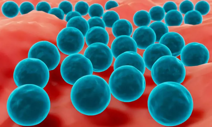
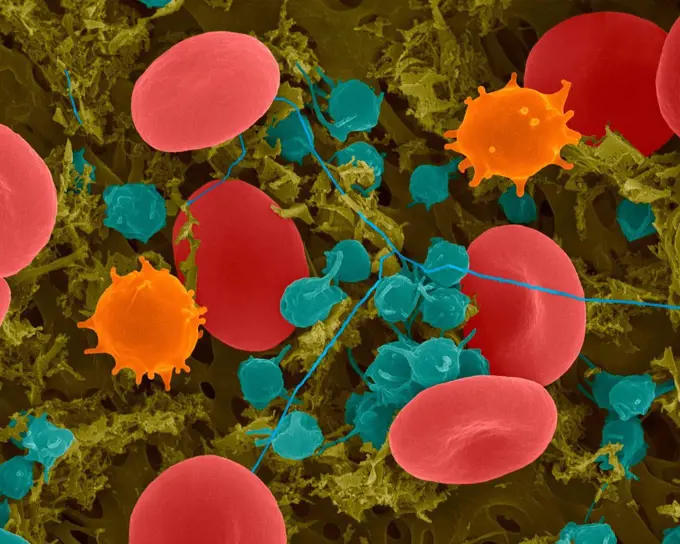
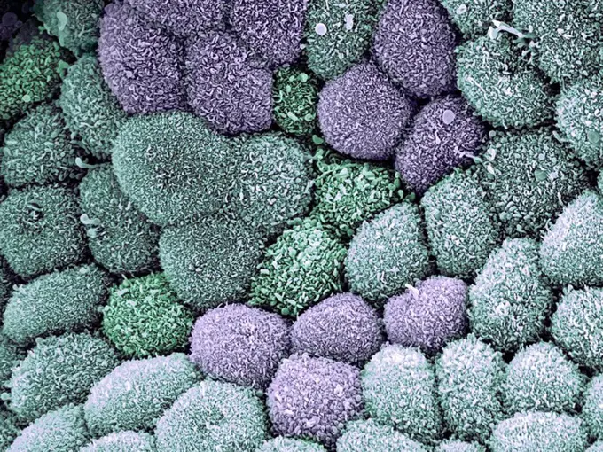
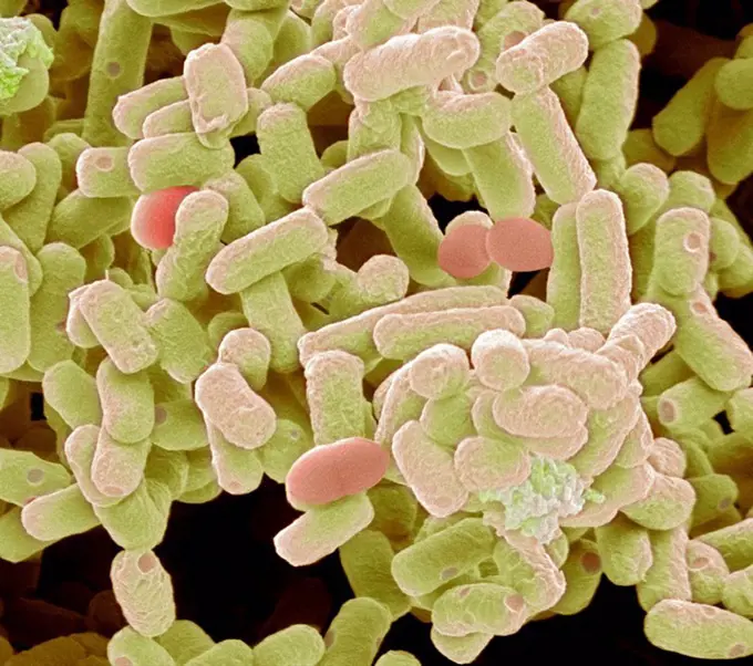
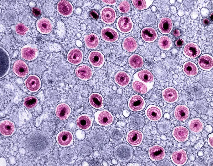
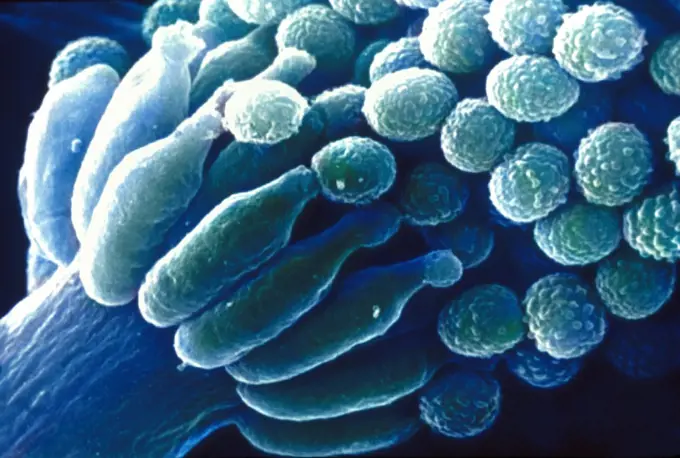
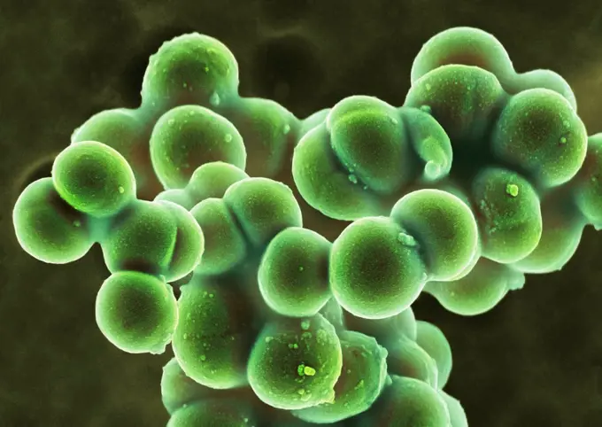
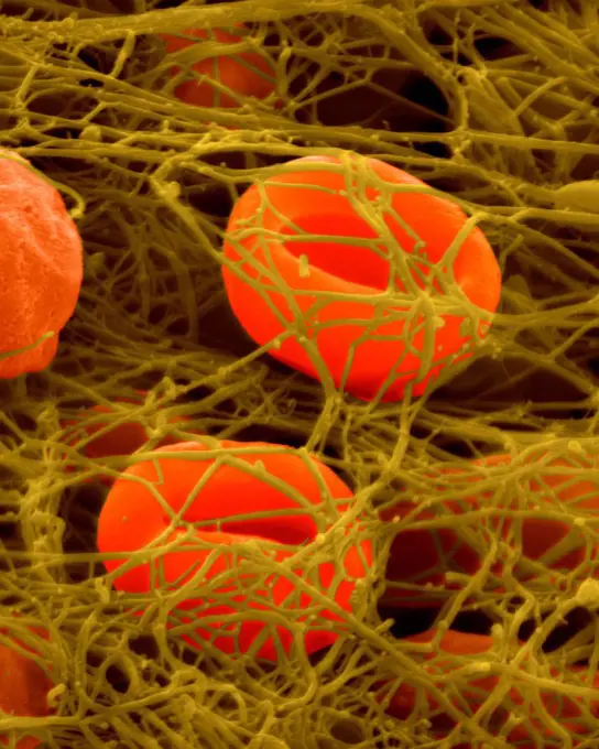
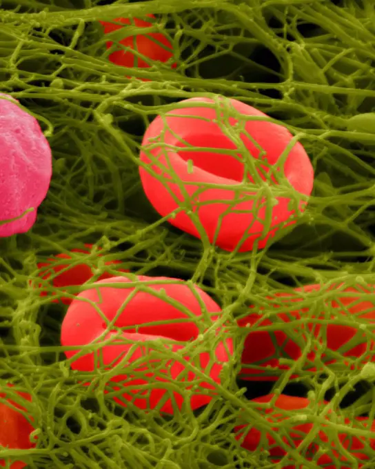
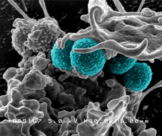
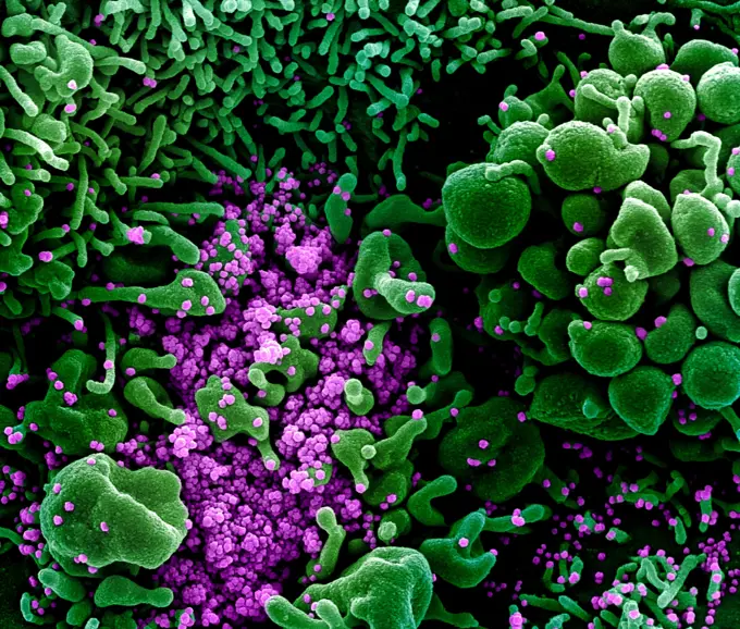
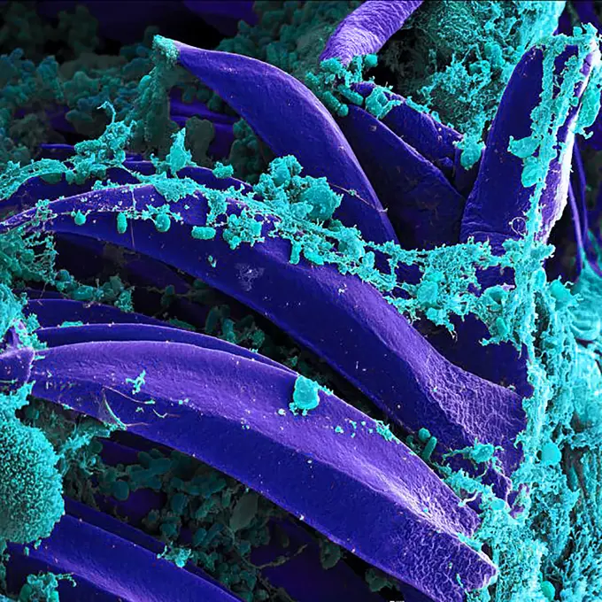
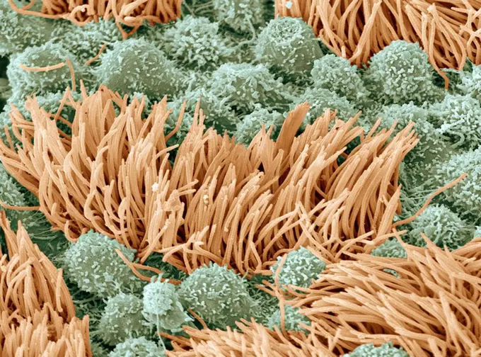
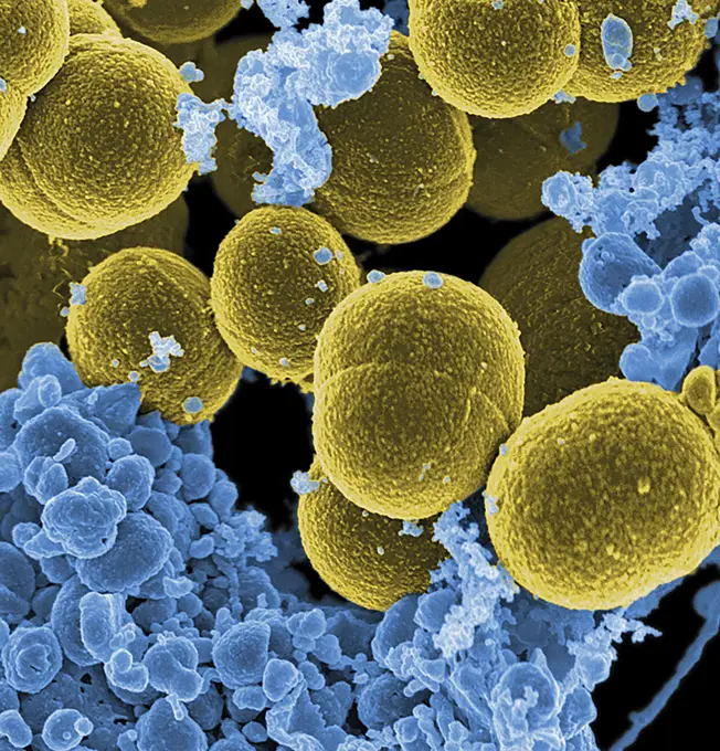
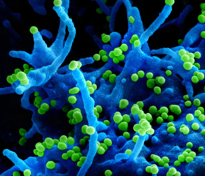
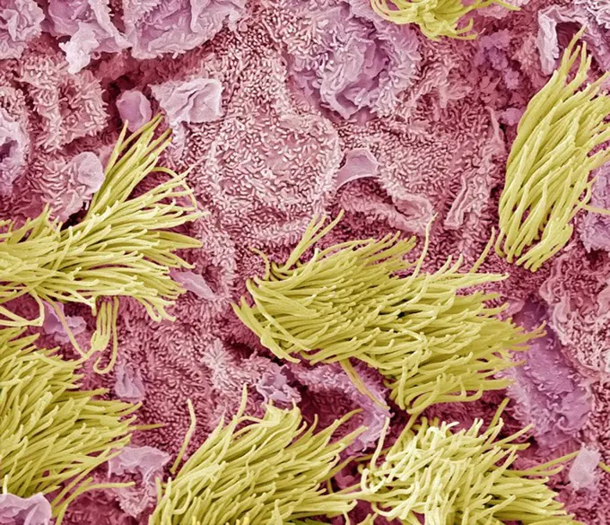
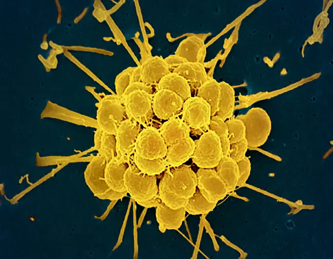
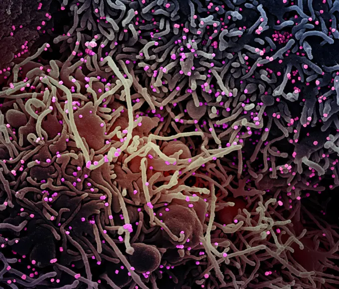
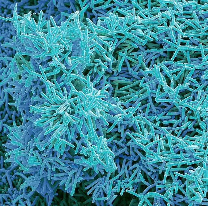
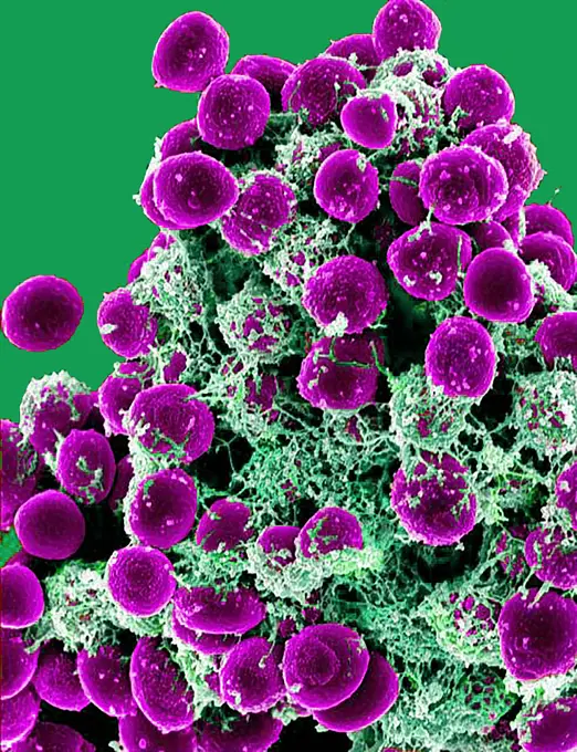
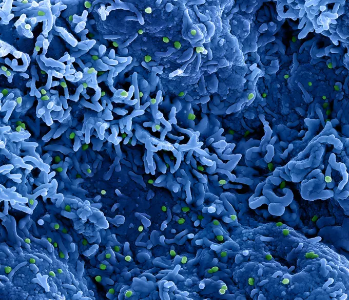
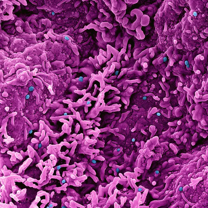
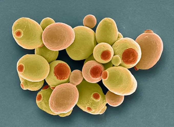
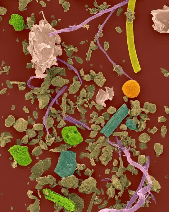
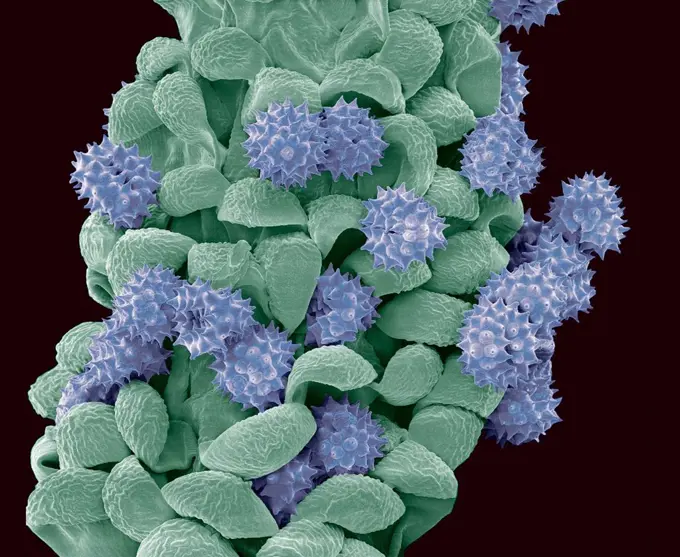
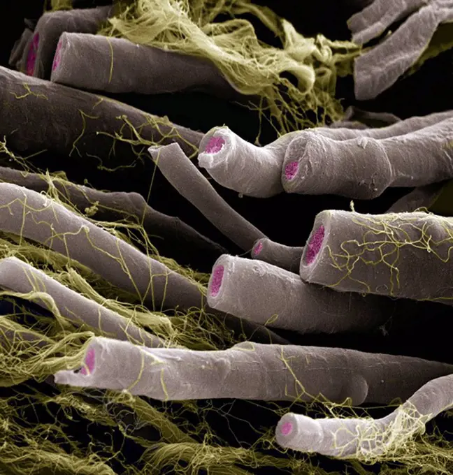
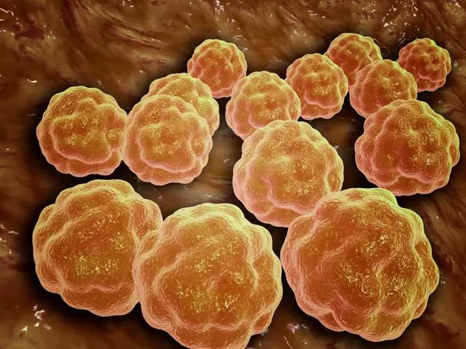
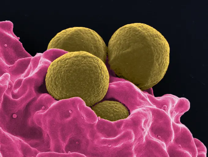
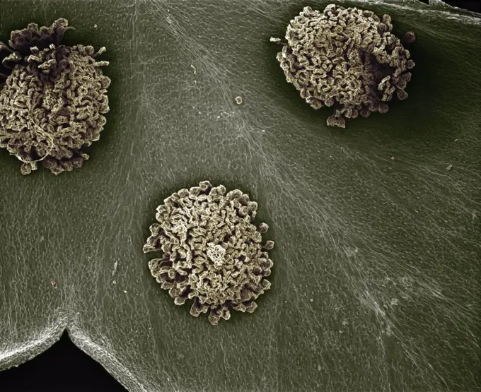
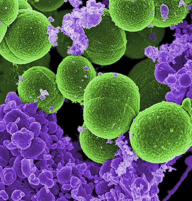
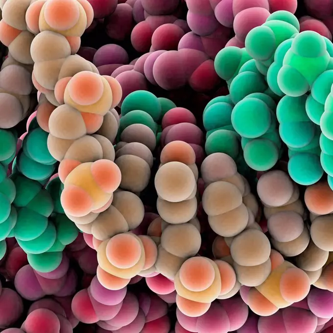
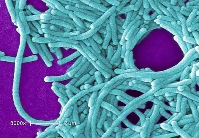
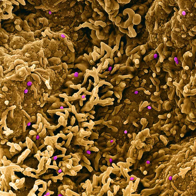
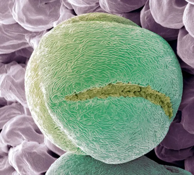
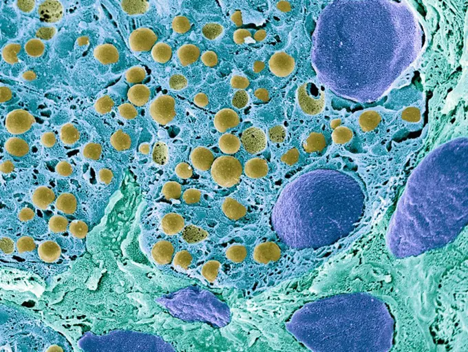
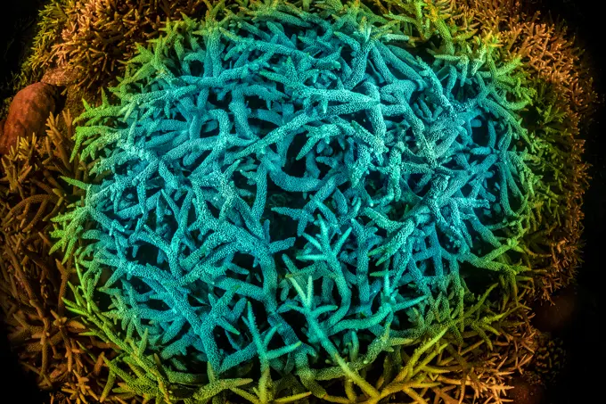
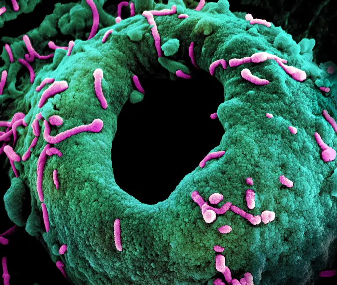
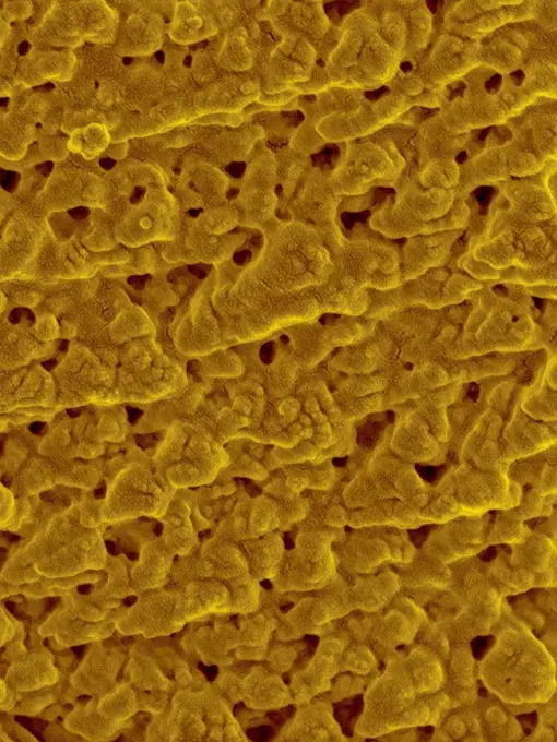
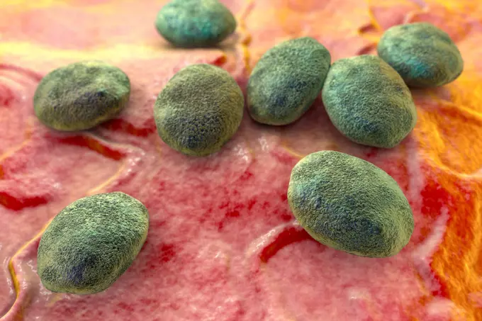
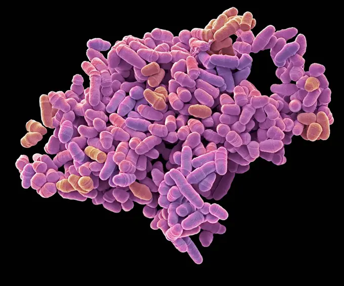
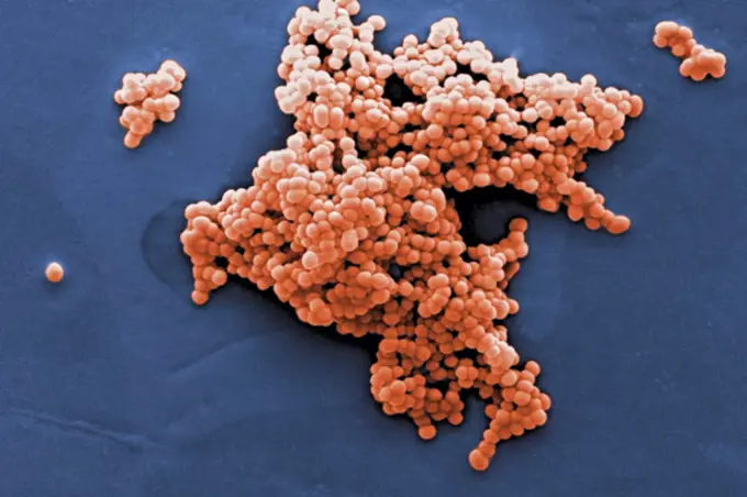
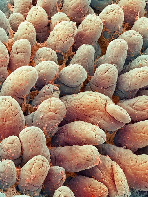
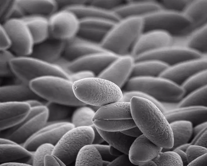
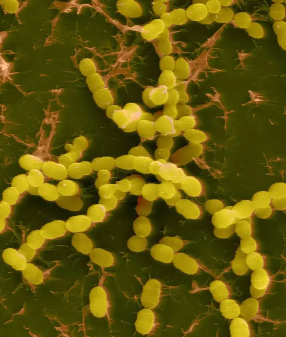
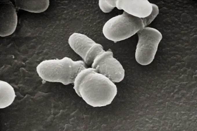
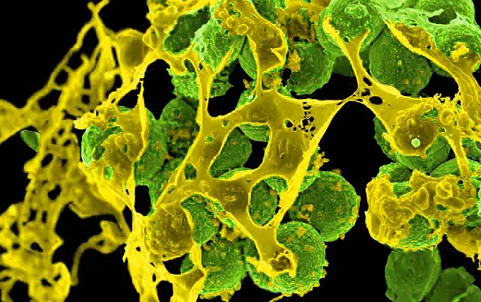
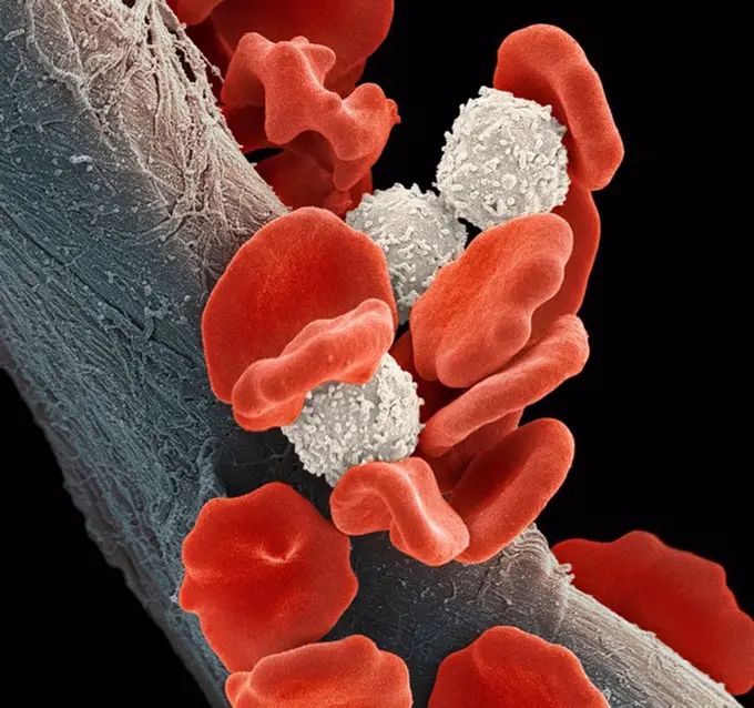
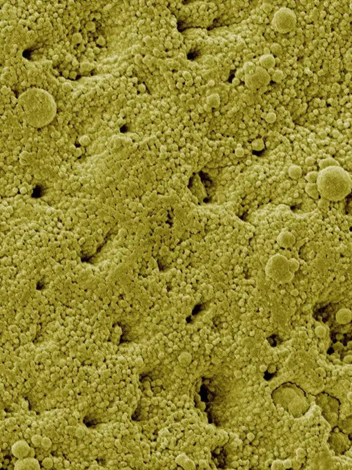
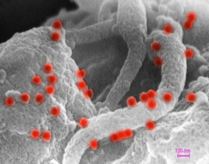
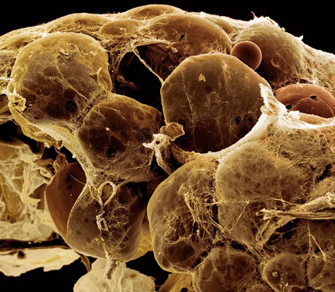
Microorganisms in Untreated Water

Colorized scanning electron micrographs of microorganisms showing bacteria, protozoa, and biofilms in untreated water specimens.

Blood clot, SEM
Blood clot, SEM
200 assets in this story
824-57659305
This digitally colorized scanning electron microscopic (SEM) image of an untreated water specimen extracted from a wild stream mainly used to control flooding during inclement weather, revealed the presence of unidentified organisms, which included bacteria, protozoa, and algae. In this particular view, a mass of gelatinous biofilm had enmeshed numbers of microorganisms, including amoebae and bacteria. CDC/ Janice Haney Carr 2009
4128-V58562034
824-63225895
Colorized scanning electron micrograph of a cell (purple) infected with SARS-CoV-2 virus particles (blue), isolated from a patient sample. Image captured at the NIAID Integrated Research Facility (IRF) in Fort Detrick, Maryland.
4128R-6954
Fallopian tube cells, SEM
824-63225879
Colorized scanning electron micrograph of an apoptotic cell (gray) heavily infected with SARS-CoV-2 virus particles (red), isolated from a patient sample. Image captured at the NIAID Integrated Research Facility (IRF) in Fort Detrick, Maryland.
1899-65662483
Scanning electron micrograph of Neisseria gonorrhoeae bacteria, which causes gonorrhea.
4269-20408841
Coloured scanning electron micrograph (SEM) of a neutrophil white blood cell (bottom) engulfing methicillin-resistant Staphylococcus aureus bacteria (MRSA, yellow).
4128R-6082
Gallstone, SEM
4128R-2752
Blood clot, SEM
4128-18680844
Staphylococcus illustration
4128R-13620338
Human red blood cells, monocytes (white blood cells), activated platelets and fibrin thread, composite coloured scanning electron micrograph (SEM). Re...
4128R-3516
Gall bladder surface, SEM
4128R-1085
Escherichia coli bacteria yellow, coloured scanning electron micrograph SEM. A few cocci bacteria pink are also present. Magnification: x40,000 when p...
1899-65662383
Transmission electron micrograph of cytomegalovirus (CMV) particles (pink) found within intracellular vesicles of an infected macrophage (purple).
824-468
Aspergillus Fumigatus
4201-66349
Daffodil (Narcissus sp) SEM close-up view of pollen on anther at 700x magnification
824-63223225
Colorized scanning electron micrograph of an apoptotic cell (blue) infected with SARS-COV-2 virus particles (red), isolated from a patient sample. Image captured at the NIAID Integrated Research Facility (IRF) in Fort Detrick, Maryland. Credit: NIAID.
4128-15659915
MRSA bacteria, SEM
824-63191206
Colorized transmission electron micrograph showing USA 300 strain of Staphlococcus aureus, shown in gold, outside a white blood cell, shown in blue.
4128R-13620357
Human red blood cells trapped in a fibrin blood clot, coloured scanning electron micrograph (SEM). Human red blood cells trapped in a fibrin blood clo...
824-63227412
Treponema pallidum, the bacterium responsible for syphilis.
4128R-15545729
Foot bacteria, SEM
4128R-11476227
Protein molecules, computer artwork.
4128R-13620359
Human red blood cells trapped in a fibrin blood clot, coloured scanning electron micrograph (SEM). Human red blood cells trapped in a fibrin blood clo...
1439-57947384
Intricate microstructures resembling a lunar landscape in monochrome
824-63191261
Interaction of MRSA with a human white cell. The bacteria shown is strain MRSA252, a leading cause of hospital-associated infections in the United States and United Kingdom.
824-63223224
Colorized scanning electron micrograph of an apoptotic cell (green) heavily infected with SARS-COV-2 virus particles (purple), isolated from a patient sample. Image captured and color-enhanced at the NIAID Integrated Research Facility (IRF) in Fort Detrick, Maryland. Credit: NIAID.
824-63194446
Scanning electron micrograph of Yersinia pestis, which causes bubonic plague, on proventricular spines of a Xenopsylla cheopis flea.
4128R-8001
Fallopian tube, SEM
824-63194632
Scanning electron micrograph of S. aureus bacteria escaping destruction by human white blood cells.
4297-1422
Scanning electron micrograph of human immunodeficiency virus (HIV), grown in cultured lymphocytes. Virions are seen as small spheres on the surface of the cells
824-63227047
Scanning electron micrograph of Lassa virus budding from a cell. Lassa virus (LASV) is an arenavirus that causes Lassa hemorrhagic fever, a type of viral hemorrhagic fever (VHF) in humans and other primates. Image purchased and colorized from the NIAID Integrated Research Facility (IRF) in Fort Detrick, Maryland. Credit: NIAID
4128R-6191
Mycobacterium chelonae bacteria, SEM
4269-20408843
Yersinia pestis bacteria that had gathered on the proventricular spines of a Xenopsylla cheopis flea. The Y. pestis bacterium is the pathogen that causes bubonic plague, scanning electron micrograph (SEM).
4269-24723
Lung. Alveoli of the lungs SEM (Scanning Electron Microscope)
4128R-10555
Uterine cancer, SEM
4269-20408842
Coloured scanning electron micrograph (SEM) of a neutrophil white blood cell (bottom) engulfing methicillin-resistant Staphylococcus aureus bacteria (MRSA, yellow).
1899-65662401
Scanning electron micrograph of Neisseria gonorrhoeae bacteria, which causes gonorrhea. Exploring a Meningitis Vaccine for Gonorrhea Prevention.
824-63223249
Colorized scanning electron micrograph of an apoptotic cell (tan) heavily infected with SARS-CoV-2 virus particles (orange), isolated from a patient sample. Image captured at the NIAID Integrated Research Facility (IRF) in Fort Detrick, Maryland. Credit: NIAID.
4128R-9301
Hyaline cartilage, SEM
4128-V58562107
824-63227330
Colorized scanning electron micrograph of a VERO E6 cell (purple) showing elongated cell projections and signs of apoptosis, after infection with SARS-COV-2 virus particles (pink), which were isolated from a specimen the patient. Image requested from the NIAID Integrated Research Facility (IRF) in Fort Detrick, Maryland.
824-63194664
Colorized scanning electron micrograph of Middle Eastern Respiratory Syndrome virus particles attached to the surface of an infected VERO E6 cell.
4128R-15545719
Plaque-forming bacteria, SEM
824-63223247
Colorized scanning electron micrograph of an apoptotic cell (green) heavily infected with SARS-CoV-2 virus particles (orange), isolated from a patient sample. Image captured at the NIAID Integrated Research Facility (IRF) in Fort Detrick, Maryland. Credit: NIAID.
824-63194643
Scanning electron micrograph of a clump of Staphylococcus epidermidis bacteria in the extracellular matrix, which connects cells and tissue.
4128R-11476175
Protein molecules, computer artwork.
824-57657568
Colorized scanning electron micrograph of monkeypox virus (green) on the surface of infected VERO E6 cells (blue).
824-65830962
Colorized scanning electron micrograph of mpox virus particles (blue) on the surface of infected VERO E6 cells (pink).
4128-15982690
Yeast cells. Coloured scanning electron micrograph (SEM) of cells of brewer's, or baker's, yeast (Saccharomyces cerevisiae). This fungus consists of s...
4128R-1257
Coloured SEM of healthy skin
824-57657677
Colorized scanning electron micrograph of monkeypox virus (green) on the surface of infected VERO E6 cells (blue).
4128R-13620323
Coloured scanning electron micrograph (SEM) of House dust. Components such as fibre, hair, pollen, insect faecal matter and scale (animal dander) caus...
4128R-8946
Common daisy anther, SEM
1525-25071553
Cells background.
4128R-2058
Nerve fibres, SEM
4239R-8255
Microscopic view of Rubella. Rubella is an acute, contagious viral infection. While the illness is generally mild in children, it has serious consequences in pregnant women causing fetal death.
824-63191226
Scanning electron micrograph of a human neutrophil ingesting MRSA.
4201-66270
Leather Fern (Arachniodes adiantiformis) SEM close-up view of sori on the underside of frond where haploid spores are formed during meiosis at 21x magnification
824-63194634
Scanning electron micrograph of S. aureus bacteria escaping destruction by human white blood cells.
4128R-11476159
Protein molecules, computer artwork.
824-63225865
Colorized scanning electron micrograph of a cell (green) infected with UK B.1.1.7 variant SARS-CoV-2 virus particles (pink), isolated from a patient sample. Image captured at the NIAID Integrated Research Facility (IRF) in Fort Detrick, Maryland.
4297-1720
SEM image of Gram-negative Legionella pneumophila bacteria
4128R-26271
MRSA bacteria. Computer artwork of methicillin-resistant Staphylococcus aureus (MRSA) bacteria.
4269-20408840
Coloured scanning electron micrograph (SEM) of a neutrophil white blood cell (bottom) engulfing methicillin-resistant Staphylococcus aureus bacteria (MRSA, yellow).
824-65830959
Colorized scanning electron micrograph of mpox virus (purple) on the surface of infected VERO E6 cells (tan).
4269-27619
Vibrio cholerae. Scanning electron micrograph (SEM) of Vibrio cholerae bacteria. This Gram-negative bacterium causes cholera. Magnified 22371x.
4128R-9541
Strawberry pollen, SEM
4128R-5412
Pancreas tissue, SEM
4070-69335081
Coral fluorescence (Acropora sp.) at night, photographed under blue light, Raja Ampat, West Papua, Ceram Sea, Pacific Ocean.
4128-V58562104
4269-27450
Red cell. Color enhanced scanning electron micrograph (SEM) of human red blood cells. Magnification 11397x.
4128-V58557935
824-65830966
Colorized scanning electron micrograph of mpox virus particles (green) on the surface of infected VERO E6 cells (blue).
1899-65662313
Colorized scanning electron micrograph of Marburg virus particles (pink) both budding and attached to the surface of an infected VERO E6 cell (green).
1525-26664763
Daisy, x370 magnification
4128-V58557940
824-57657585
Colorized transmission electron micrograph of monkeypox virus particles (orange) found within an infected cell (green), cultured in the laboratory.
4128R-13620177
Coloured scanning electron micrograph (SEM) Emu eggshell surface with tiny pores (Dromaius novaehollandiae). Calcium carbonate crystals make up the Em...
4128-24795774
Nematode worm eggs, illustration
4128R-15465081
Dividing yeast cells, SEM
4384-113
Under a moderate magnification of 2,969X, this scanning electron micrograph (SEM) revealed a number of clusters of Gram-positive, beta-hemolytic Group C Streptococcus sp.  bacteria. The genus Streptococcus is comprised of microaerophilic cocci (round), which are not motile and occur in chains or pairs. The genus is defined by a combination of antigenic, hemolytic, and physiological characteristics into Groups A, B, C, D, F, and G. Groups A and D can be transmitted to humans via food. Photo by Ja
824-63225868
Colorized scanning electron micrograph of a cell infected with SARS-CoV-2 virus particles (yellow), isolated from a patient sample. Image captured at the NIAID Integrated Research Facility (IRF) in Fort Detrick, Maryland.
4128R-11476233
Protein molecules, computer artwork.
4128-V58557877
4128R-2637
Intestinal lining, SEM
4201-66318
Mediterranean Fan Palm (Chamaerops humilis) SEM close-up view of pollen at 270x magnification
4128R-13620051
Coloured scanning electron micrograph (SEM) of Streptococcus thermophilus, Gram-positive, coccoid prokaryote (bacterium). Streptococcus thermophilus (...
4128R-5426
Tomato leaf surface, SEM
4128R-4688
Blood vessel in the lung, SEM
4384-310
Malassezia lipophilis
824-63194423
Scanning electron micrograph of methicillin-resistant Staphylococcus aureus (MRSA, green) surrounded by cellular debris. MRSA resists treatment with many antibiotics.
4128R-1898
Leukaemia blood cells, SEM
4128R-13622337
Coloured scanning electron micrograph (SEM) Chicken eggshell surface (Gallus gallus domesticus). Shown here are the surface mineral crystals that form...
1899-65662513
A colorized scanning electron microscope image of Group A Streptococcus (orange) during phagocytic interaction with a human neutrophil (teal).
4297-1419
This scanning electron micrograph shows human immunodeficiency virus (HIV-1), (spherical in appearance) co-cultivated with human lymphocytes.
824-63227071
This scanning electron microscope image shows SARS-CoV-2 (round gold particles) emerging from the surface of a cell cultured in the laboratory. SARS-CoV-2, also known as 2019-nCoV, is the virus that causes COVID-19. Image captured and colorized at Rocky Mountain Laboratories in Hamilton, Montana. Credit: NIAID
4128R-1452
Fat tissue, SEM
824-63225923
Colorized scanning electron micrograph of a cell (blue) infected with a variant strain of SARS-CoV-2 virus particles (green), isolated from a patient sample. Image captured at the NIAID Integrated Research Facility (IRF) in Fort Detrick, Maryland.
4128-V58559208
PREVIOUS
of 2
NEXT
2401 S. Ervay, Suite 206
Dallas, Texas 75215
United States
Get Started
Free ResearchMy BoardsMy Cart
For Creators
How To License Your ContentContributor PortalFrame of Mind
Resources
API accessPricing
Contact
+1 866 236 0087help@viewpictures.co.uk Contact form
©2026 View Pictures. All Rights Reserved. -A
This site is protected by reCAPTCHA and Google Privacy Policy and Terms of Service apply.