My Boards
View Pictures
  • My Boards

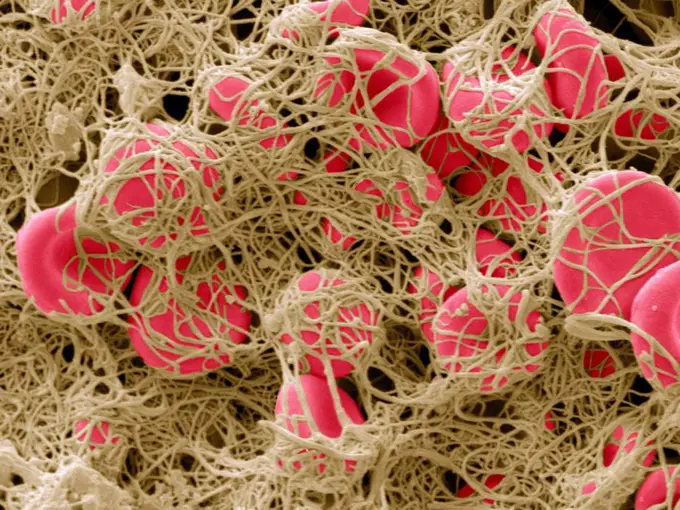
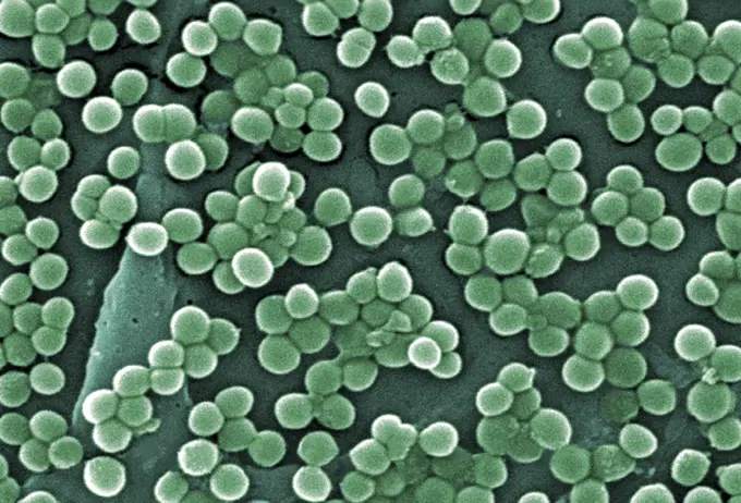
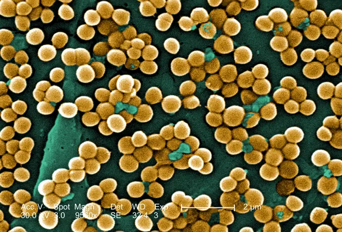
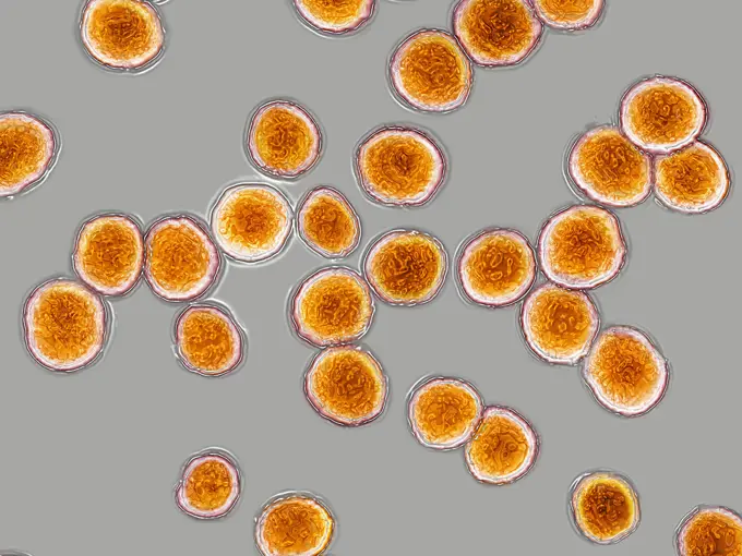
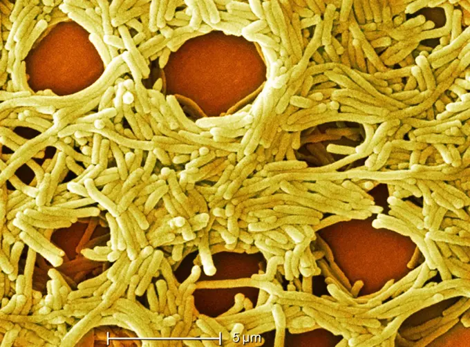
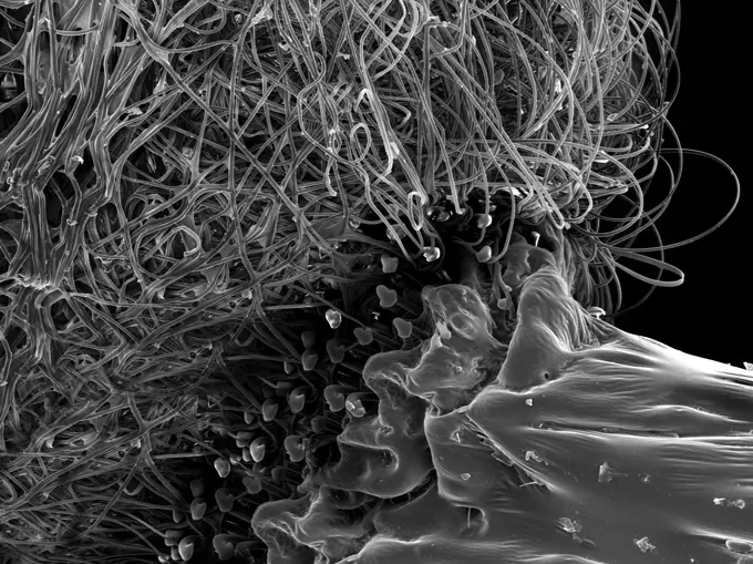
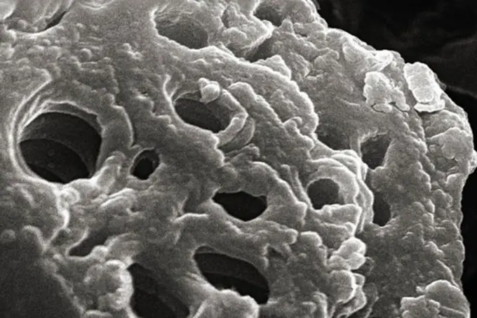
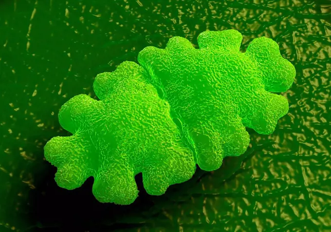
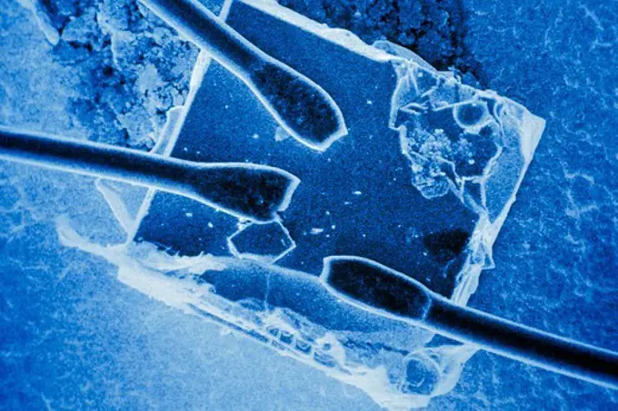
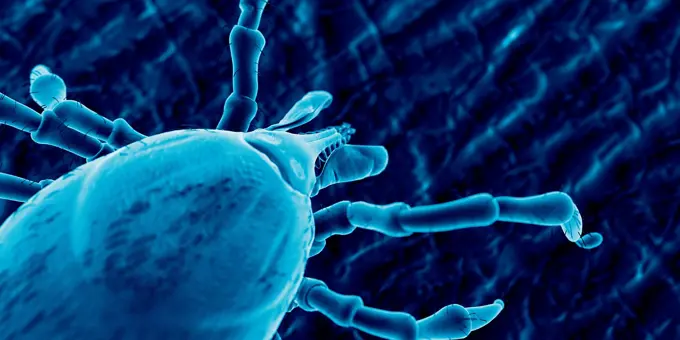
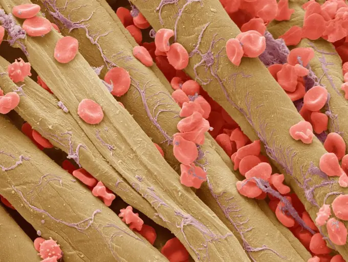
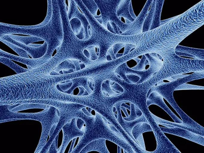
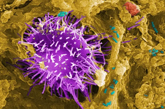
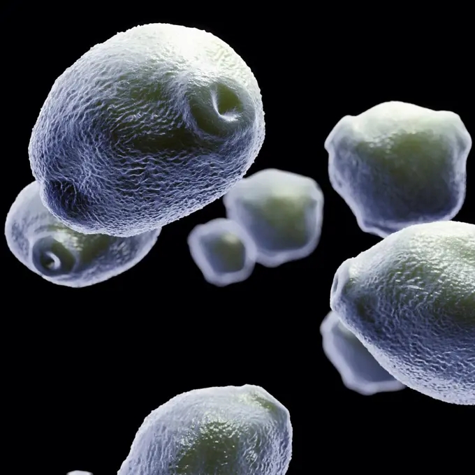
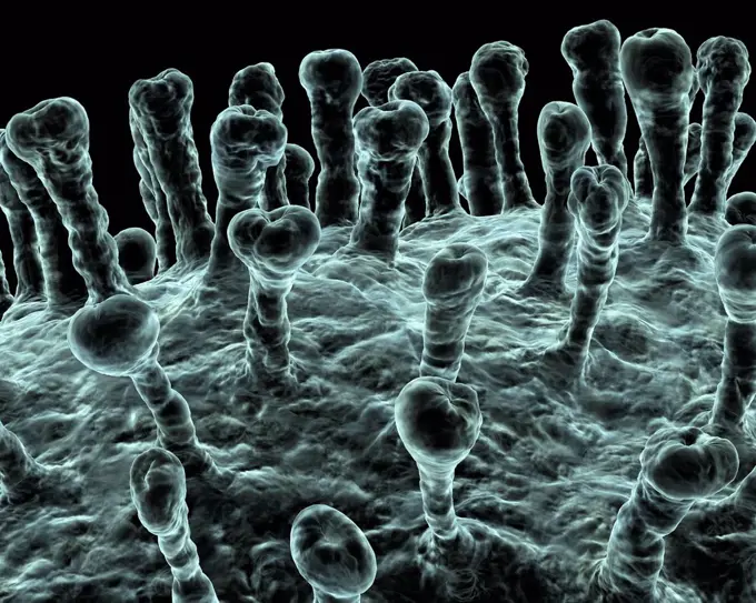
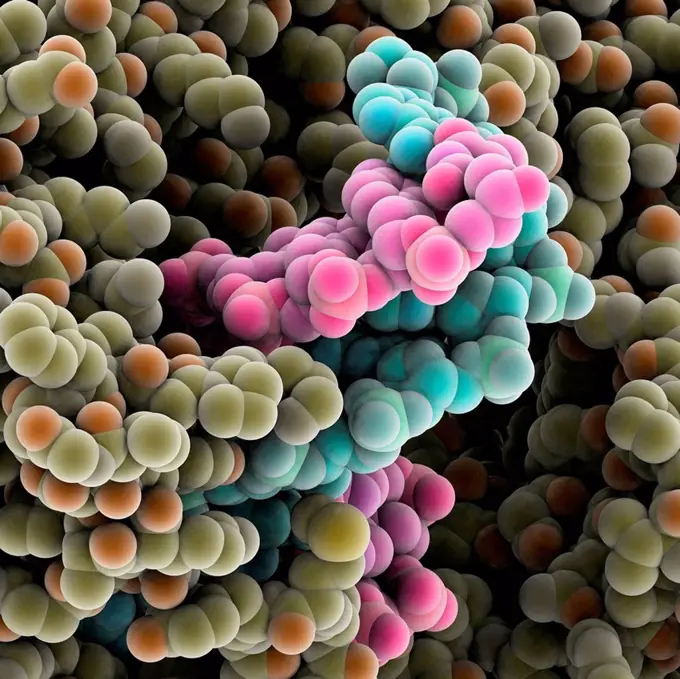
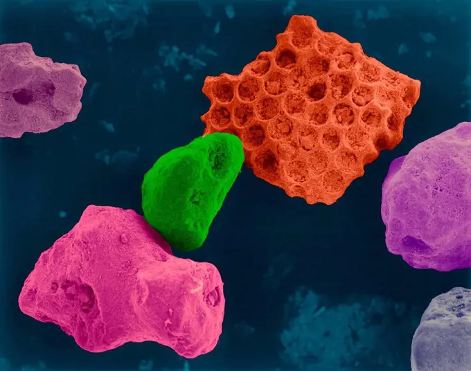
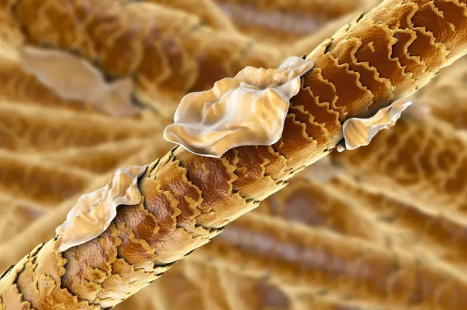
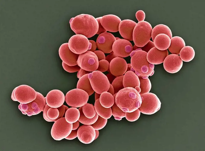
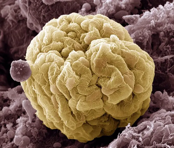
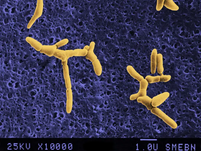
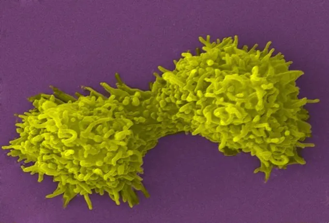
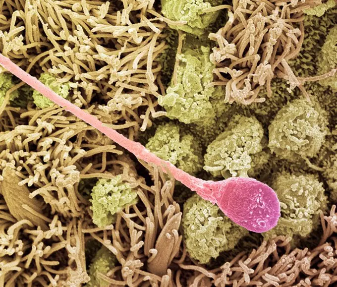
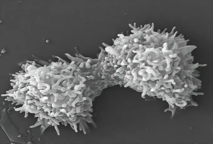
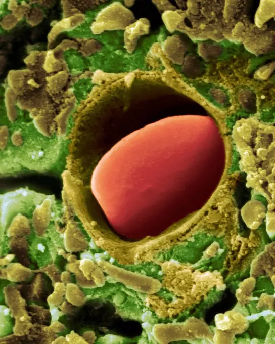
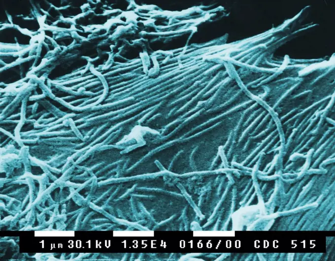
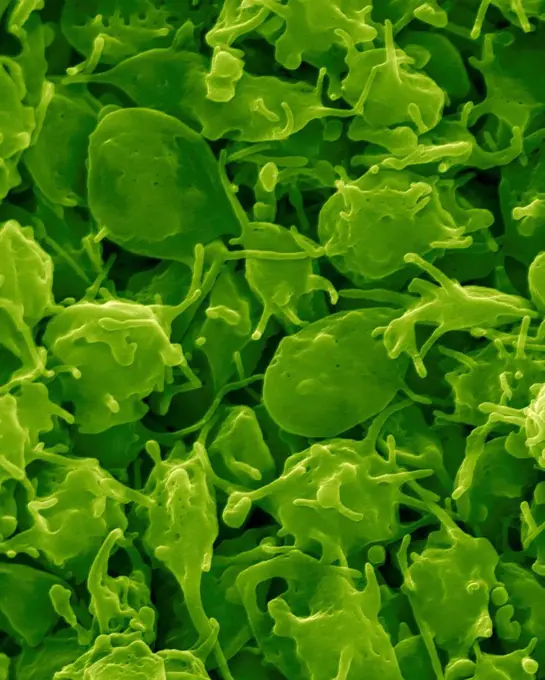
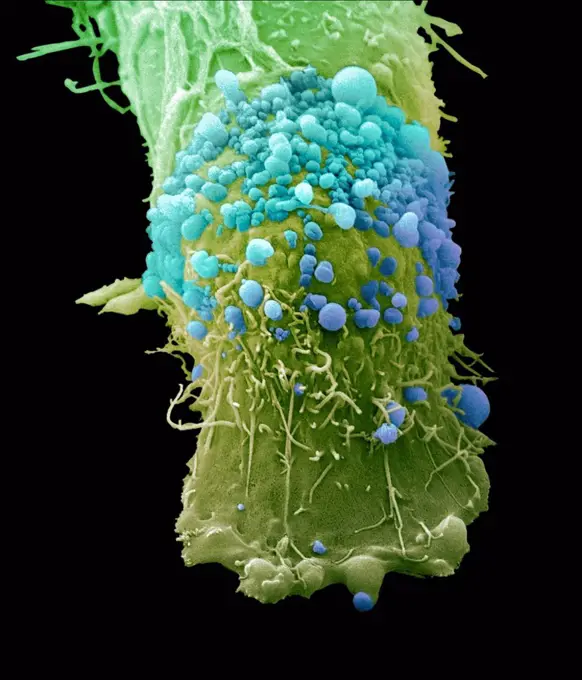
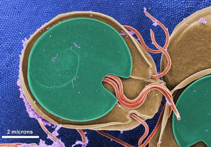
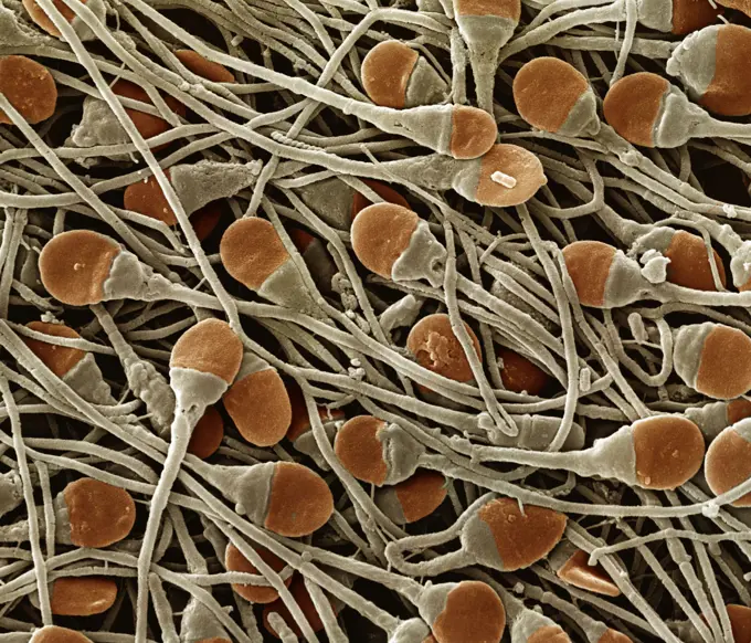
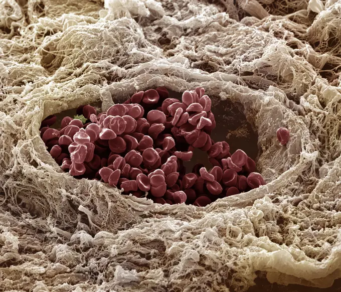
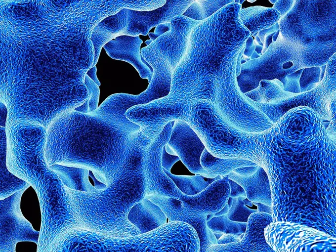
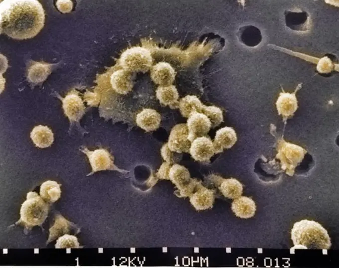
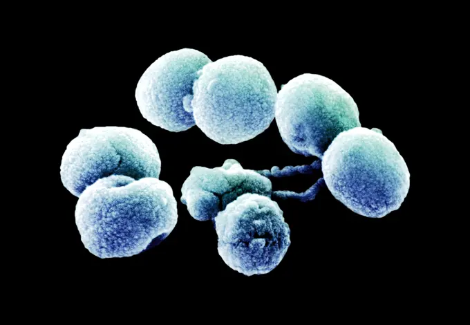
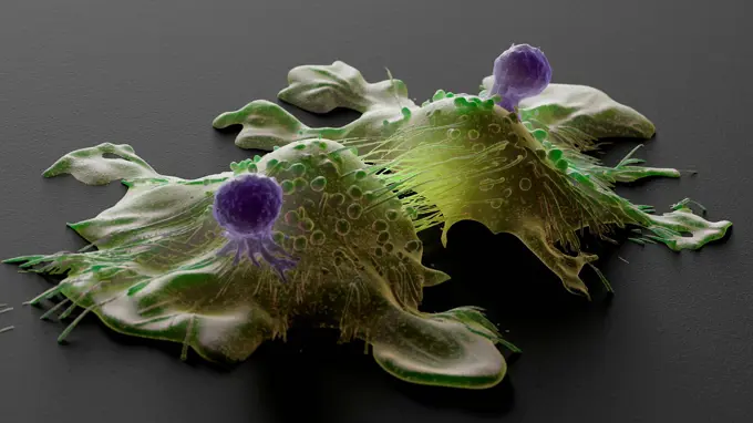
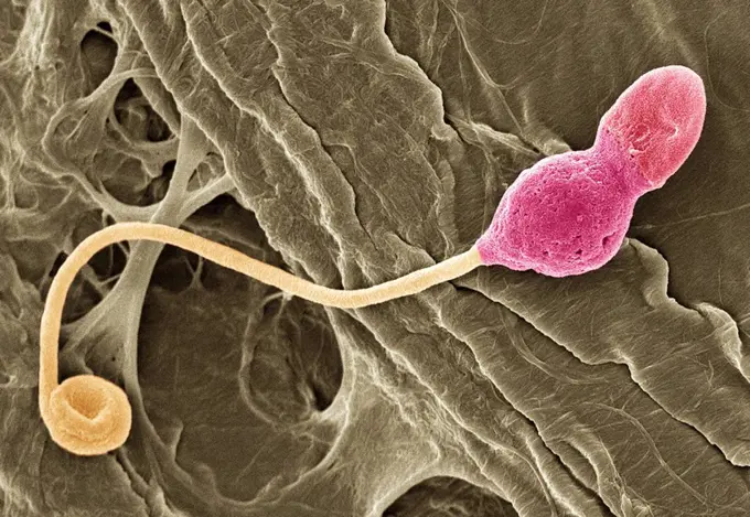
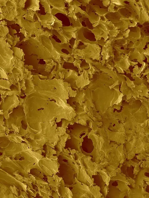
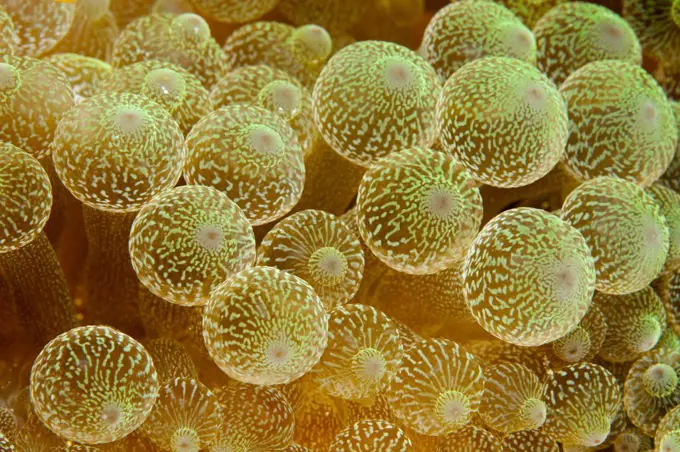
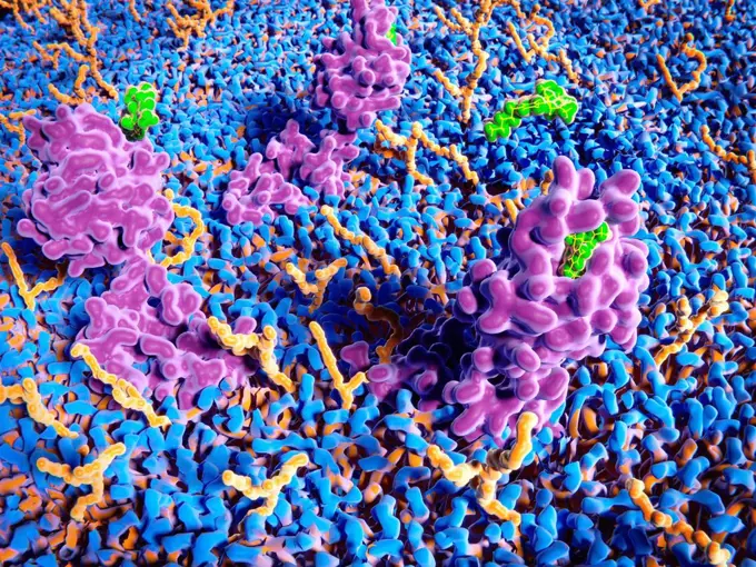
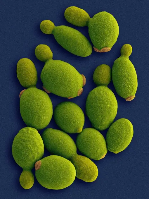
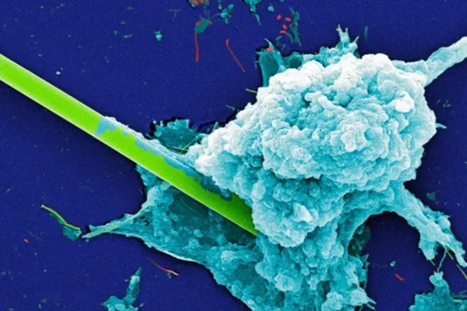
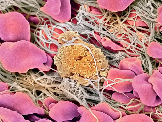
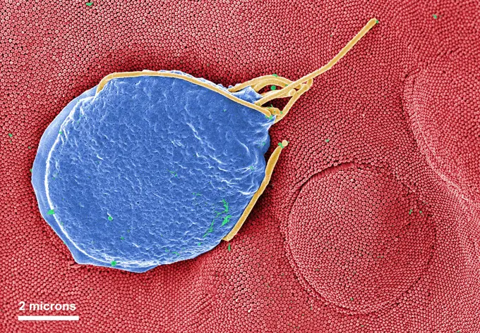
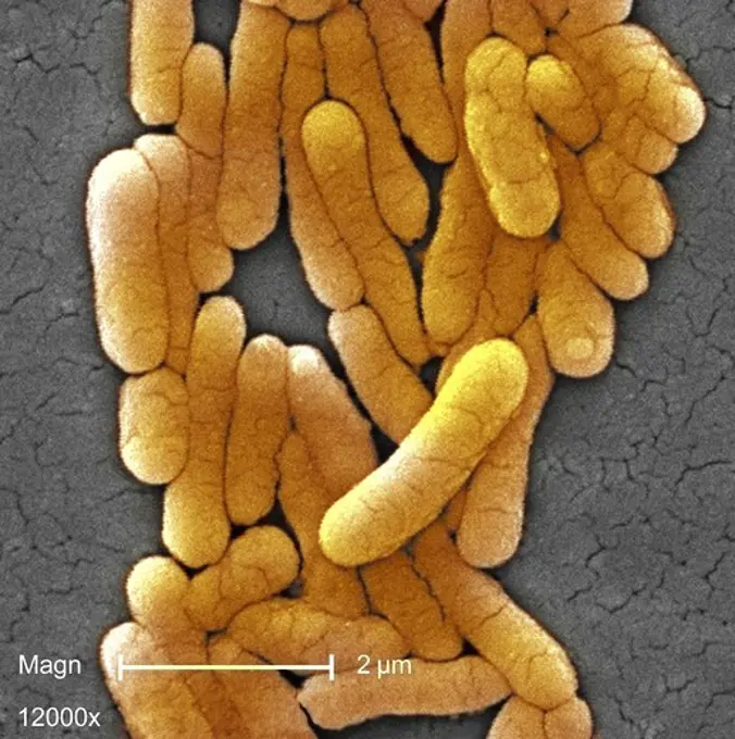
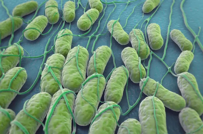
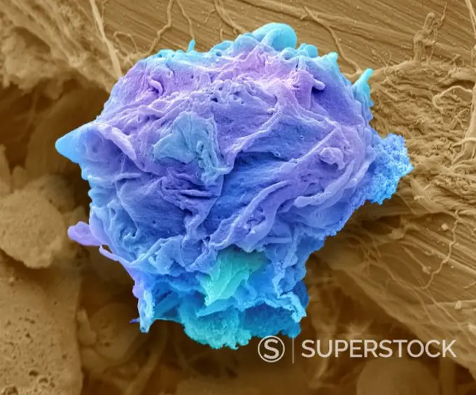
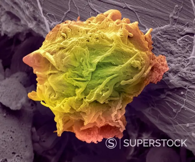
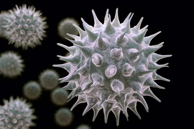
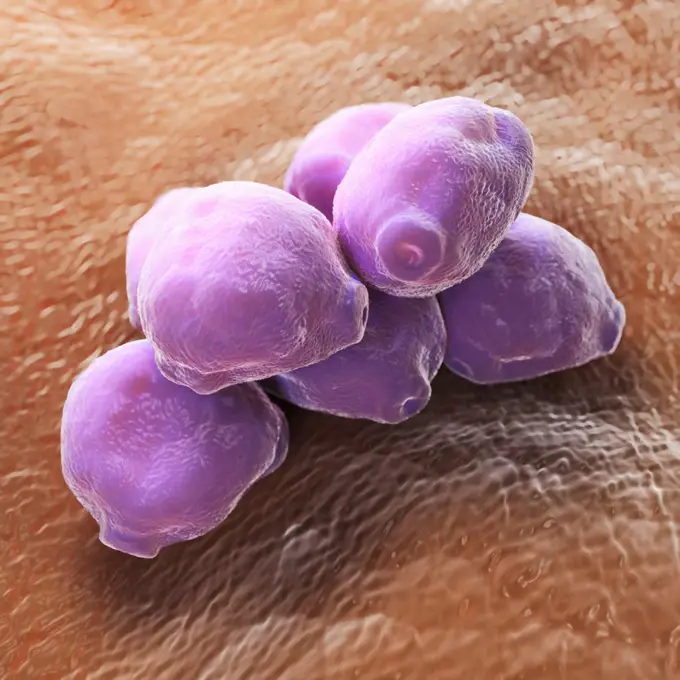
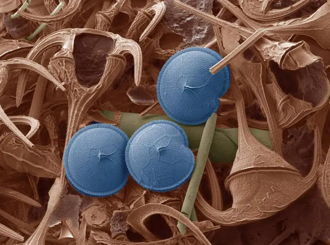
Microorganisms in Untreated Water

Colorized scanning electron micrographs of microorganisms showing bacteria, protozoa, and biofilms in untreated water specimens.

Blood clot, SEM
Blood clot, SEM
200 assets in this story
824-63225926
Colorized scanning electron micrograph of chronically infected and partially lysed cells (brown) infected with a variant strain of SARS-CoV-2 virus particles (purple), isolated from a patient sample. Image captured at the NIAID Integrated Research Facility (IRF) in Fort Detrick, Maryland.
4269-27384
Methicillin-Resistant Staphylococcus Aureus. Scanning electron micrograph (SEM) of methicillin-resistant Staphylococcus aureus bacteria (MRSA); Magnification 9560x. This strain of Gram-positif bacterium has developed wide-ranging antibiotic resistance such as methicillin, and are hospital pathogens.
824-63194408
This scanning electron micrograph (SEM) depicted numerous clumps of methicillin-resistant Staphylococcus aureus bacteria, commonly referred to by the acronym, MRSA, Magnified 9560x. Recently recognized outbreaks, or clusters of MRSA in community settings have been associated with strains that have some unique microbiologic and genetic properties, compared with the traditional hospital-based MRSA strains, which suggests some biologic properties, eg virulence factors like toxins, may allow the community strains to spread more easily, or cause more skin disease.
824-63190266
Staphylococcus aureus is the bacterium responsible for food poisoning, localised skin diseases, respiratory diseases and in some extreme cases, septicemia. Image taken with an optical microscope X 1000.
1773-97830
Electron micrograph of Legionella bacteria, 6500x
4297-1786
Scanning Electron Micrograph (SEM) of methicillin-resistant Staphylococcus aureus bacteria, commonly known as MRSA
4128R-13818100
MRSA bacteria. Computer artwork of methicillin-resistant Staphylococcus aureus (MRSA) bacteria.
1439-57941499
Intricate microstructures weaving a complex web
4384-414
Microscopic Detail of Dandelion
4128R-12923035
Euastrum oblongum
4377-206
Superconducting Ceramic Compound
4378-4460
Cilia of the Respiratory Tract
4128-30418013
Tick on human skin, illustration
1899-65662312
Colorized scanning electron micrograph of Marburg virus particles (yellow) both budding and attached to the surface of an infected VERO E6 cell (blue).
4128R-8235
Used surgical swab, SEM
824-63128359
Surface of the colon mucosa. SEM x 200.
4128R-2575
Abstract computer artwork
824-63194563
This colorized transmission electron micrograph (TEM) revealed the presence of a number of Novel H1N1 virus virions in this tissue culture sample. Novel H1N1 (referred to as swine flu early on) is a new influenza virus causing illness in people. This new virus was first detected in people in the United States in April 2009. This virus is spreading from person-to-person worldwide, probably in much the same way that regular seasonal influenza viruses spread. On June 11, 2009, the World Health Organization (WHO) reported that a pandemic of novel H1N1 flu was underway.
4128-V58557889
4384-171
Under a moderately-high magnification of 2500X, this digitally-colorized scanning electron micrograph (SEM) of an untreated water specimen extracted from a wild stream mainly used to control flooding during inclement weather, revealed the presence of unidentified organisms, which included bacteria, protozoa, and algae. In this particular view, a microorganism is featured, the exterior of which is covered by numerous projections imparting an appearance of a sea urchin. This microscopic pin cushi
4128R-12285315
Fat tissue. Coloured scanning electron micrograph (SEM) of a sample of fat tissue, showing fat cells (adipocytes, blue/purple) surrounded by fine stra...
1899-61460370
Colorized scanning electron micrograph of monkeypox virus (green) on the surface of infected VERO E6 cells (red)
4269-27453
Enterococcus. Scanning electron micrograph (SEM) of Enterococcus faecalis (formerly known as Streptococcus faecalis). This Gram-positive bacterium is commonly found in human intestine. It can also cause urinary tract and  skin infections. E. faecalis is resistant to a number of antibiotics and is often involved in nosocomial diseases.
1899-65662309
Colorized scanning electron micrograph of Marburg virus particles (yellow) both budding and attached to the surface of an infected VERO E6 cell (blue).
824-65830616
Colorized scanning electron micrograph of Marburg virus particles (yellow) both budding and attached to the surface of an infected VERO E6 cell (blue).
4378-4538
Birch Tree Pollen
4128R-28139
Human coronavirus. Computer artwork of a Human coronavirus particle. Coronaviruses primarily infect the upper respiratory and gastrointestinal tract a...
4128R-11476169
Protein molecules, computer artwork.
4128R-13620306
Coloured scanning electron micrograph (SEM) of Coral sand (Kailua, Hawaii). Beach sand contains both rock minerals and skeletons that have been derive...
4128R-15366461
Human hair with dandruff, illustration
4128-20239392
Microsporum canis fungus, illustration
4128R-8370
Yeast cells, SEM
4128R-14150
Dense connective tissue, coloured scanning electron micrograph SEM. The main component of this tissue is collagen. Magnification: x1500 when printed a...
4128R-4089
Kidney glomerulus, SEM
824-63128416
Listeria denitrificans seen at SEM x 10,000. See image n 0218605 on the site.
4048-16067038
Novel Coronavirus SARS-CoV-2, causes 'coronavirus disease-2019', abbreviated as COVID-19. Scanning electron microscope image shows SARS-CoV-2 (yellow) emerging from the surface of cells (pink) isolated from a US patient. Colors are artificially assigned to the monotone image for clarity. Image by National Institute of Allergy and Infectious Disease (NIAID)  (BSLOC_2020_2_244)
4297-1727
SEM of two Acanthamoeba polyphaga protozoa interacting
824-63219651
This scanning electron microscope image shows SARS-Cov-2 also known as 2019-nCoV, the virus that causes COVID-19 isolated from a patient in the United States, emerging from the surface of cells cultured in laboratory.
4128R-3182
Sperm cell, SEM
824-63194433
Scanning electron micrograph of a murine macrophage infected with Francisella tularensis strain LVS. Macrophages were dry-fractured by touching the cell surface with cellophane tape after critical point drying to reveal intracellular bacteria. Bacteria (colorized in pink) are located either in the cytosol or within a membrane-bound vacuole.
1899-61460843
This scanning electron microscope (SEM) image revealed a protozoan Acanthamoeba polyphaga, as it was about to complete the process of cell division, known as mitosis, becoming two separate organisms. Note the numerous pseudopodia, projecting from the surfaces of these organisms. These pseudopodia allow amoebas to move and grasp objects in their environment. CDC/Catherine Armbruster Margaret Williams 2009
824-63206959
Meningococcus seen under a scanning electron microscope.
4128-15659920
Heart capillary, SEM
6145-44718530
A scanning electron microscope photograph of iron crystals which grow in a small vug or cavity in a recrystallized breccia (fragmented rock) from the Apollo 15 Hadley-Apennino lunar landing site. The largest crystal is three microns across. Perfectly developed crystals such as these indicate slow formation from a hot vapor as the rock was cooling. The crystals are resting on an interlocking lattice of pyroxene (calsium-magnesium-iron silicate).
1899-53513326
Colorized scanning electron micrograph of Ebola virus particles (green) in extracellular space between infected African green monkey kidney cells. Image captured and color-enhanced at the NIAID Integrated Research Facility in Ft. Detrick, Maryland.
4297-1785
Scanning Electron Micrograph (SEM) of methicillin-resistant Staphylococcus aureus bacteria, commonly known as MRSA
4269-27455
Enterococcus. Scanning electron micrograph (SEM) of Enterococcus faecalis (formerly known as Streptococcus faecalis). This Gram-positive bacterium is commonly found in human intestine. It can also cause urinary tract and  skin infections. E. faecalis is resistant to a number of antibiotics and is often involved in nosocomial diseases.
824-63194416
Under a very high magnification of 25000X, this colorized scanning electron micrograph (SEM) revealed the presence of a single Gram-negative Salmonella typhimurium bacterium, which was imaged right at the point where it was undergoing the process of cell division, resulting in the formation of two separate organisms. This dividing bacterium had been isolated from a pure culture.
1439-57944417
A closeup view of a purple spherical object with craterlike textures on its surface.
4128R-30469
Fractured tendon. Coloured scanning electron micrograph (SEM) of a fractured flexor tendon of the finger showing the collagen fibres. The parallel arr...
4048-6171
Ebola virions (virus particles). Ebola hemorrhagic fever is a severe, often-fatal disease in humans, monkeys, gorillas, and chimpanzees that has appeared sporadically since its initial recognition in 1976.
4128R-485
Moth proboscis, SEM
4128-V58562053
824-63123763
This scanning electron micrograph depicts a grouping of Gram-negative Flexispira rappini bacteria, magnified 13,951x. Its name F. rappini is considered provisional, for it was never formally proposed or accepted. Subsequently determined to be closely related to Helicobacter spp. it is referred to as Helicobacter sp. flexispira in the literature.
4128R-8571
Dental plaque, SEM
4128R-13619965
Activated platelets with channels of the open canalicular system (OCS), coloured scanning electron micrograph (SEM). Platelets are blood cell fragment...
4128R-12699832
Coloured scanning electron micrograph (SEM) of fungal cells. The round structures are sporangia, which house the fungus' reproductive cells (spores). ...
4128R-13047384
Skin cancer cell, coloured scanning electron micrograph (SEM). Cultured melanoma cell showing the numerous blebs and microvilli characteristic of canc...
824-63194912
This scanning electron micrograph (SEM) revealed the ventral surface of a Giardia muris trophozoite that had settled atop the mucosal surface of a rat's intestine. Note the microvilli, which can be seen in the background, as tiny rounded structures that are approximately 0.15 microns in diameter. These microvilli cover the surface of each intestinal epithelial cell. The Giardia's ventral adhesive disk resembles a suction cup, where overlapping microtubules in the cytoplasm form a number-6-shaped figure. The edge of the suction cup, called the ventrolateral flange, partially encircles the adhesive disk and is absent posteriorly where a ventral pair of flagella emerges from above, dorsal to the disk. Giardia muris has four pairs of flagella that are responsible for the organism's motility. The adhesive disk facilitates adhesion to the intestinal surface. The protozoan Giardia causes the diarrheal disease called giardiasis.
4269-27686
Lung trachea epithelium. Scanning electron micrograph (SEM) of the lung trachea epithelium which consists of ciliated cells and non-ciliated cells.
4128R-15514869
Antibody blocking programmed cell death, illustration
4220-21334743
Scanning Electron Micrograph (SEM): human sperm.     Date:
4220-21334663
Scanning Electron Micrograph (SEM): Human skin section across vein showing red blood cells.     Date:
824-63225327
Microsporum canis is a species of fungus of the genus Microsporum responsible for the majority of cases of ringworm in dogs and cats, and in humans. X 1000 optical microscopy.
824-63223123
Coxiella burnetii, the bacteria that cause Q fever. Credit: NIAID.
4128R-12576723
Osteoporosis. Computer artwork of the trabeculae in the cancellous (spongy) bone tissue affected by osteoporosis. The cancellous tissue fills the inte...
4297-1453
Under a moderately-high magnification of 8000X, this scanning electron micrograph (SEM) depicted a grouping of Gram-negative Legionella pneumophila bacteria
1899-54026852
Meningococcus seen under a scanning electron microscope.
824-63225850
Colorized scanning electron micrograph of a cell (blue) infected with SARS-CoV-2 virus particles (pink), isolated from a patient sample. Image captured at the NIAID Integrated Research Facility (IRF) in Fort Detrick, Maryland.
824-63223172
Colorized scanning electron micrograph of Marburg virus particles (blue) both budding and attached to the surface of infected VERO E6 cells (orange). Image captured and color-enhanced at the NIAID Integrated Research Facility in Fort Detrick, Maryland. Credit: NIAID.
4128-28515065
Bacteria cultured on Petri dish
4297-1743
SEM image of the Death of a Cancer Cell (step 2 of a 6 step sequence)
4269-7090
Scanning Electron Micrograph (SEM) of Streptococcus pneumoniae bacteria. This Gram-negative bacterium is a leading cause of pneumonia. It also causes many other pathologies like middle ear infections (otitis media), bacteremia and meningitis.
4128-V58557948
4128-28970694
CAR-T cell therapy, illustration
4128R-732
Deformed sperm cell, SEM
824-63225921
Colorized scanning electron micrograph of a cell (blue) infected with a variant strain of SARS-CoV-2 virus particles (yellow), isolated from a patient sample. Image captured at the NIAID Integrated Research Facility (IRF) in Fort Detrick, Maryland.
4128R-13620254
Coloured scanning electron micrograph (SEM) of White bread surface. Shown here is a slice of white sandwich bread, revealing the large airspaces that ...
4070-21284568
Bulb tentacle sea anemone (Entacmaea quadricolor) close up detail, Maldives, Indian Ocean
4128R-15046068
THC binding to cannabinoid receptors, illustration
4128R-13620407
Coloured scanning electron micrograph (SEM) of Komagataella phaffii - methylotrophic yeast. Komagataella phaffii is a newly identified yeast that has ...
4384-256
Scanning Electron Micrograph of Untreated Water Sample
4128R-1736
Blood clot, SEM
824-63194914
This digitally-colorized scanning electron micrograph (SEM) depicted a Giardia muris protozoan adhering itself to the microvillous border of an intestinal epithelial cell. Each small circular profile under the protozoan represents the rounded tip of a single microvillous, and it is estimated that 2000 to 3000 microvilli cover the surface of a single intestinal epithelial cell. The two circular lesions on the right side of the photograph are impressions made by the ventral adhesive disk of other G. muris organisms. This disk acts like a suction cup, facilitating the organism's attachment to the intestinal surface. The protozoan Giardia causes the diarrheal disease called giardiasis.
4297-1459
Scanning photomicrograph of clustered Gram-negative Salmonella typhimurium bacteria at a high magnification of 12000X
4378-3089
A culture of Salmonella bacteria on a plane surface. Salmonella is a genus of rod-shaped, Gram-negative bacteria.
4128R-12573050
Lymphoma cancer cell. Coloured scanning electron micrograph (SEM) of a lymphoma cell. A lymphoma is a cell of the immune system that has become cancer...
4128R-12573023
Lymphoma cancer cell. Coloured scanning electron micrograph (SEM) of a lymphoma cell. A lymphoma is a cell of the immune system that has become cancer...
4378-4458
Sunflower Pollen
1848-58228860
Fluoresce hard coral, Coral fluorescenc
4269-27367
Vibrio parahaemolyticus. Scanning electron micrograph (SEM) of Vibrio parahaemolyticus. bacteria. This Gram-negative bacterium causes gastointestinal infection to man. It is closely related to Vibrio cholerae, the  agent of cholera. Magnified 19058x.
4378-4634
Birch Tree Pollen
4128-V58561165
4201-66316
Mite (Blattisocius tarsalis) SEM close-up view of individual predating on eggs of Indian Meal Moth (Plodia interpunctella), this species is used to control population sizes of the moth, seen at 70x magnification
4128-V58557881
1439-57947383
Intricate crystal formations in monochrome
1848-61071915
Marine sponge in reef, Porifera, Kimbe Bay, New Britain, Papua New Guinea, Oceania
1848-57203766
Fishing nets
4128R-341
Prorocentrum dinoflagellates, SEM
6145-45246389
Epithelial and fibroblast cell coculture Long-term growth human mammary epithelial cells (HMEC) admixed in coculture with fibroblast from the same initial breast tissue grown as 3-dimenstional constructions in the presence of attachment beads in the NASA Bioreactor. A A typical constrct about 2.0 mm in diameter without beads on the surface. The center of these constrcts is hollow, and beads are organized about the irner surface. Although the coculture provides smaller constructs than the monoculture, the metabolic of the organized cells is about the same. B, C, D Closer views of cells showing that the shape of cells and cell-to-cell interactions apprear different in the coculture than in the monoculture constructs. NASA's Marshall Space Flight Center (MSFC) is sponsoring research with Bioreactors, rotating wall vessels designed to grow tissue samples in space, to understand how breast cancer works. This ground-based work studies the growth and assembly of human mammary epithelial cell
PREVIOUS
of 2
NEXT
2401 S. Ervay, Suite 206
Dallas, Texas 75215
United States
Get Started
Free ResearchMy BoardsMy Cart
For Creators
How To License Your ContentContributor PortalFrame of Mind
Resources
API accessPricing
Contact
+1 866 236 0087help@viewpictures.co.uk Contact form
©2026 View Pictures. All Rights Reserved. -A
This site is protected by reCAPTCHA and Google Privacy Policy and Terms of Service apply.