My Boards
View Pictures
  • My Boards

Microscopic Medical Images

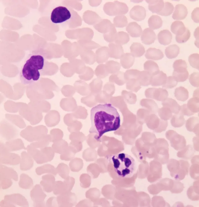
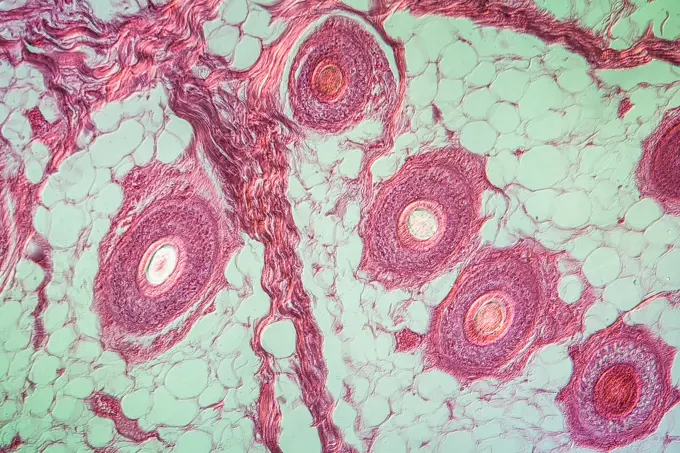
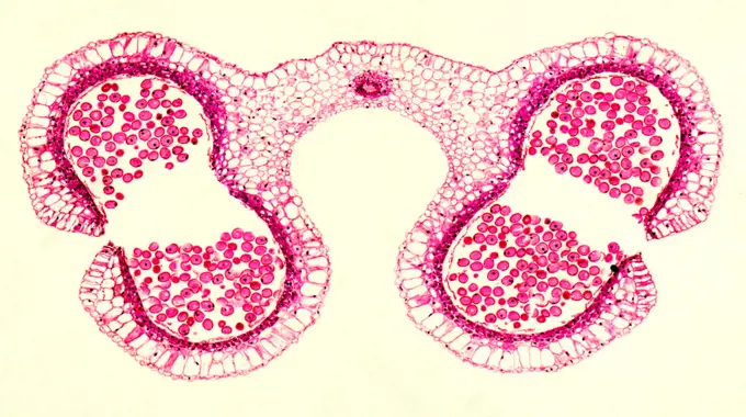
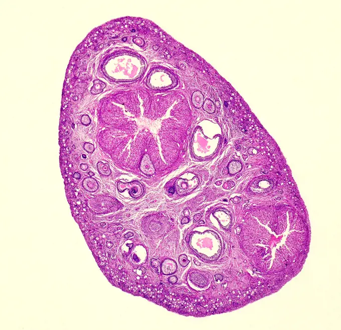
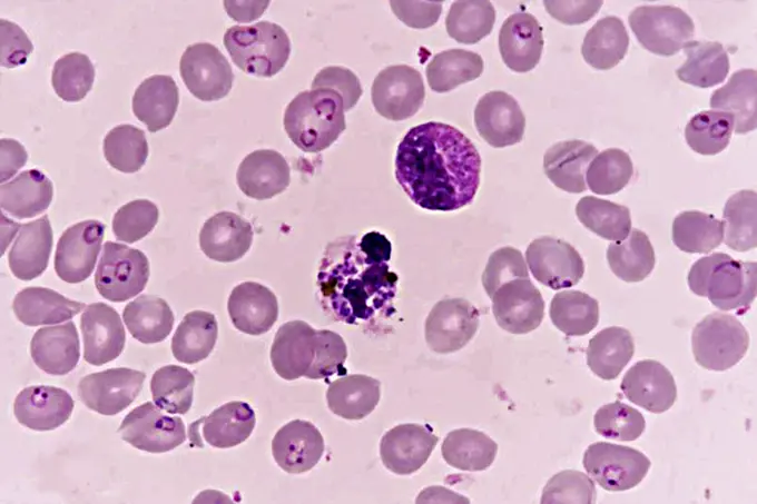
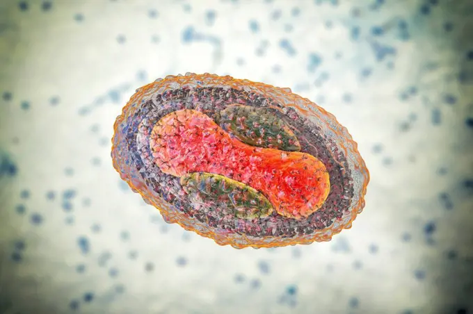
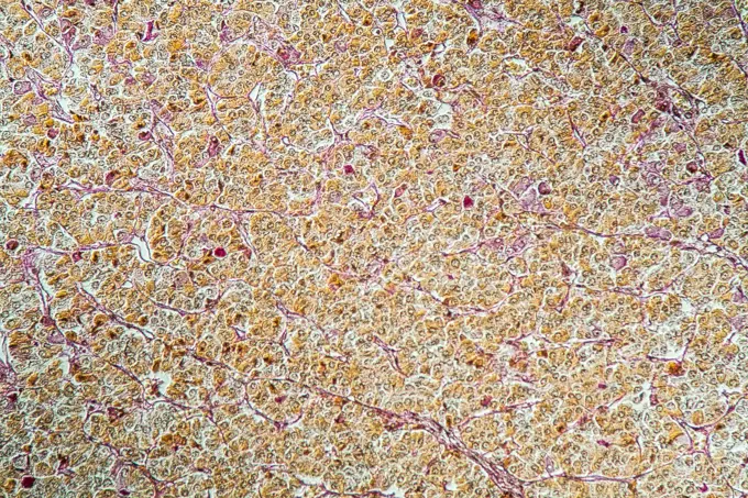
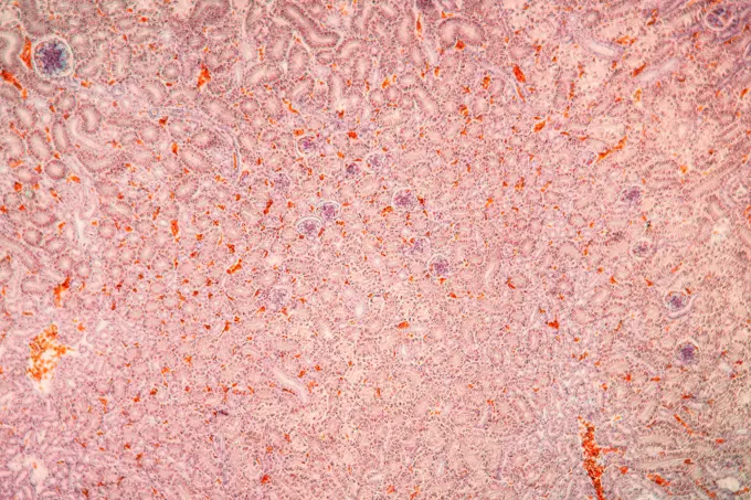
Detailed microscopic views of various medical conditions and tissues. Pink and purple hues highlight cellular structures and abnormalities.

Kidney chicken kidney chicken tissue under the microscope 100x copyright: xzoonar.com/DR.XNORBERBERTXLANGEX 13656601
312 assets in this story
1788-111173883
Circulating human blood showing lymphocytes seen under a microscope, at x600 magnification.
6188-55643875
Kopfhaut Kopfhaut Haare quer 100x Copyright: xZoonar.com/Dr.xNorbertxLangex 13660656
4141-111428913
Light micrograph transverse section through the mature anther of a lily flower anther (Lilium species), UK
6145-29267375
Visceral leishmaniasis: gastrointestinal tract
4141-111428909
Light micrograph transverse section through a mature ovary of a rabbit showing the Graafian follicles. TS, UK
4128-18929742
Spinal column. LM
1773R-87672
Close up of invasive colon cancer cells
1899-53511532
Plasmodium falciparum is a malaria-causing parasite or malaria, transmitted by the bite of female anopheles which releases merozoites into the blood which enter red blood cells. Optical microscope view.
6145-45245371
Dr. Lisa E. Freed of the Massachusetts Institute of Technology and her colleagues have reported that initially disc-like specimens of cartilage tend to become spherical in space, demonstrating that tissues can grow and differentiate into distinct structures in microgravity. The Mir Increment 3 (Sept. 16, 1996 - Jan. 22, 1997) samples were smaller, more spherical, and mechanically weaker than Earth-grown control samples. These results demonstrate the feasibility of microgravity tissue engineering and may have implications for long human space voyages and for treating musculoskeletal disorders on earth. Constructs grown on Mir (A) tended to become more spherical, whereas those grown on Earth (B) maintained their initial disc shape. These findings might be related to differences in cultivation conditions, i.e., videotapes showed that constructs floated freely in microgravity but settled and collided with the rotating vessel wall at 1g (Earth's gravity). In particular, on Mir the construct
4128-20044932
Molluscum contagiosum virus, illustration
6188-55644465
Hypophyse Hypophyse Hirnanhangsdrüse unter dem Mikroskop 100x Copyright: xZoonar.com/Dr.xNorbertxLangex 13728187
6145-29248847
Diagnostic test for Fragile X, using presence (normal) or absence (Fragile X syndrome) of FMR-1 protein. FMR-1 protein expression in blood cells has been made visible with antibodies against the FMR-1 protein. The presence of FMR-1 protein is made visible as red staining. a: red staining in cells of normal individual. b/c: absence of staining in male patient. d: female patient; one cell is showing staining and in the other cell there is an absence of staining - this individual is a carrier.
PREVIOUS
of 4
NEXT
2401 S. Ervay, Suite 206
Dallas, Texas 75215
United States
Get Started
Free ResearchMy BoardsMy Cart
For Creators
How To License Your ContentContributor PortalFrame of Mind
Resources
API accessPricing
Contact
+1 866 236 0087help@viewpictures.co.uk Contact form
©2026 View Pictures. All Rights Reserved. -A
This site is protected by reCAPTCHA and Google Privacy Policy and Terms of Service apply.