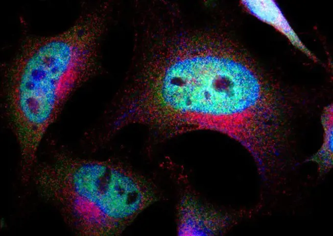
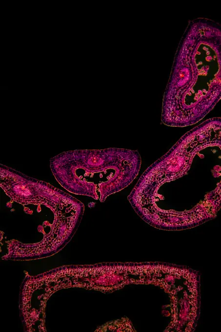
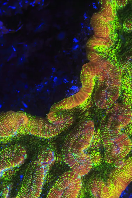
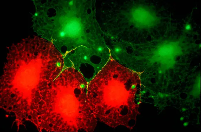
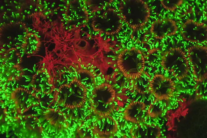
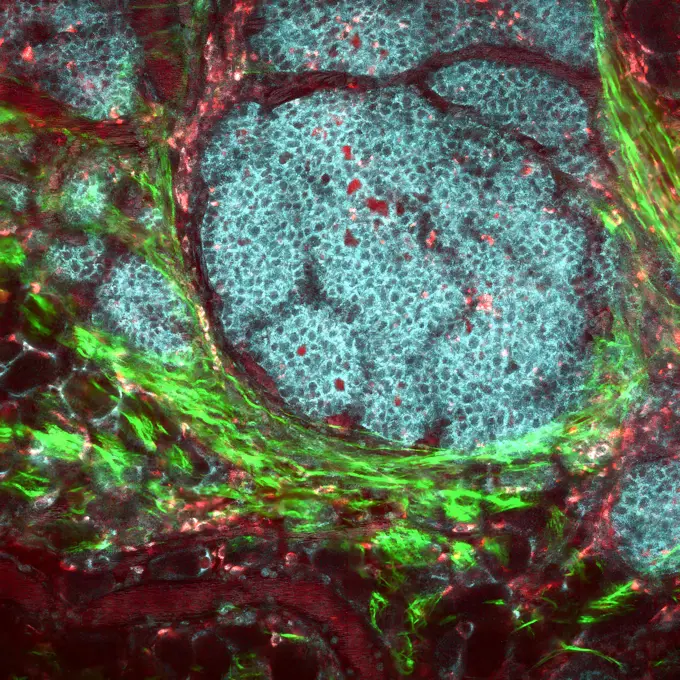
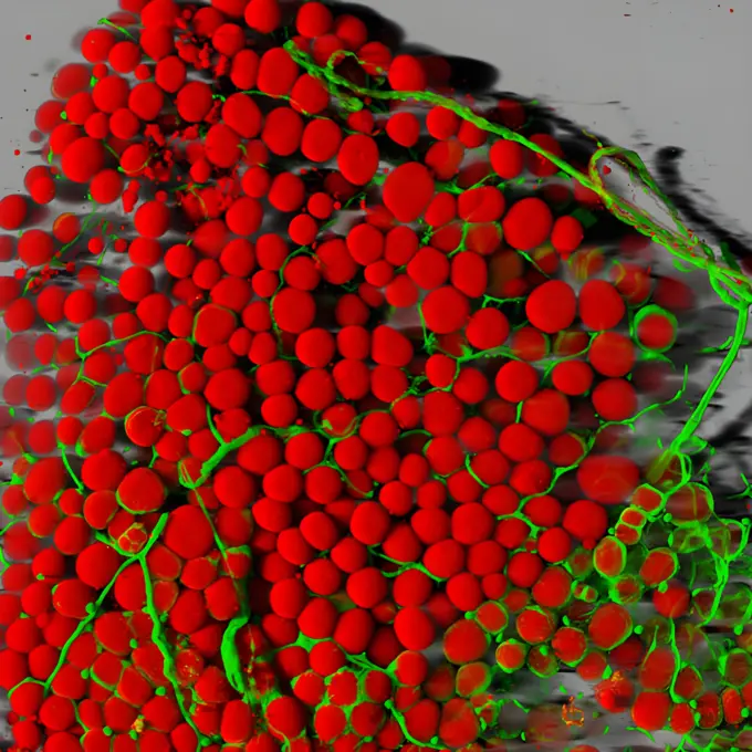
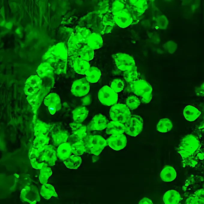
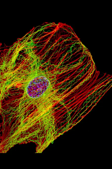
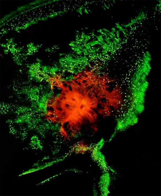
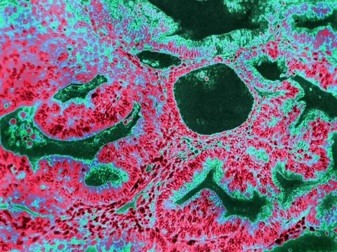
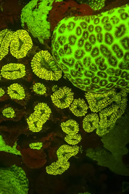
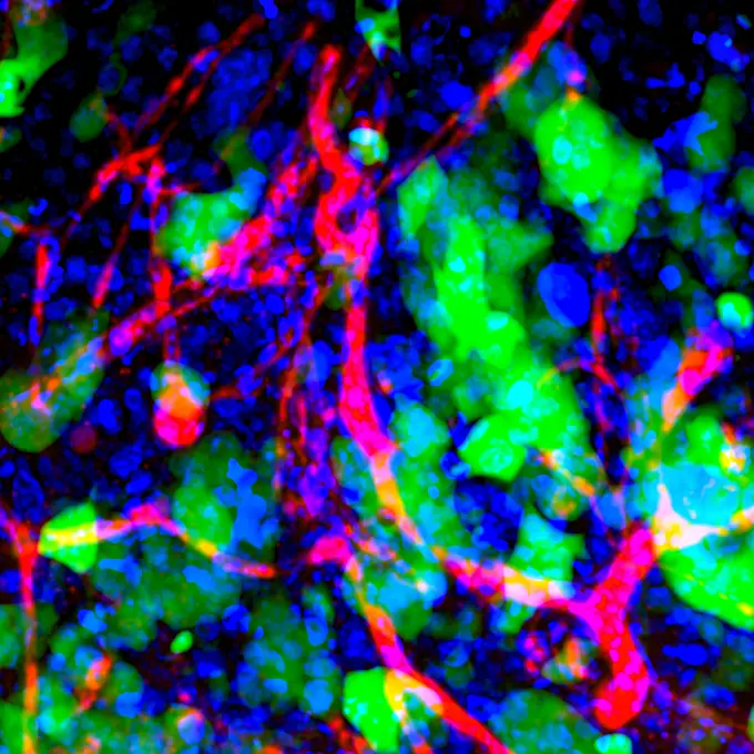
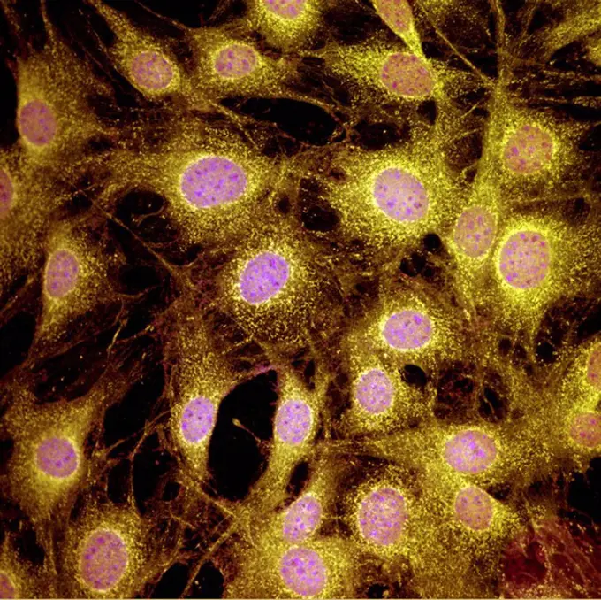
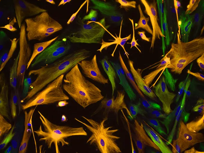
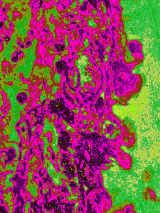
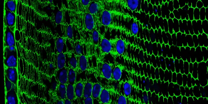
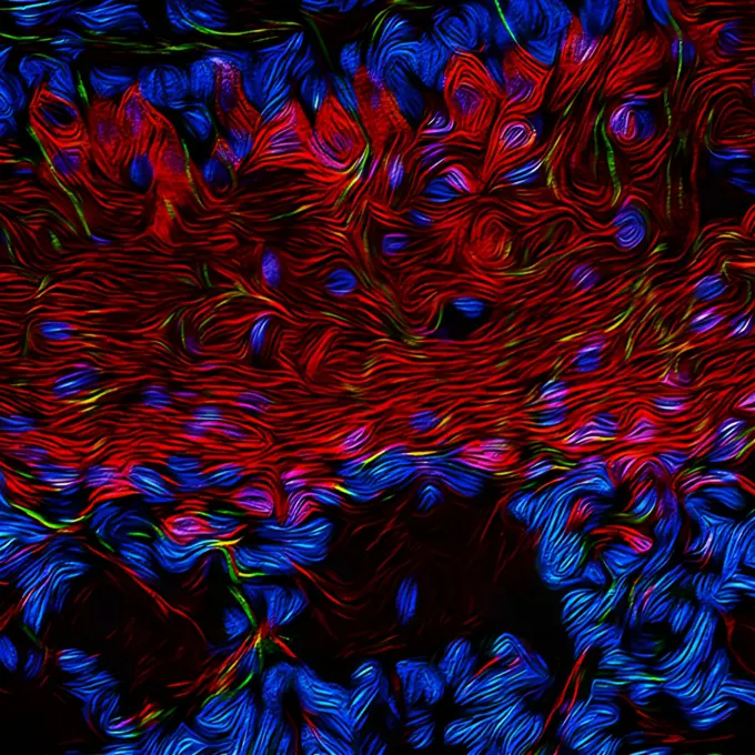
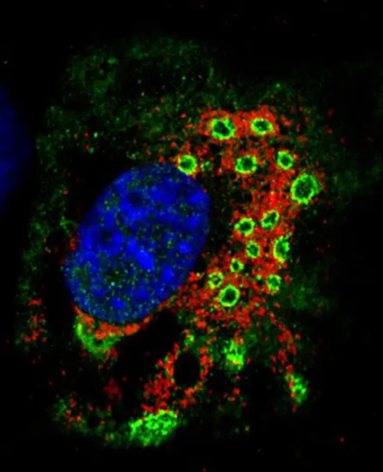
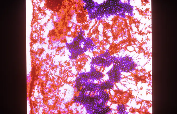
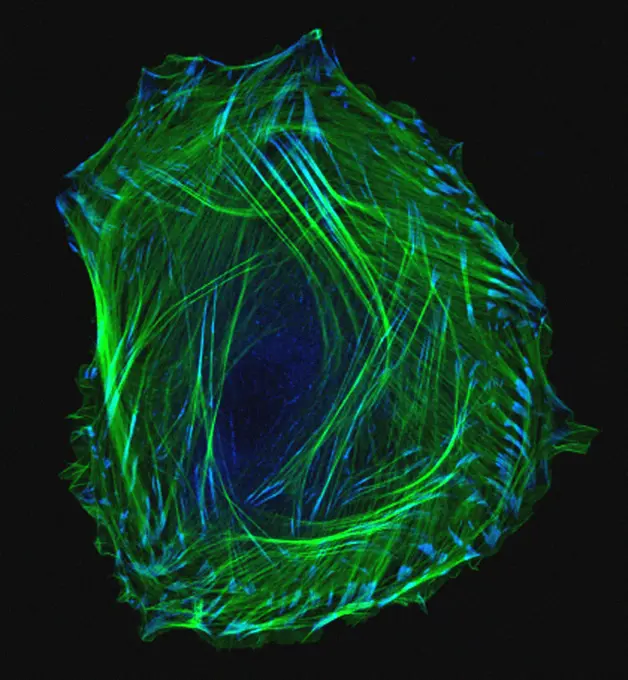
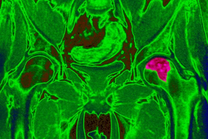
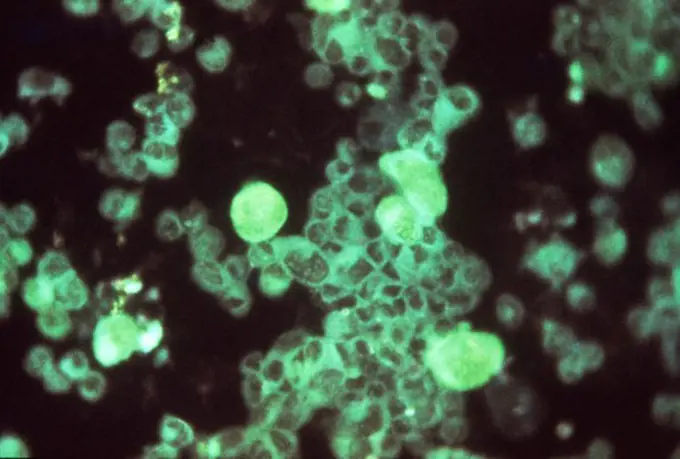
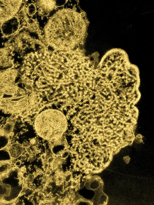
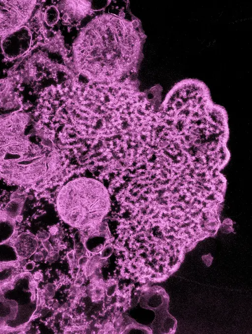
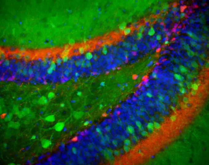
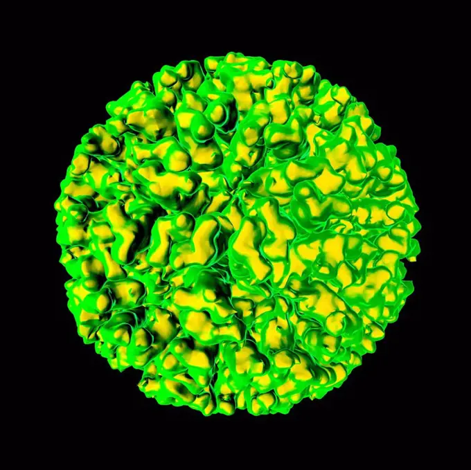
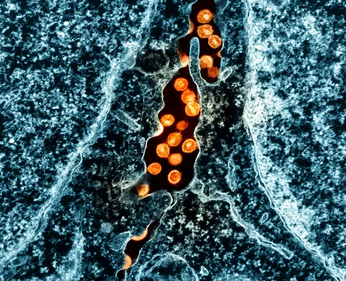
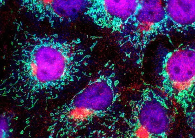
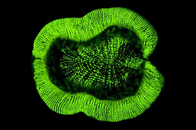
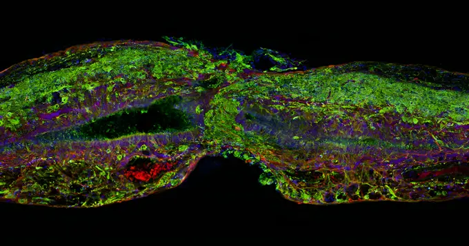
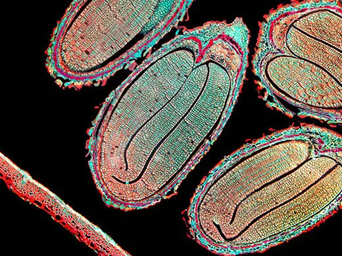
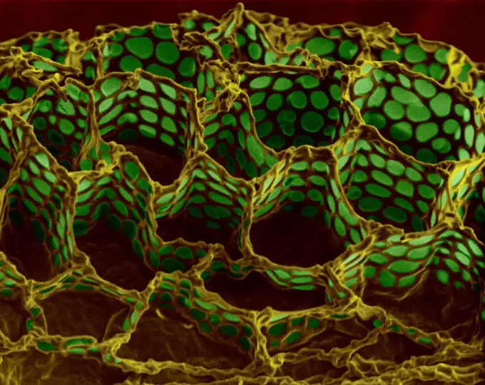
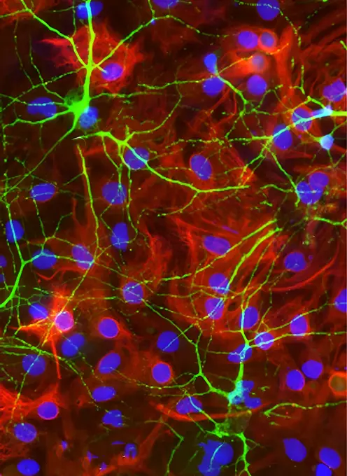
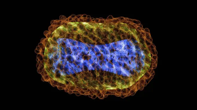
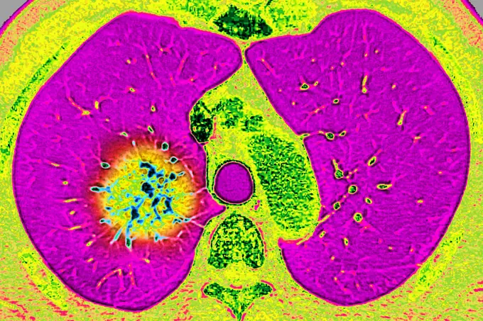
My Boards
View Pictures
  • My Boards

Microscopic Views of Cells

Microscopic images show various cell types, including cancer cells and HeLa cells. Colorful visuals display cellular structures and interactions.

Immortal Cells:  HeLa Cells, Microscopic View
Immortal Cells: HeLa Cells, Microscopic View
146 assets in this story
824-63179029
Invasive basal cell carcinoma, cancer of the uterus. Microscopy of a histological section x 500.
4341-103
Immortal Cells:  HeLa Cells, Microscopic View
824-63223122
Microscopy of cells infected with Langat virus. The viral protein NS5 (red) interacts with prolidase (green) to prevent cells from mounting an antiviral response. Blue denotes the cell nucleus, a structure where genetic information is stored. Langat virus (LGTV) is a virus of the genus Flavivirus. It was first isolated in Malaysia in 1956 from a hard tick of the Ixodes genus. Credit: NIAID.
6188-55643017
Crowberry leaves cross section 100x crowberry leaves cross section 100x copyright: xzoonar.com/dr.xlangeex 13391238
824-63227224
Human colon cancer cells with cell nuclei stained red and E-cadherin protein stained green. E-cadherin is a cell adhesion molecule and its loss signals a process known as epithelial-mesenchymal transition in which cells acquire the ability to migrate and become invasive.
824-63227270
Immunofluorescent staining of Merkel cell carcinoma tumor tissue illustrating expression of CD200 (green) on the surface of tumor cells. CD200 plays a role in immunosuppression. Endothelial marker CD31 (red) highlights blood vessels. Merkel cell carcinoma is a rare and aggressive skin cancer.
7203-70647340
Night dive at Barrier Reef near Saint Georges Caye, Fluorescence emitted at night, exposed with special UV blocking filters, Belize, Central America
6214-V61192508
6145-29295482
Injected fibroblast cells in culture
1899-61460545
Acanthamoeba is a microscopic, free-living amoeba, or amoeba (single-celled living organism), that can cause rare but serious eye, skin, and central nervous system infections. The amoeba is found worldwide in the environment in water and soil. The amoeba can spread to the eyes through the use of contact lenses, cuts or wounds in the skin, or by being inhaled into the lungs. Most people will be exposed to Acanthamoeba in their lifetime, but very few will become ill from this exposure
7203-70647370
Natural occurring Fluorescence emitted at night using special uv blocking filters. carpeting Cup Coral (Tubastraea sp.) Galapagos Islands National Park, Ecuador
824-63227129
This image of a breast cancer tumor and its microenvironment was obtained from a live mouse model using multiphoton microscopy and endogenous fluorescence. That is, the image was obtained without fluorophores, stains or dyes, using only the metabolic cofactors of NADH and FAD, which are already inside the cells, as well as second harmonic generation to see the collagen. This technique has significant clinical potential for patients who need label-free imaging and can lead to more effective diagnoses and treatments. Tumor cells are displayed in cyan, macrophages in red, collagen fibers in green.
824-63227187
The fat cells of a mouse (red) are shown surrounded by a network of blood vessels (green). Fat cells store and release energy, protect organs and nervous tissues, insulate us from the cold and help us absorb important vitamins
824-57657922
Acanthamoeba is a microscopic, free-living amoeba, or amoeba (single-celled living organism), that can cause rare but serious eye, skin, and central nervous system infections. The amoeba is found worldwide in the environment in water and soil. The amoeba can spread to the eyes through the use of contact lenses, cuts or wounds in the skin, or by being inhaled into the lungs. Most people will be exposed to Acanthamoeba in their lifetime, but very few will become ill from this exposure.
824-63227306
This cross section of regenerated muscle shows muscle stem cells (red) in their niche along muscle fibers (green). The blue dots are the DNA in the fiber nuclei. Researchers have found that injecting the molecule prostaglandin E2 into muscles after injury induces the division of muscle stem cells and accelerates regeneration. Prostaglandin E2 is an inflammatory molecule released in response to muscle injury or rigorous exercise.
824-63227239
Prohibited parvalbumin-containing interneurons from mouse hippocampus. Credit: McBain Laboratory, NICHD/NIH
6214-V61192459
824-66066035
Transmission electron micrograph of West Nile virus particles (yellow/red) replicating within the cytoplasm of an infected VERO E6 cell (teal). Image captured at the NIAID Integrated Research Facility (IRF) in Fort Detrick, Maryland. Credit: NIAID.
824-63227137
This image shows a normal fibroblast, a cell type common in connective tissue and frequently examined in research laboratories. Unlike the spiky version, this cell has a healthy skeleton composed of actin (red) and microtubules (green). Actin fibers contain like muscles to create tension and microtubules contain like bones to resist compression.
824-63193293
This fluorescence microscopy image shows the distribution of two nanoparticles pumped gently into the brain. One particle (red) bound to brain tissue near the site of infusion while the other particle penetrated outwards several millimeters (green). Nanoparticles can be filled with either drugs or DNA for treatment of diseases including brain tumors, Alzheimer's, and Parkinson's. Visual studies like this help determine what properties are best suited to deliver therapy to these diseases. Source: Frank Szoka, Jr. and JA MacKay, University of California at San Francisco
1525-22204438
Close-up of a pattern
4239-69148202
Detail of a fluorescing coral colony growing on a shallow reef.
824-63179015
Cancer of the uterus. Adeno-acanthoma (glandular tumor). Cancer of the endometrium develops from the membrane lining the uterus. Microscopy of a histological section.
4269-25301
Ebola Virus. Color enhanced Transmission Electron Micrograph (TEM) of the ebola virus.
1899-53511450
Angiography of myocardial infarction caused by atheromatous plaque in the left coronary artery. Angiographic visualization is produced by injection of contrast product.
824-63227349
Most tumors are made up of many different types of cancer cells (tumor heterogeneity), which often harbor unique genetic mutations. Researchers want to determine how different subgroups of cancer cells within a tumor exhibit the ability of cancer cells to metastasize or develop resistance to therapeutic agents. This image of a mouse model of pancreatic cancer shows a multicolor lineage (and ampquotconfetti and ampquot) plot and demonstrates that a significant number of metastases presented from clusters of heterogeneous cells rather than outgrowth of single cells.
1899-54027684
Pulmonary infarction (pulmonary embolism) seen on a radial cross-section chest CT scan.
824-63227171
To better understand the role of bacteria in health and disease, National Institutes of Health researchers administered viruses to fruit flies and monitored the lifetime activity of hundreds of genes that scientists traditionally thought control aging. To their surprise, they not only extended the lives of flies, but also drastically altered the activity of many of these genes. Their results reveal that only about 30% of genes are traditionally associated with aging determining an animal's internal clock, while the others are affected by the body's response to bacteria.
7203-70647287
Natural occurring red and green Fluorescence in Stony Corals and Encrusting Corals, captured with special ultraviolet light-blocking filters, Palau, Micronesia, Rock Islands, World Heritage Site, Western Pacific
824-63227311
Cancer that has spread (metastasized) to the brain is normally incurable because the protective blood-brain barrier blocks most treatments from entering. Nanoparticles capable of transporting drugs and crossing the barrier can help deliver life-saving therapies to these tumors. This image shows blood vessels (red), cell nuclei (blue) and metastatic human breast cancer cells (green) in the brain of a mouse, after intravenous administration of experimental nanoparticles capable of crossing the blood-brain barrier.
4128-18929775
Gall bladder, LM
4128R-6343
Fibroblast cells
1899-61460382
Colorized transmission electron micrograph of monkeypox particles (yellow) found within an infected cell (blue), cultured in the laboratory
824-66065992
In a study of flies, NIH scientists showed how the immune system may be a culprit in the damage caused by aging brain disorders. More information:.
6145-29268494
Fibroblasts showing actin & Golgi apparatus
824-63191246
Human neural progenitor cells were isolated under selective culture conditions from the developing human brain and directed through lineage differentiation to GFAP+ (glial fibrillary acid protein) astrocytes. Following 3 weeks in astrocyte selective medium, cells were fixed and stained with antibodies to intermediate filament proteins that characterize the cells as either astrocytes (GFAP in orange) or neural progenitor cells (nestin in green). Nuclei are stained with DAPI in blue. Microscopic examination over the course of differentiation showed loss of progenitor cells (nestin down regulation) as cell population increasingly became astrocytes (GFAP up regulation). Photomicrograph was taken on Axiovert 200M Zeiss inverted microscope at 200 x magnification.
824-63179019
Cervical cancer. Cellular anomalies seen by the microscopy of a histological section of the endocervical epithelium.
824-63191216
Multiphoton fluorescence image of HeLa cells stained with the actin binding toxin phalloidin (red), microtubules (cyan) and cell nuclei (blue). Nikon RTS2000MP custom laser scanning microscope.
824-63227186
Here you can see the lens development process at work in a cross section of tissue from an adult mouse. In mice, as in humans, a single layer of rod-like epithelial cells (far left, blue/green) gives rise to specialized lens cells (middle, blue/green) throughout life. The new cells resemble their progenitor cells, with the nuclei (blue) and cytoskeletal protein actin (green). But soon these cells will produce large amounts of water-soluble proteins, called crystalline, to improve their transparency, while gradually breaking down their nuclei to eliminate light-scattering mass. What remains are fully differentiated, enucleated, non-replicating lens fibrous cells (right, green), which refract light onto the retina at the back of the eye.
824-63217795
The human coronavirus is the cause of respiratory infections (colds), and gastroenteritis. View made from a transmission electron microscopy view. Viral diameter approximately: 80 to 160 nm.
1899-53511457
Angiography of myocardial infarction affected by atheromatous plaque in the left coronary artery. Angiographic visualization is produced by injection of contrast product.
824-63227343
This confocal microscope image was awarded by Jeremy McIntyre, an NIH-supported researcher who studies the biology of smell at the University of Florida School of Medicine.
824-63195708
Produced by the National Institute of Allergy and Infectious Diseases (NIAID), this photomicrograph of a fluorescent-stained cell reveals numbers of internalized green-glowing vacuoles containing numerous rotovirus particles undergoing replications. Note that the cell nucleus stained blue. Rotavirus is a contagious virus that can cause gastroenteritis (inflammation of the stomach and intestines). Symptoms include severe watery diarrhea, often with vomiting, fever, and abdominal pain. Infants and young children are most likely to get rotavirus disease. They can become severely dehydrated and need to be hospitalized and can even die.
4269-7069
Transmission electron micrograph (TEM) of the Foot and mouth disease virus.
1899-65662385
Colorized transmission electron micrograph of HIV-1 virus particles (pink) budding and replicating from an H9 T cell (teal). The virus particles are in various stages of maturity, which accounts for differences in shape.
824-63227123
An NIH-funded study on mice provided a detailed blueprint of the brain circuitry behind thirst and satiety.
824-63198854
Degenerative disk disease. Dorsal vertebrae osteoarthritis seen on a side x-ray of the thorax.
824-63225916
Transmission electron micrograph of SARS-CoV-2 virus particles (gold) within endosomes of a heavily infected nasal Olfactory Epithelial Cell. Image captured at the NIAID Integrated Research Facility (IRF) in Fort Detrick, Maryland.
824-63191203
Embryonic smooth muscle cell. Immuno-fluorescently labeled actin cytoskeleton (green) and vinculin in cell adhesions (blue). Laser scanning confocal microscopy.
6214-V61192449
824-63206943
Osteoarthritis of the left fermoral head, seen on a frontal pelvic MRI scan.
4297-1753
Immunofluorescent staining technique showing cytomegalovirus in human embryonic lung
824-63195685
This transmission electron micrograph (TEM) depicted a number of Nipah virus virions that had been isolated from a patient's cerebrospinal fluid (CSF) specimen. Nipah virus is a member of the family Paramyxoviridae, and is related, but not identical to Hendra virus. Nipah virus was initially isolated in 1999 upon examining samples from an outbreak of encephalitis and respiratory illness among adult men in Malaysia and Singapore. Infection with Nipah virus was associated with an encephalitis (inflammation of the brain) characterized by fever and drowsiness and more serious central nervous system disease, such as coma, seizures, and inability to maintain breathing. Illness with Nipah virus begins with 3-14 days of fever and headache. This is followed by drowsiness and disorientation characterized by mental confusion. These signs and symptoms can progress to coma within 24-48 hours. Some patients have had a respiratory illness during the early part of their infections.
824-63227395
These skin cancer cells show from a mouse, an animal commonly used to study human diseases (including many types of cancer) and to test the effectiveness of drugs. The two cells contained here are linked by actin (green), a cell skeleton protein. Although actin is required by many cells for normal movement, it also allows cancer cells to spread to other parts of the body.
6188-60022202
A flash of electrical energy. Electric discharge on a black background. Magical light effect. A flash of electrical energy. Electric discharge on a black background. Magical light effect Copyright: xZoonar.com/BrehovxAntonx 14940202
6188-60022210
A flash of electrical energy. Electric discharge on a black background. Magical light effect. A flash of electrical energy. Electric discharge on a black background. Magical light effect Copyright: xZoonar.com/BrehovxAntonx 14940567
824-63195686
This transmission electron micrograph (TEM) depicted a number of Nipah virus virions that had been isolated from a patient's cerebrospinal fluid (CSF) specimen. Nipah virus is a member of the family Paramyxoviridae, and is related, but not identical to Hendra virus. Nipah virus was initially isolated in 1999 upon examining samples from an outbreak of encephalitis and respiratory illness among adult men in Malaysia and Singapore. Infection with Nipah virus was associated with an encephalitis (inflammation of the brain) characterized by fever and drowsiness and more serious central nervous system disease, such as coma, seizures, and inability to maintain breathing. Illness with Nipah virus begins with 3-14 days of fever and headache. This is followed by drowsiness and disorientation characterized by mental confusion. These signs and symptoms can progress to coma within 24-48 hours. Some patients have had a respiratory illness during the early part of their infections.
824-63191202
The hilus of the dentate gyrus from an Nr4a1-eGFP (green) transgenic mouse stained for calretinin (red) and DAPI (blue) to show nuclei.
4128R-11473908
Feline calicivirus, computer artwork.
1788-111174040
Human kidney tissue seen under a microscope using fluorescence.
824-63227372
Scientists at the NIH's National Eye Institute have developed a promising gene therapy strategy for a form of Leber congenital amaurosis (ACL), a rare disease that causes severe vision loss in children. The scientists tested their approach using retinal tissue made in the lab from patient cells, called retinal organoids, one of which is pictured here.
1899-65662405
Colorized transmission electron micrograph of HIV-1 virus particles (orange) budding and replicating from an H9 T cell (teal). The virus particles are in various stages of maturity, which accounts for differences in shape. A Study Shows an HIV Vaccine Can Generate Key Antibody Response in People.
4128-111493679
Biofilm, conceptual illustration.
4341-120
Purple Nucei and Aquamarine Mitochondria, Microscopic View
824-63227194
All creatures secrete vital proteins. Researchers at NIH's National Institute for Dental and Craniofacial Research are studying fruit fly glands (like the one pictured here) and cell packets known as secretory granules (red) that store proteins for secretion . This work may help scientists better understand how secretion goes wrong, and how it might be treated, in conditions such as salivary gland disorders, which cause dry mouth.
824-63227207
These cells get their name from the hair-like structures that extend from them into the fluid-filled tube of the inner ear. When sound reaches the ear, the hairs bend and the cells convert this movement into signals that are relayed to the brain. When we crank up the music in our cars or join tens of thousands of cheering fans at a football stadium, the noise can cause hair to pinch and break, leading to long-term hearing loss.
4359-296
Molecular Modelling
4269-25094
Ebola Virus. Color enhanced Transmission Electron Micrograph (TEM) of the ebola virus (Filoviridae).
824-63227136
On the right, two cells (greenish yellow) are in the process of forming bubbles, or plasma membrane vesicles (PMV). During this process of being, a cell's membrane temporarily dissociates from its underlying cytoskeleton, forming a tiny pocket which, over the course of about 30 minutes, is
824-63194674
This fluorescent acid-fast stained (Smithwick) photomicrograph revealed the presence of Mycobacterium tuberculosis bacteria in a sputum smear. M. tuberculosis is an acid-fast bacterium (AFB), and is therefore, undetectable when stained using a Gram-stain technique. However, using the Smithwick method, which employs acridine orange as the specific stain, the M. tuberculosis bacteria glow yellow under ultraviolet light microscopy.
824-63225919
Transmission electron micrograph of SARS-CoV-2 virus particles (gold) within endosomes of a heavily infected nasal Olfactory Epithelial Cell. Image captured at the NIAID Integrated Research Facility (IRF) in Fort Detrick, Maryland.
7203-70646216
Natural occurring fluorescence in underwater bubble coral (Euphyllia ancora). Night dive near Lewolin Village, Illi Api Island, Selat Boleng Strait, Indonesia
4070-21284682
Fluorescent corals (Trachyphyllia geoffroyi) Great Barrier Reef, Australia
1525-22737164
Star field in space a nebulae and a gas congestion. Elements of this image furnished by NASA . 3D rendering. Star field in space and a nebulae. 3D rendering
4298-1028
Marchantia liverwort antheridium, magnification 40x, image enhanced
824-63194518
This colorized negative-stained transmission electron micrograph (TEM) depicts the ultrastructural details of a number of influenza virus particles, or virions. A member of the taxonomic family Orthomyxoviridae, the influenza virus is a single-stranded RNA organism. The flu is a contagious respiratory illness caused by influenza viruses. It can cause mild to severe illness, and at times can lead to death. The best way to prevent this illness is by getting a flu vaccination each fall.
4128R-10088
Tuberculosis bacteria, artwork
1899-53511613
Stress fractures on the shins, visualized by scintigraphy.
824-63227324
Zebrafish spinal cord tissue section regenerating after injury. Glial cells (red) first cross the space between the cut ends. Neural cells (green) soon follow. Cell nuclei are stained blue and purple.
4298-1003
Capsella (Schefflera) embryo in ovary, magnification 40x, imaged enhanced
4141-28551
copepod parasitic in salps sapphirina sp. great barrier reef males have transparent exoskeletal plates that split
4128R-13620169
Foxglove seed surface (Digitalis purpurea), coloured scanning electron micrograph (SEM). This image shows the seed coat (or testa) of a foxglove seed ...
824-63227323
Researchers at the University of Texas have looked at how immune cells called neutrophils can help protect the brain from damage caused by intracerebral hemorrhagic stroke.
4298-1035
Microscopic view of filamentous algae Oedogonium showing oogonium, magnification 200x, image enhanced
4128-19489278
Monkeypox virus, illustration
824-63227432
An NIH-funded mouse study is the first to show that visual stimulation helps rewire the visual system and partially restores sight. This image shows regeneration of mouse retinal ganglion cell axons (magenta and green) extending from the site of optic nerve injury (left).
824-63191201
The CA1 region of the hippocampus from an Nr4a1-eGFP (green) transgenic mouse stained for the CB1 cannabinoid receptor (red) and DAPI to show nuclei (blue). Epifluorescence.
1899-65662347
Transmission electron micrograph of West Nile virus particles (gold) replicating within the cytoplasm of an infected VERO E6 cell (pink/purple/blue).
824-63224523
Pulmonary arteritis causing progressive occlusion of the bronchial arteries, seen by a radial CT scan of the lungs (right lung).
824-63183645
Pox virus. Image produced using high-dynamic-range imaging (HDRI) from an image taken with transmission electron microscopy. Viral diameter ranges from around 100 to 300 nm.
824-63227093
A mouse neural stem cell (blue and green) sits in a lab dish, on top of a special gel containing a mat of synthetic nanofibers (purple). The cell grows and sends out spindly appendages, called axons (green), in an effort to reestablish connections with other nearby nerve cells.
824-63227318
Neural circuits in the mouse retina. Cone photoreceptors (red) allow color vision bipolar neurons (magenta) to relay information further down the circuit and a subtype of bipolar neuron (green) helps process signals detected by other photoreceptors in the penumbra.
1899-54027129
Transluminal agioplasty of the right, subclavian artery, seen on a frontal chest x-ray.
824-63191262
Hippocampus from an Nr4a1-eGFP (green) transgenic mouse stained for calbindin (red) and DAPI to show nuclei (blue). Epifluorescence.
1848-58228094
Fluoresce hard coral, Coral fluorescenc
1899-53511332
Atheroma reducing the internal diameter of an artery. View of a histological section of an artery suffering from atherosclerosis.
824-63224533
Atheroma reducing the internal diameter of an artery. View of a histological section of an artery suffering from atherosclerosis.
824-65830656
Transmission electron micrograph of West Nile virus particles (teal) replicating within the cytoplasm of an infected VERO E6 cell (brown).
824-63227264
Mutations in the LRRK2 gene have been linked to about three percent of Parkinson's disease cases. Researchers have now found evidence that LRRK2 protein activity may be affected in many more patients with Parkinson's disease, even when the LRRK2 gene itself is not mutated. The study could lead researchers to rethink how to treat the disorder.
6145-44915106
This picture shows a lab demonstration of the measurement chamber inside the Tunable Laser Spectrometer, an instrument that is part of the Sample Analysis at Mars investigation on NASA's Curiosity rover.
PREVIOUS
of 2
NEXT
2401 S. Ervay, Suite 206
Dallas, Texas 75215
United States
Get Started
Free ResearchMy BoardsMy Cart
For Creators
How To License Your ContentContributor PortalFrame of Mind
Resources
API accessPricing
Contact
+1 866 236 0087help@viewpictures.co.uk Contact form
©2026 View Pictures. All Rights Reserved. -A
This site is protected by reCAPTCHA and Google Privacy Policy and Terms of Service apply.