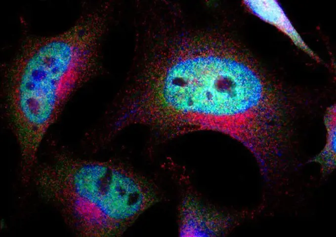
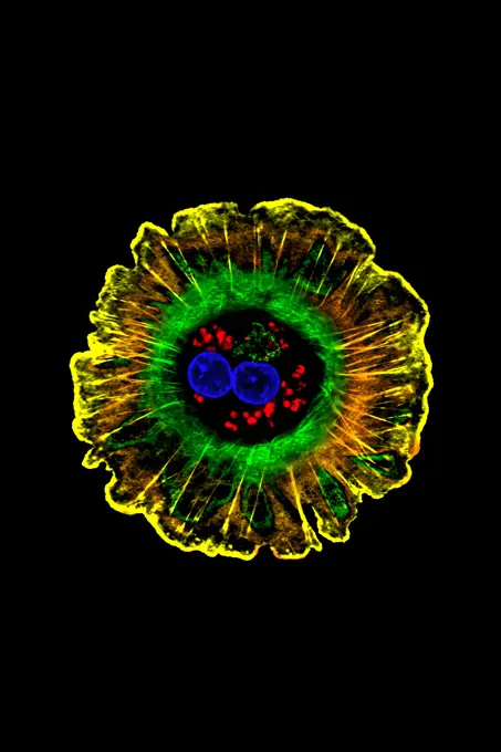
My Boards
View Pictures
  • My Boards

Microscopic Views of Cells

Microscopic images show various cell types, including cancer cells and HeLa cells. Colorful visuals display cellular structures and interactions.

Immortal Cells:  HeLa Cells, Microscopic View
Immortal Cells: HeLa Cells, Microscopic View
146 assets in this story
1848-77402165
3D Illustration Concept of Human Respiratory System Lungs Anatomy
824-63184372
Fracture of the medial cuneiform bone in the foot. Radial scan of the right foot.
1899-61460378
Colorized transmission electron micrograph of monkeypox particles (yellow) found within an infected cell (red), cultured in the laboratory
4128R-10082
Tuberculosis bacteria, artwork
4128R-12577656
Multicoloured lights, computer illustration.
824-63227227
Hepatocytes, like the one shown here, are the most abundant cell type in the human liver. They play an important role in building proteins produced from bile, a fluid that helps digest fats and chemically process molecules normally found in the body, such as hormones, as well as foreign substances such as drugs and alcohol.
4128-V58569357
7203-70646183
Natural occurring red fluorescing sponges (Leucetta sp.) and green hard coral (Acropora sp.). Night dive at Wetar Island, Banda Sea, Indonesia
6188-55645527
Radiolarien unter dem Mikroskop 100x Radiolarien unter dem Mikroskop 100x Copyright: xZoonar.com/Dr.xNorbertxLangex 13889514
824-63227050
Scanning electron micrograph of a macrophage. Credit: NIAID
4269-26761
Red blood cells production. In vitro production of human mature and functional red blood cells from stem blood cells called CD34, Professor Luc Douay's research team, Saint Antoine Faculty of Medicine, Paris. Confocal micrograph showing red blood cells.
824-63225801
Cross-section of skin at steady state.
6188-55643040
Electron microscopy electron microscopic intake of goldfish
4298-1013
Japanese holly fern (Cyrtomium sp) sporangia, magnification 200x, image enhanced
4128R-2255
Marsh marigold carpel. Light micrograph of a transverse section through the carpel of a marsh marigold Caltha palustris flower. The ovules dark green ...
4379-1864
Fluorescing Mushroom coral (Fungia Sp.) underwater, Tunku Abdul Rahman Marine Park, Kota Kinabalu, Sabah State, Malaysia
4197-66703966
Graphics, background and blue veins for blood system, illustration or design isolated in studio. Capillary, black backdrop and varices for human anatomy, fractal texture or abstract glow of wallpaper
7203-70646178
Natural occurring red fluorescing sponges (Leucetta sp.) and green hard coral (Acropora sp.). Night dive at Wetar Island, Banda Sea, Indonesia
7203-70647309
Night dive at Barrier Reef near Saint Georges Caye, Fluorescence emitted at night, exposed with special UV blocking filters, Belize, Central America
1525-26913198
Star field in space a nebulae and a gas congestion. Elements of this image furnished by NASA . 3D rendering. Star field in space and a nebulae. 3D rendering
6188-64561317
Cosmic images computer processed images of galaxies Copyright: xAshaxAdatia/DinodiaxPhotox
6145-44935118
Caption This is an image of magnetic loops on the sun, captured by NASA's Solar Dynamics Observatory on July 18, 2012. It has been processed to highlight the edges of each loop to make the structure more clear. A series of loops such as this is known as a flux rope, and these lie at the heart of eruptions on the sun known as coronal mass ejections (CMEs.) This is the first time scientists were able to discern the timing of a flux rope's formation.
824-63195628
Abdominal CT scan showing the adrenal glands.
1848-49528452
Hydrozincite on Calcite seen under UV Light (Blue)
824-63227165
In these images from Gabrielle Kardon's NIH-supported lab at the University of Utah, Salt Lake City, you see the developing forelimb of a healthy mouse strain (top) compared to that of a mutant mouse strain with a stiff, abnormal gait (bottom).
4220-21080553
Calcite (red) and Willamite (green) under flourescent light     Date:
4269-27852
Intestine tumor. Three-dimensional computed tomographic reconstruction scan of the small intestine showing a Gastrointestinal Stromal Tumor (GIST), seen as a yellow spot in the jejunum. In red, the abdominal aorta.
1848-66800972
Map of Comoros, Polygonal mesh line map, modern map
4128R-11543364
Nematode, or roundworm, computer illustration.
4128R-11543366
Nematode, or roundworm, computer illustration.
4128-20239425
Rabies viruses, illustration
6145-45248714
Left image The x-ray data from the Chandra X-Ray Observatory (CXO) has revealed a bright central star surrounded by a cloud of multimillion-degree gas in the planetary nebula known as the Cat's Eye. This CXO image, where the intensity of the x-ray emission is correlated to the brightness of the orange coloring, captures the expulsion of material from a star that is expected to collapse into a white dwarf in a few million years. The intensity of x-rays from the central star was unexpected, and it is the first time astronomers have seen such x-ray emission from the central star of a planetary nebula. Right image An image of Cat's Eye taken by the Hubble Space Telescope (HST). By comparing the CXO data with that from the HST, researchers are able to see where the hotter, x-ray emitting gas appears in relation to the cooler material seen in optical wavelengths by the HST. The CXO team found that the chemical abundance in the region of hot gas (its x-ray intensity is shown in purple) was no
1848-64384807
Map of Comoros, Polygonal mesh line map, flag map
4128R-13022946
Insulin binding to insulin receptor, illustration. The insulin receptor (violet) is a transmembrane protein, that is activated by insulin (green). Ins...
6145-45265403
Taken on April 24, 2019, this rainbow-colored image shows the Martian moon Phobos, as viewed by NASA's 2001 Mars Odyssey orbiter using its infrared camera, Thermal Emission Imaging System (THEMIS). Each color represents a different temperature range, with the warmest in the center and coolest on the outer edge. This was the first time THEMIS was used to observe Phobos while in a full moon phase, which offers scientists a much better view for studying the composition of the Martian moon. Previous half-moon views, which can be seen here, were better for studying surface textures.
4197-V71961830
4297-1013
Colorized scanning electron microscope image of dill pollen. The pollen has been acetolyzed to remove cytoplasm and pollenkit in order to reveal the intricate wall structure. This image shows a single grain of dill pollen. Family: Apiaceae, Anethum graveolens
6188-63890538
Solar Cell Clean and renewable energy from solar cell Copyright: xZoonar.com/MarkoxBericx 12542867
4269-2464
Fluorescent light micrograph of healthy human bronchus cells made for researches on mucoviscidosis (3 different magnifications). The cells lining the lumen of the bronchus (black area at right) are rich in CFTR protein (Cystic Fibrosis Transmembrane Conductance Regulator) which appears in green, particularly in the walls of the cells in direct contact with the lumen (bright green line).
1848-64382426
Map of Cape Verde, Polygonal mesh line map, flag map
6145-44708673
Microscopic Image of Martian Surface Material on a Silicone Substrate
1899-60981
Ultraviolet Image Of The Crab Nebula Supernova Remnant.
824-63223222
Transmission electron micrograph of SARS-CoV-2 virus particles, isolated from a patient. Image captured and color-enhanced at the NIAID Integrated Research Facility (IRF) in Fort Detrick, Maryland. Credit: NIAID.
1848-69291247
Fish salmon in scientific high-tech thermal imager on black background isolated
6145-45248764
Dr. Lisa E. Freed of the Massachusetts Institute of Technology and her colleagues have reported that initially disc-like specimens tend to become spherical in space, demonstrating that tissues can grow and differentiate into distinct structures in microgravity. The Mir Increment 3 (Sept. 16, 1996 - Jan. 22, 1997) samples were smaller, more spherical, and mechanically weaker than Earth-grown control samples. These results demonstrate the feasibility of microgravity tissue engineering and may have implications for long human space voyages and for treating musculoskeletal disorders on earth. Final samples from Mir and Earth appeared histologically cartilaginous throughout their entire cross sections (5-8 mm thick), with the exception of fibrous outer capsules. Constructs grown on Earth (A) appeared to have a more organized extracellular matrix with more uniform collagen orientation as compared with constructs grown on Mir (B), but the average collagen fiber diameter was similar in the two
6145-29294467
Molecular model of alpha-haemolysin, stereo
PREVIOUS
of 2
NEXT
2401 S. Ervay, Suite 206
Dallas, Texas 75215
United States
Get Started
Free ResearchMy BoardsMy Cart
For Creators
How To License Your ContentContributor PortalFrame of Mind
Resources
API accessPricing
Contact
+1 866 236 0087help@viewpictures.co.uk Contact form
©2026 View Pictures. All Rights Reserved. -A
This site is protected by reCAPTCHA and Google Privacy Policy and Terms of Service apply.