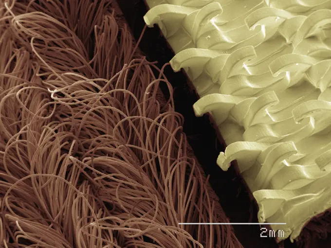
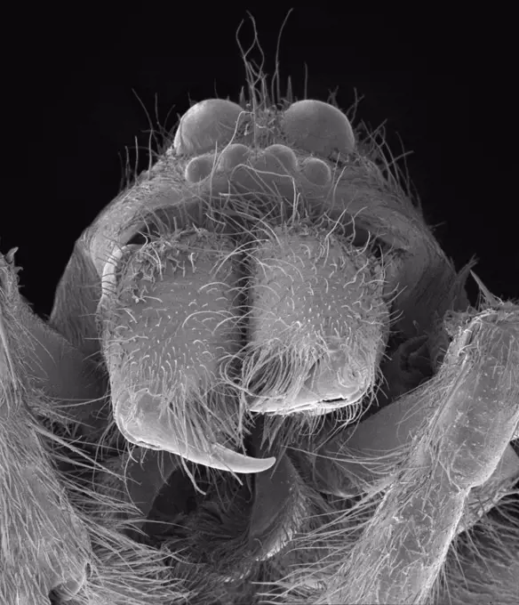
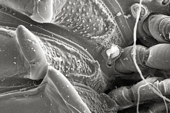
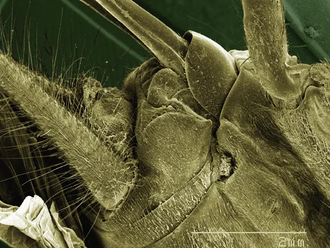
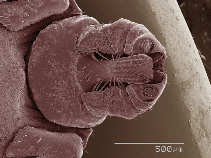
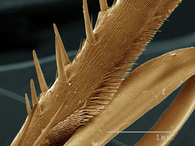
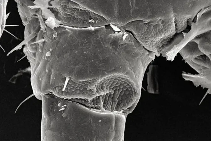
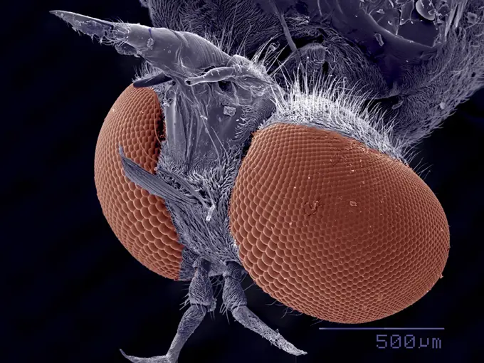
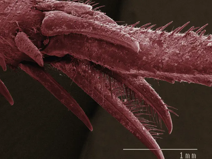
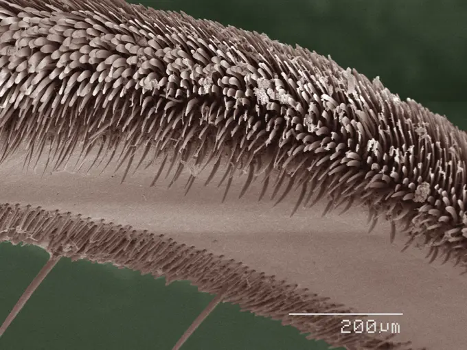
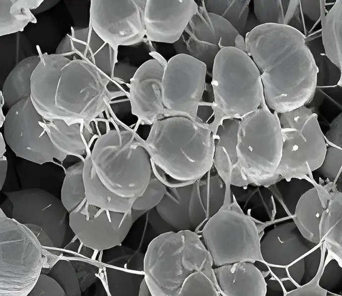
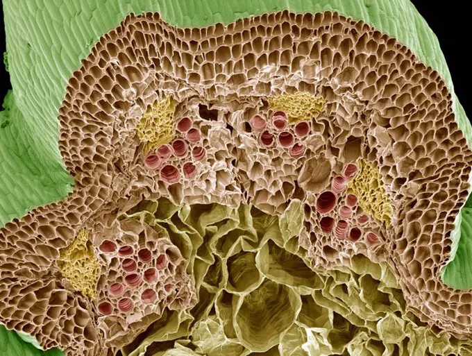
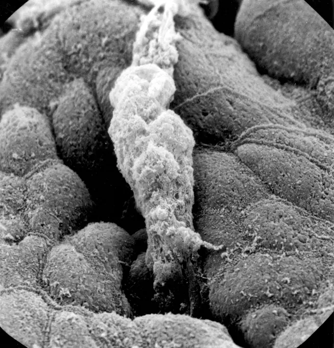
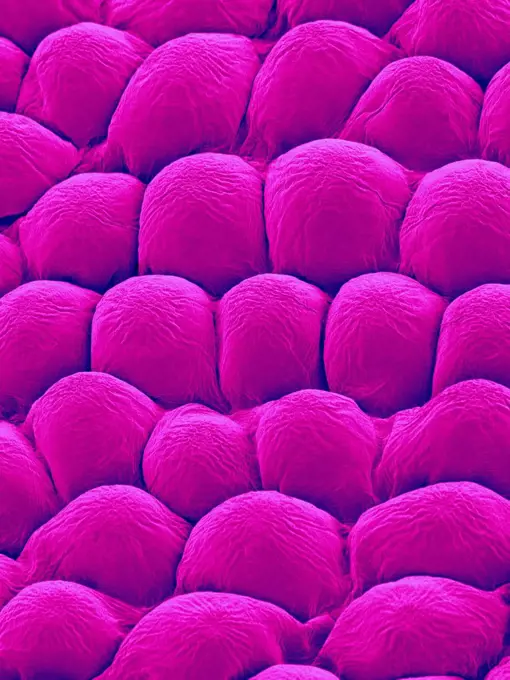
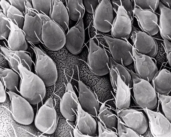
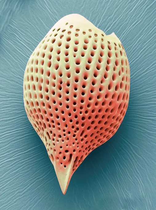
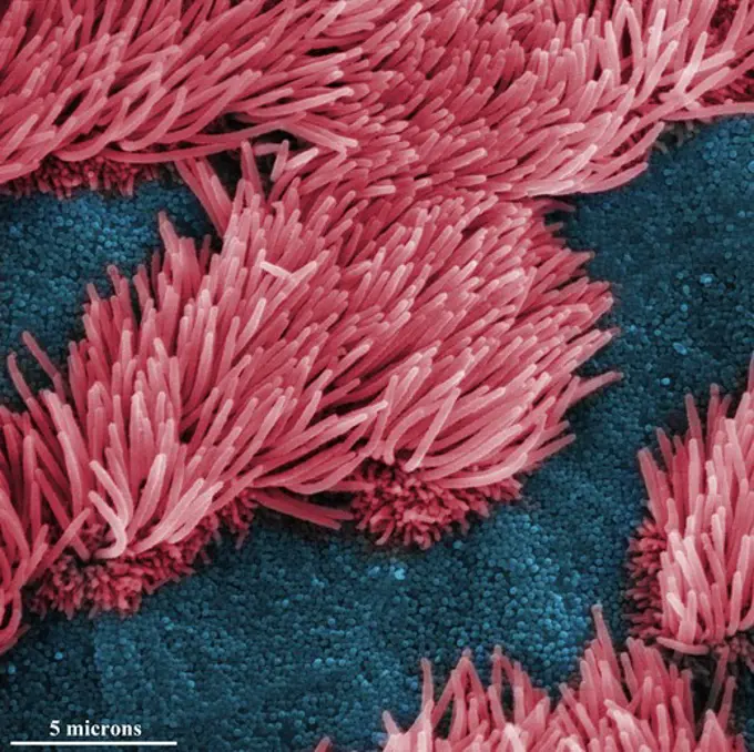
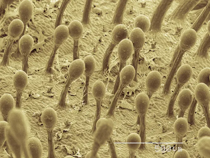
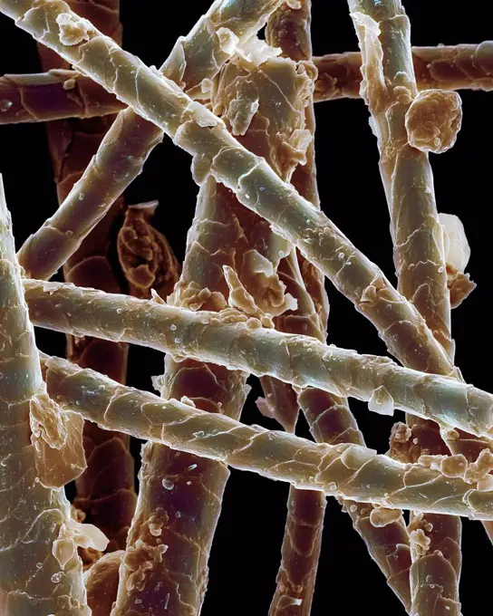
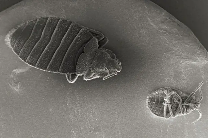
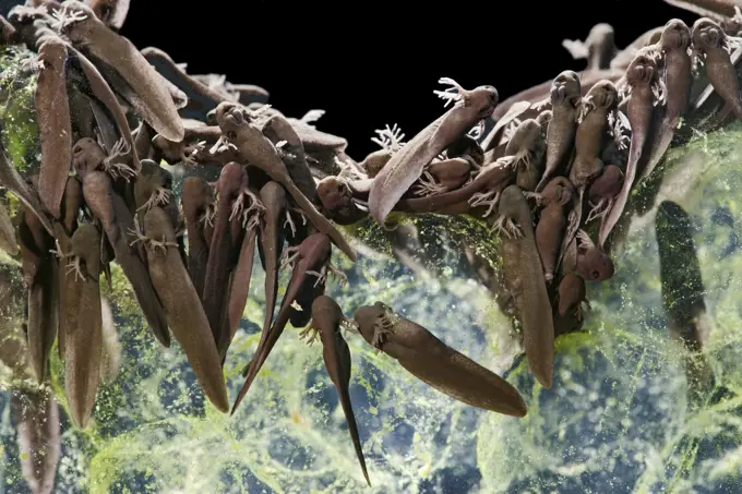
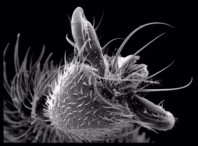
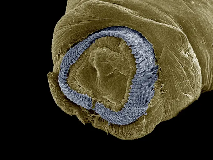
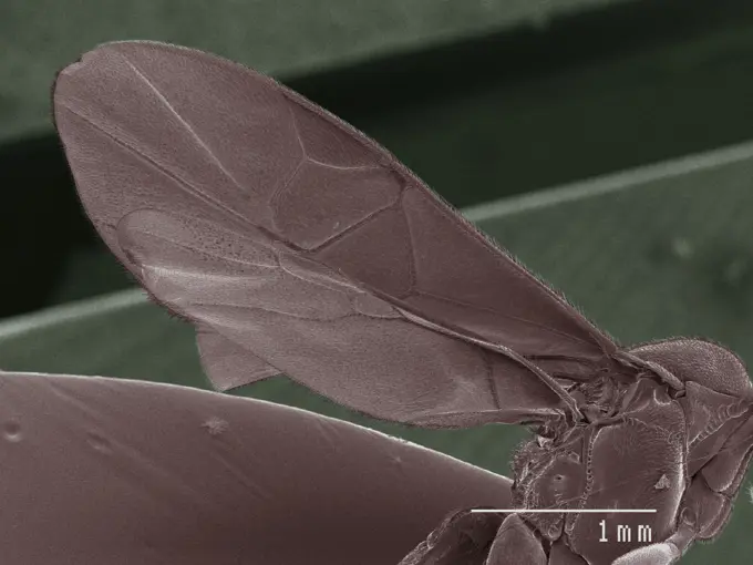
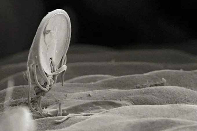
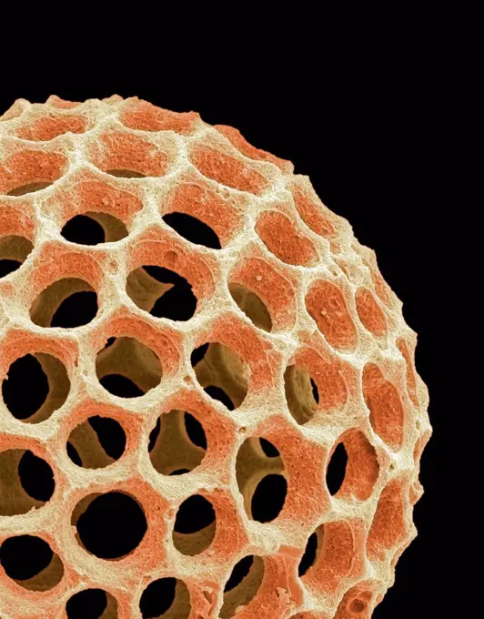
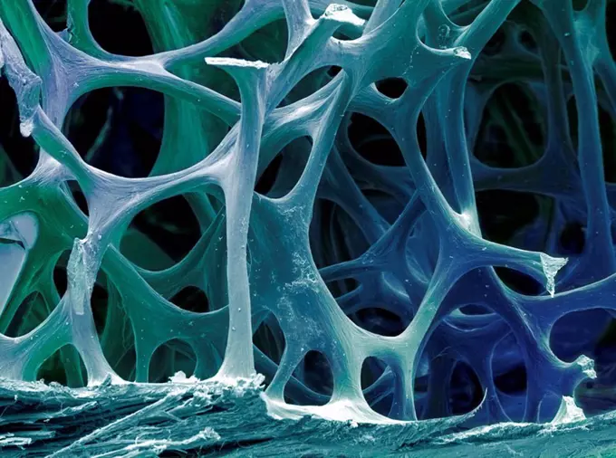
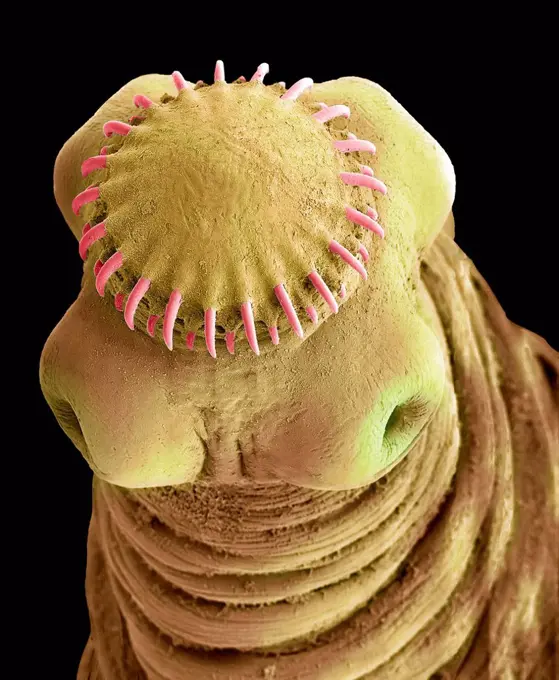
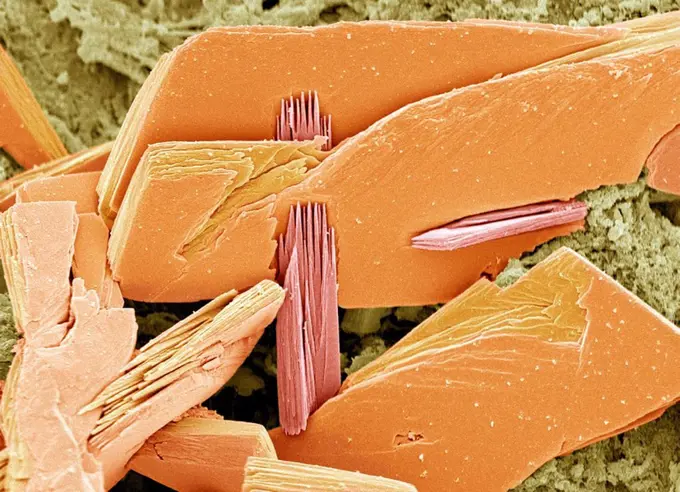
My Boards
View Pictures
  • My Boards

Microscopic Wonders of Biology

Extreme close-ups of organic structures and insect appendages, showcasing intricate textures and colors using advanced microscopy techniques.

Extreme closeup of insect appendage under the microscope
160 assets in this story
1439-57940165
Extreme closeup of insect appendage under the microscope
1439-57940001
Microscopic closeup of organic structures with fine textures
1439-57940068
Intricate microscape of scientific wonder
4128R-15307
Asparagus Asparagus officinalis pollen grains, coloured scanning electron micrograph SEM. Magnification: x2000 when printed at 10 centimetres wide.
1439-57939986
Magnified view of textured surfaces resembling an alien landscape
4201-82341
Wolf Spider (Lycosa tarantula) SEM close-up view of face at 14x magnification
4384-378
Microscopic Detail of a Millipede
4384-383
Microscopic Detail of a Wasps' Nest
1439-57940210
Closeup view of an insect under the microscope
1439-57939990
Closeup of a mite under scanning electron microscopy
1439-57940008
Closeup view of a bee's leg under the microscope
4384-430
Microscopic Detail of Body Louse's Leg Joint
4384-214
Scanning Electron Micrograph of a Beetle's Antenna
4128R-15306
Asparagus Asparagus officinalis pollen grains, coloured scanning electron micrograph SEM. Magnification: x2000 when printed at 10 centimetres wide.
1439-57939980
Closeup view of a microscopic landscape revealing intricate textures and colors
1439-57940078
A closeup view of red blood cells within a vessel
1439-57940040
Closeup view of a microscopic landscape featuring a detailed structure.
1439-57943306
Closeup of a dust mite in incredible detail
1439-57940115
Intricate closeup of muscle tissue under a microscope
4384-108
Under a magnification of 40X, this scanning electron micrograph (SEM) revealed some of the ultrastructural architecture found in a brown recluse spiders (Loxesceles reclusa) web that had entrapped an unidentified ant. Though the prey had been enwrapped in a silk cocoon, there were still some of its body parts that were visible such as one of its two compound eyes, and one of its two segmented antennae (background).  The antenna is composed of three main regions: scape, pedicle, and flagellum.
6145-29257473
Deiter's cells & outer hair cells, cochlea
1439-57943308
An upclose sepiatoned micrograph of an insect claw
4384-409
Microscopic Detail of Mosquito's Exoskeletal Surface
1439-57940064
Extreme closeup of a microstructure under an electron microscope.
4384-369
Microscopic Detail of Dragonfly
1439-57940079
Closeup of a microscopic structure resembling a landscape
4201-66325
SEM close-up view of gnathosoma at 140x magnification
4128R-7971
Human hair, SEM
4384-386
Microscopic Detail of a Wasps' Nest
1439-57943372
Closeup of an insect claw gripping a fiber
4128R-1656
Wood, SEM
1439-57940122
A magnified view of a surface with fine hairlike structures.
1439-57939984
Closeup of microscopic structures in sepia tones
1439-57943316
A detailed closeup of an insect's head under a scanning electron microscope
1439-57940218
Closeup of an ant's jaw viewed under a microscope
1439-57940166
Upclose view of a textured surface under a microscope
824-63225937
Phenotypic variants of Neisseria gonorrhoeae bacteria, scanning electron microscope image.
4128R-443
Nasturtium stem, SEM
4269-6806
Scanning electron micrograph (SEM) of stomach wall lined with mucus producing epithelial cells. At center, a drop of secretion from a fundus gland. Maginification 3000x.
1439-57940142
Closeup of a mite under the microscope in striking detail.
1439-57940035
A microscopic view of a seed resting on a leaf's surface.
1439-57940097
Intricate closeup of a microscopic landscape
4128R-13620154
Papillae on the upper surface of a rose flower petal (Rosa sp), coloured scanning electron micrograph (SEM). Papillae are projections from epidermal c...
1439-57952412
Closeup view of textured surfaces under a microscope
1439-57949019
An upclose view of an insect leg under a scanning electron microscope, revealing intricate details
1439-57952639
Extreme closeup of an ant's head
4384-368
Small, Unidentified Insect
4384-139
This scanning electron micrograph (SEM) depicted the mucosal surface of the small intestine of a gerbil infested with Giardia sp. protozoa.  The intestinal epithelial surface is almost entirely obscured by the attached Giardia trophozoites.  The protozoan  Giardia  causes the diarrheal disease called giardiasis.  Giardia species exist as free-swimming (by means of flagella) trophozoites, and as egg-shaped cysts. It is the cystic stage, which facilitates the survival of these organisms under hars
1439-57940206
Extreme closeup of an insect head captured through microscopy
1439-57940147
Closeup of an ant under electron microscope
1439-57949026
Microscopic wonderland showcasing a closeup of organic structures
4128R-12573038
Radiolarian. Coloured scanning electron micrograph (SEM) of a radiolarian. Radiolarians are amoeboid protozoa that produce intricate mineral skeletons...
4297-1257
Scanning electron microscopic image of lung trachea epithelium
1439-57939994
A microscopic view of fungal spores
4220-21334673
Scanning Electron Micrograph (SEM): Human Retina showing photoreceptors rod and cone cells.     Date:
1439-57949066
Extreme closeup of a surface with unique texture and seeds or spores attached
4128-15659919
Dander on dog hair, SEM
1439-57952419
Mouthparts of groundhog flea (Phthiraptera: Polyplacidae Linognathoides Marmotae)
4384-258
Scanning Electron Micrograph of Bed Bugs
6145-44718355
PHOTOMICRO
6145-44716255
S70-20417 (December 1969) --- Enlarged view shows hypervelocity impact of cosmic dust on broken glass particles, taken during the examination of Apollo 11 lunar material by Dr. G. J. Wasserberg, J. DeVaney and K. Evans at California Institute of Technology. The photograph is enlarged 4,850 times actual size.
4201-21258171
Violet (Viola sp) pollen, seen under SEM, 2535x magnification
1439-57940076
Up close and personal with a caterpillar's microscopic world
6145-44718313
PHOTOMICRO
1439-57943339
Closeup view of a microscopic structure with intricate details
4220-20056281
Common frog Tadpoles - emerging, 1 x at 35mm (Rana temporaria). Bedfordshire, UK.
1439-57949085
Microscopic landscape of bacteria intertwined
1439-57952642
A closeup of a butterfly wing with stunning texture and detail
1439-57940029
Upclose view of a microorganism under a scanning electron microscope
1773-209551
SEM of Bee toe
1439-57947640
Intricate microlandscape captured by an electron microscope
4128-V58557932
1439-57940012
Extreme closeup of butterfly wing scales
1439-57952411
Extreme closeup of a microstructure resembling a bead on a helical rod
4128-V58557945
824-63123720
An electron micrograph of a wasp's head region and the insect's compound eye, magnified 35x. The wasps compound eye is actually made up of many identical receptor units called ommatidia. Each receptor contains its own lens and light sensitive cells, and
4201-66307
Assassin Bug (Reduviidae) SEM close-up view of eye, which is made up of multiple facets, at 45x magnification
1439-57943333
Extreme closeup of a microscopic structure in shades of gold and blue
1439-57940170
Closeup view of an insect wing under a microscope.
4128R-5020
Osteoporotic bone, SEM
4239-18641379
Microscopic view of phospholipids. Phospholipids are a major component of all cell membranes as they can form lipid bilayers.
4128-V58573768
1439-57949064
Microscopic marvel in monochrome
4128R-165
Trachea mucous membrane, SEM
4128R-15047063
Microstructures on nickel, SEM
4239-18641575
Microscopic visualization of a group of dust mites.
4384-305
Giardia Intestinal Protozoan
4128R-11310474
Acrospaera radiolarian. Coloured scanning electron micrograph (SEM) of the shell of an Acrospaera sp. radiolarian. Radiolaria are single-celled protoz...
1439-57939985
Closeup view of microstructures resembling a textured wave pattern
4128R-6102
Bird bone tissue, SEM
4128R-15545737
Tapeworm cysticercus, SEM
4128R-5754
Albumen crystals, SEM
1439-57952644
Upclose encounter with the microscopic world.
1439-57940034
A highly magnified micrograph of an intricate organism
4384-322
Textured Surface of Honeydew Melon
824-57659298
Magnified 3592X, this 2002 scanning electron microscope (SEM) image revealed some of the ultrastructural morphology exhibited by five Hartmannella vermiformis amoeba trophozoites. The trophozoite stage of an amoeba's life cycle is its vegetative phase, devoted to feeding, movement, and reproduction. This free-living protozoan moves in response to chemical cues in its environment by extending pseudopodia, or false feet, a number of which are visible in this image. CDC/Dr Rodney Donlan 2002
4201-66351
Confused Flour Beetle (Tribolium confusum) SEM close-up view of pair eating flour at 42x magnification
4128R-13620202
Coloured scanning electron micrograph (SEM) Chicken eggshell cross section (Gallus gallus domesticus). Shown from left to right in the image. hard min...
4201-66251
Common European Scorpion (Buthus occitanus) SEM close-up view of the articulation between two segments of the tail at 56x magnification
4384-437
Microscopic Detail of a Rayon Swab
PREVIOUS
of 2
NEXT
2401 S. Ervay, Suite 206
Dallas, Texas 75215
United States
Get Started
Free ResearchMy BoardsMy Cart
For Creators
How To License Your ContentContributor PortalFrame of Mind
Resources
API accessPricing
Contact
+1 866 236 0087help@viewpictures.co.uk Contact form
©2026 View Pictures. All Rights Reserved. -A
This site is protected by reCAPTCHA and Google Privacy Policy and Terms of Service apply.